
This report contains the collective views of an international group of experts and does not necessarily represent the decisions or the stated policy of the United Nations Environment Programme, the International Labour Organization, or the World Health Organization.
Concise International Chemical Assessment Document 50
First draft prepared by Dr J.F. Risher, Agency for Toxic Substances and Disease Registry (ATSDR), Atlanta, Georgia, USA
Published under the joint sponsorship of the United Nations Environment Programme, the International Labour Organization, and the World Health Organization, and produced within the framework of the Inter-Organization Programme for the Sound Management of Chemicals.
World Health Organization
Geneva, 2003
The International Programme on Chemical Safety (IPCS), established in 1980, is a joint venture of the United Nations Environment Programme (UNEP), the International Labour Organization (ILO), and the World Health Organization (WHO). The overall objectives of the IPCS are to establish the scientific basis for assessment of the risk to human health and the environment from exposure to chemicals, through international peer review processes, as a prerequisite for the promotion of chemical safety, and to provide technical assistance in strengthening national capacities for the sound management of chemicals.
The Inter-Organization Programme for the Sound Management of Chemicals (IOMC) was established in 1995 by UNEP, ILO, the Food and Agriculture Organization of the United Nations, WHO, the United Nations Industrial Development Organization, the United Nations Institute for Training and Research, and the Organisation for Economic Co-operation and Development (Participating Organizations), following recommendations made by the 1992 UN Conference on Environment and Development to strengthen cooperation and increase coordination in the field of chemical safety. The purpose of the IOMC is to promote coordination of the policies and activities pursued by the Participating Organizations, jointly or separately, to achieve the sound management of chemicals in relation to human health and the environment.
WHO Library Cataloguing-in-Publication Data
Elemental mercury and inorganic mercury compounds : human health aspects.
(Concise international chemical assessment document ; 50)
1.Mercury - adverse effects 2.Mercury compounds - adverse effects 3.Risk assessment
4.Environmental exposure 5.Occupational exposure I.International Programme on Chemical Safety II.Series
ISBN 92 4 153050 2 (NLM Classification: QV 293)
ISSN 1020-6167
©World Health Organization 2003
All rights reserved. Publications of the World Health Organization can be obtained from Marketing and Dissemination, World Health Organization, 20 Avenue Appia, 1211 Geneva 27, Switzerland (tel: +41 22 791 2476; fax: +41 22 791 4857; email: ). Requests for permission to reproduce or translate WHO publications — whether for sale or for noncommercial distribution — should be addressed to Publications, at the above address (fax: +41 22 791 4806; email: permissions@who.int).
The designations employed and the presentation of the material in this publication do not imply the expression of any opinion whatsoever on the part of the World Health Organization concerning the legal status of any country, territory, city or area or of its authorities, or concerning the delimitation of its frontiers or boundaries. Dotted lines on maps represent approximate border lines for which there may not yet be full agreement.
The mention of specific companies or of certain manufacturers’ products does not imply that they are endorsed or recommended by the World Health Organization in preference to others of a similar nature that are not mentioned. Errors and omissions excepted, the names of proprietary products are distinguished by initial capital letters.
The World Health Organization does not warrant that the information contained in this publication is complete and correct and shall not be liable for any damages incurred as a result of its use.
The Federal Ministry for the Environment, Nature Conservation and Nuclear Safety, Germany, provided financial support for the printing of this publication.
|
5. ENVIRONMENTAL TRANSPORT, DISTRIBUTION, AND TRANSFORMATION |
|
7. COMPARATIVE KINETICS AND METABOLISM IN LABORATORY ANIMALS AND HUMANS |
Concise International Chemical Assessment Documents (CICADs) are the latest in a family of publications from the International Programme on Chemical Safety (IPCS) — a cooperative programme of the World Health Organization (WHO), the International Labour Organization (ILO), and the United Nations Environment Programme (UNEP). CICADs join the Environmental Health Criteria documents (EHCs) as authoritative documents on the risk assessment of chemicals.
International Chemical Safety Cards on the relevant chemical(s) are attached at the end of the CICAD, to provide the reader with concise information on the protection of human health and on emergency action. They are produced in a separate peer-reviewed procedure at IPCS. They may be complemented by information from IPCS Poison Information Monographs (PIM), similarly produced separately from the CICAD process.
CICADs are concise documents that provide summaries of the relevant scientific information concerning the potential effects of chemicals upon human health and/or the environment. They are based on selected national or regional evaluation documents or on existing EHCs. Before acceptance for publication as CICADs by IPCS, these documents undergo extensive peer review by internationally selected experts to ensure their completeness, accuracy in the way in which the original data are represented, and the validity of the conclusions drawn.
The primary objective of CICADs is characterization of hazard and dose–response from exposure to a chemical. CICADs are not a summary of all available data on a particular chemical; rather, they include only that information considered critical for characterization of the risk posed by the chemical. The critical studies are, however, presented in sufficient detail to support the conclusions drawn. For additional information, the reader should consult the identified source documents upon which the CICAD has been based.
Risks to human health and the environment will vary considerably depending upon the type and extent of exposure. Responsible authorities are strongly encouraged to characterize risk on the basis of locally measured or predicted exposure scenarios. To assist the reader, examples of exposure estimation and risk characterization are provided in CICADs, whenever possible. These examples cannot be considered as representing all possible exposure situations, but are provided as guidance only. The reader is referred to EHC 170.1
While every effort is made to ensure that CICADs represent the current status of knowledge, new information is being developed constantly. Unless otherwise stated, CICADs are based on a search of the scientific literature to the date shown in the executive summary. In the event that a reader becomes aware of new information that would change the conclusions drawn in a CICAD, the reader is requested to contact IPCS to inform it of the new information.
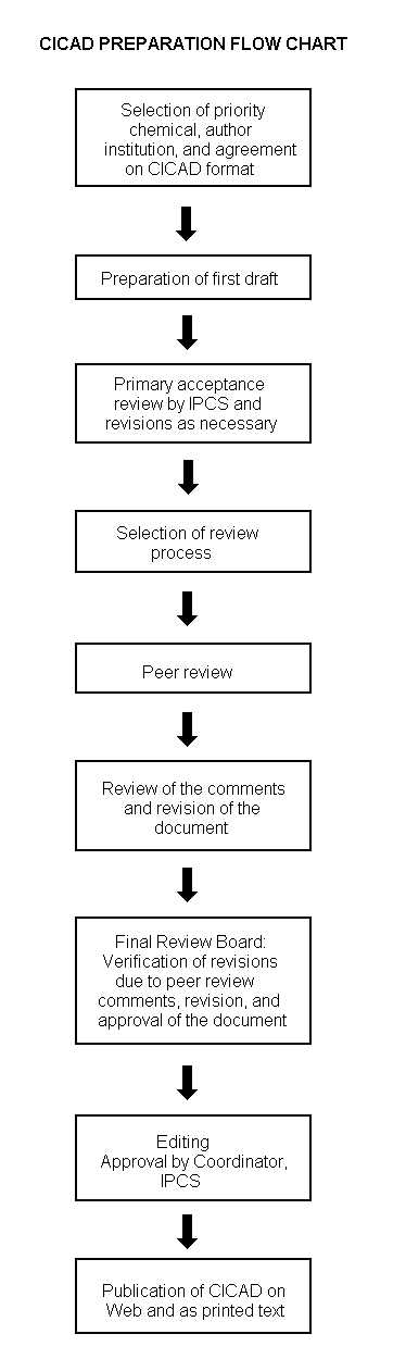
The flow chart on page 2 shows the procedures followed to produce a CICAD. These procedures are designed to take advantage of the expertise that exists around the world — expertise that is required to produce the high-quality evaluations of toxicological, exposure, and other data that are necessary for assessing risks to human health and/or the environment. The IPCS Risk Assessment Steering Group advises the Coordinator, IPCS, on the selection of chemicals for an IPCS risk assessment based on the following criteria:
Thus, it is typical of a priority chemical that
The Steering Group will also advise IPCS on the appropriate form of the document (i.e., EHC or CICAD) and which institution bears the responsibility of the document production, as well as on the type and extent of the international peer review.
The first draft is based on an existing national, regional, or international review. Authors of the first draft are usually, but not necessarily, from the institution that developed the original review. A standard outline has been developed to encourage consistency in form. The first draft undergoes primary review by IPCS to ensure that it meets the specified criteria for CICADs.
|
Advice from Risk Assessment Steering Group Criteria of priority:
Thus, it is typical of a priority chemical that
Special emphasis is placed on avoiding duplication of effort by WHO and other international organizations. A prerequisite of the production of a CICAD is the availability of a recent high-quality national/regional risk assessment document = source document. The source document and the CICAD may be produced in parallel. If the source document does not contain an environmental section, this may be produced de novo, provided it is not controversial. If no source document is available, IPCS may produce a de novo risk assessment document if the cost is justified. Depending on the complexity and extent of controversy of the issues involved, the steering group may advise on different levels of peer review:
|
The second stage involves international peer review by scientists known for their particular expertise and by scientists selected from an international roster compiled by IPCS through recommendations from IPCS national Contact Points and from IPCS Participating Institutions. Adequate time is allowed for the selected experts to undertake a thorough review. Authors are required to take reviewers’ comments into account and revise their draft, if necessary. The resulting second draft is submitted to a Final Review Board together with the reviewers’ comments. At any stage in the international review process, a consultative group may be necessary to address specific areas of the science.
The CICAD Final Review Board has several important functions:
Board members serve in their personal capacity, not as representatives of any organization, government, or industry. They are selected because of their expertise in human and environmental toxicology or because of their experience in the regulation of chemicals. Boards are chosen according to the range of expertise required for a meeting and the need for balanced geographic representation.
Board members, authors, reviewers, consultants, and advisers who participate in the preparation of a CICAD are required to declare any real or potential conflict of interest in relation to the subjects under discussion at any stage of the process. Representatives of nongovernmental organizations may be invited to observe the proceedings of the Final Review Board. Observers may participate in Board discussions only at the invitation of the Chairperson, and they may not participate in the final decision-making process.
The source document upon which this CICAD is based is the Toxicological profile for mercury (update), published by the Agency for Toxic Substances and Disease Registry of the US Department of Health and Human Services (ATSDR, 1999). Data identified as of January 1999 were considered in the source document. Data identified as of November 1999 were considered in the preparation of this CICAD. Information on the availability and the peer review of the source document is presented in Appendix 1. Information on the peer review of this CICAD is presented in Appendix 2. This CICAD was considered at a meeting of the Final Review Board, held in Helsinki, Finland, on 26–29 June 2000 and approved as an international assessment by mail ballot of the Final Review Board members on 27 September 2002. Participants at the Final Review Board meeting are presented in Appendix 3. The International Chemical Safety Cards for elemental mercury and six inorganic mercury compounds, produced by the International Programme on Chemical Safety, have also been reproduced in this document.
Mercury is a metallic element that occurs naturally in the environment. There are three primary categories of mercury and its compounds: elemental mercury, which may occur in both liquid and gaseous states; inorganic mercury compounds, including mercurous chloride, mercuric chloride, mercuric acetate, and mercuric sulfide; and organic mercury compounds. Organic mercury compounds are outside the scope of this document.
Elemental mercury is the main form of mercury released into the air as a vapour by natural processes.
Exposure to elemental mercury by the general population and in occupational settings is primarily through inhaling mercury vapours/fumes. The average level of atmospheric mercury is now approximately 3–6 times higher than the level estimated for preindustrial ambient air.
Dental amalgam constitutes a potentially significant source of exposure to elemental mercury, with estimates of daily intake from amalgam restorations ranging from 1 to 27 µg/day, the majority of dental amalgam holders being exposed to less than 5 µg mercury/day. Mercuric chloride, mercuric oxide, mercurous acetate, and mercurous chloride are, or have been, used for their antiseptic, bactericidal, fungicidal, diuretic, and/or cathartic properties. A less well documented use of elemental mercury among the general population is its use in ethnic or folk medical practices. These uses include the sprinkling of elemental mercury around the home and automobile. No reliable data are currently available to determine the extent of such exposure.
Analytical methods exist for the specific assessment of organic and inorganic mercury compounds; however, most available information on mercury concentrations in environmental samples and biological specimens refers to total mercury.
Intestinal absorption varies greatly among the various forms of mercury, with elemental mercury being the least absorbed form (<0.01%) and only about 10% of inorganic mercury compounds being absorbed. For elemental mercury, the main route of exposure is by inhalation, and 80% of inhaled mercury is retained. Inorganic mercury compounds may be absorbed through the skin in toxicologically relevant quantities.
Elemental mercury is lipid soluble and easily penetrates biological membranes, including the blood–brain barrier. Metabolism of mercury compounds to other forms of mercury can occur within the tissues of the body. Elemental mercury can be oxidized by the hydrogen peroxide–catalase pathway in the body to its inorganic divalent form. After exposure to elemental mercury or inorganic mercury compounds, the main route of excretion is via the urine. Determination of concentrations in urine and blood has been extensively used in the biological monitoring of exposure to inorganic forms of mercury; hair mercury levels do not reliably reflect exposure to elemental mercury or inorganic mercury compounds.
Neurological and behavioural disorders in humans have been observed following inhalation of elemental mercury vapour, ingestion or dermal application of inorganic mercury-containing medicinal products, such as teething powders, ointments, and laxatives, and ingestion of contaminated food. A broad range of symptoms has been reported, and these symptoms are qualitatively similar, irrespective of the mercury compound to which one is exposed. Specific neurotoxic symptoms include tremors, emotional lability, insomnia, memory loss, neuromuscular changes, headaches, polyneuropathy, and performance deficits in tests of cognitive and motor function. Although improvement in most neurological dysfunctions has been observed upon removal of persons from the source of exposure, some changes may be irreversible. Acrodynia and photophobia have been reported in children exposed to excessive levels of metallic mercury vapours and/or inorganic mercury compounds. As with many effects, there is great variability in the susceptibility of humans to the neurotoxic effects of mercury.
The primary effect of long-term oral exposure to low amounts of inorganic mercury compounds is renal damage. Inorganic forms of mercury have also been associated with immunological effects in both humans and susceptible strains of laboratory rodents, and an antibody-mediated nephrotic syndrome has been demonstrated through a variety of exposure scenarios. However, conflicting data from occupational studies preclude a definitive interpretation of the immunotoxic potential of inorganic forms of mercury.
Mercuric chloride has been shown to demonstrate some carcinogenic activity in male rats, but the data for female rats and for mice have been equivocal or negative. There is no credible evidence that exposure of humans to either elemental mercury or inorganic mercury compounds results in cancer.
There is convincing evidence that inorganic mercury compounds can interact with and damage DNA in vitro. Data from in vitro studies indicate that inorganic mercury compounds may induce clastogenic effects in somatic cells, and some positive results have also been reported in in vivo studies. The combined results from these studies do not suggest that metallic mercury is a mutagen.
Parenteral administration of inorganic mercury compounds is embryotoxic and teratogenic in rodents at sufficiently high doses. Animal data from studies in which the exposure pattern was similar to human exposure patterns and limited human data do not indicate that elemental mercury or inorganic mercury compounds are developmental toxicants at dose levels that are not maternally toxic.
Several studies are in agreement that mild subclinical signs of central nervous system toxicity can be observed among people who have been exposed occupationally to elemental mercury at a concentration of 20 µg/m3 or above for several years. Extrapolating this to continuous exposure and applying an overall uncertainty factor of 30 (10 for interindividual variation and 3 for extrapolation from a lowest-observed-adverse-effect level, or LOAEL, with slight effects to a no-observed-adverse-effect level, or NOAEL), a tolerable concentration of 0.2 µg/m3 was derived. In a 26-week study, a NOAEL for the critical effect, nephrotoxicity, of 0.23 mg/kg body weight was identified for oral exposure to mercuric chloride. Adjusting to continuous dosage and applying an uncertainty factor of 100 (10 for interspecific extrapolation and 10 for interindividual variation), a tolerable intake of 2 µg/kg body weight per day was derived. Use of a LOAEL of 1.9 mg/kg body weight in a 2-year study as a starting point yields a similar tolerable intake.
The chemical and physical properties vary with the form of mercury. Physical/chemical properties additional to those given below may be found in the International Chemical Safety Cards reproduced in this document (IPCS, 2000a–g): mercury (ICSC 0056); mercuric acetate (ICSC 0978); mercuric chloride (ICSC 0979); mercurous chloride (ICSC 0984); mercuric nitrate (ICSC 0980); mercuric oxide (ICSC 0981); and mercuric sulfate (ICSC 0982).
Elemental mercury (Hg0) (CAS No.
Elemental mercury is the most volatile form of mercury. It has a vapour pressure of 0.3 Pa at 25 °C and transforms into the vapour phase at typical room temperatures. It is relatively insoluble in water (56 µg/litre at 25 °C). Elemental mercury is soluble in lipids and nitric acid, soluble in pentane (2.7 mg/litre), insoluble in hydrochloric acid, and soluble in sulfuric acid upon boiling.
Inorganic mercury occurs as salts of its divalent and monovalent cationic forms. Of the large number of existing inorganic mercury compounds, those that have been extensively used in toxicology testing or that are in widespread use are briefly described below.
Mercuric chloride (HgCl2; CAS No.
Mercurous chloride (Hg2Cl2; CAS No.
Mercuric sulfide (HgS; CAS No.
Mercuric acetate (HgC4H6O4; CAS No.
The concentration of mercury can be accurately determined in air, water, soil, and biological samples (blood, urine, tissue, hair, breast milk, and breath) by a variety of analytical methods. Most of these methods are total mercury (inorganic plus organic mercury compounds) methods based on wet oxidation followed by a reduction step, but methods also exist for the separate quantification of inorganic mercury compounds and organic mercury compounds. Some analytical methods also require the predigestion of the sample prior to the reduction to elemental mercury. Since mercury is relatively volatile, care must be taken to avoid its loss during sample preparation and analysis. Labware should be thoroughly cleaned and acid-leached prior to use for trace-level analysis of mercury and its compounds, and due care should be taken to preclude the possibility of contamination by naturally occurring environmental mercury. Mercury readily forms amalgams with other metals (e.g., silver, zinc, tin), which can possibly contribute to mercury loss during analysis.
Mercury concentrations in humans and other mammals have been determined in blood, urine, body tissues, hair, breast milk, and umbilical cord blood. Most methods use atomic absorption spectrometry (AAS), atomic fluorescence spectrometry (AFS), or neutron activation analysis (NAA), although mass spectrometry (MS), spectrophotometry, and anodic stripping voltammetry (ASV) have also been employed. The most commonly used method is cold vapour (CV) AAS (ATSDR, 1999). Through CVAAS, mercury concentrations below the microgram per litre or microgram per kilogram level can be reliably (>76% recovery) measured through either direct reduction of the sample or reduction subsequent to predigestion. Electrothermal AAS has also been demonstrated to be highly sensitive and to produce excellent accuracy (ATSDR, 1999). Sub-microgram per litre or microgram per kilogram range sensitivity and excellent accuracy have also been demonstrated with gas chromatography (GC)/microwave-induced plasma atomic emission detection (Bulska et al., 1992). Recovery of >90% and high precision have also been obtained with AFS when the samples were predigested in a closed container in a microwave oven (Vermeir et al., 1991a,b). ASV and isotope-dilution spark source MS, which also require predigestion of the sample, have also produced high precision and accuracy (recoveries >90%). Inductively coupled plasma–atomic emission spectroscopy (ICP-AES) and ICP-MS can also be used to accurately (>90% recovery) determine total mercury in blood and urine with sub-microgram per litre sensitivity, but with less precision. In the case of blood mercury analysis, methods exist for the separation of organic and inorganic mercury (ATSDR, 1999). For analysis of urine mercury levels, expression of urinary mercury in units of micrograms of mercury per gram of creatinine is useful in adjusting for the variability in urine output or urine concentration.
As with the biological samples, a number of analytical methods can be used to determine mercury levels in air, water, soils, sediments, pharmaceuticals, and fish and other foods. In the case of complex samples, decomposition of the matrix and reduction of the mercury to its elemental form are required.
CVAAS and CVAFS have been shown to be sensitive (detection at low- to mid-nanogram-per-cubic-metre levels), accurate, and precise methods for monitoring mercury in air in the form of both vapours and suspended particulates (ATSDR, 1999). AFS, partially due to its low-nanogram-per-cubic-metre sensitivity and high accuracy and precision, is gaining in popularity (Horvat, 1996). The combination of AFS, AAS, and GC has been shown to be effective in speciating different organic and inorganic forms of mercury (Bloom & Fitzgerald, 1988).
Detection and quantification of mercury in aqueous media can be accomplished through a number of analytical methods. CVAAS, ASV, ICP-MS, ICP-AES, microwave-induced plasma AES, NAA, GC/AFS, high-performance liquid chromatography (HPLC) with ultraviolet detection, HPLC with electron capture detection, and spectrophotometry have all been successfully employed to quantify mercury in drinking-water, surface water, groundwater, snow, seawater, and wastewater effluents (ATSDR, 1999). CVAAS, because of its high sensitivity (sub-nanogram per litre) for mercury and high reliability, is the method preferred by the US Environmental Protection Agency (US EPA, 1994a,b) and the Association of Official Analytical Chemists (AOAC, 1984). While water samples generally do not require predigestion, mercury is usually reduced to the elemental state and preconcentrated prior to the actual analysis. As with samples from other media, a colorimetric method based on the formation of a coloured complex in the presence of mercury (Cherian & Gupta, 1990) may be used as a quick and simple field screen to detect mercury at mid-microgram-per-litre concentrations; however, without a predigestion method, organically bound mercury might not be fully measurable.
CVAAS, a sensitive and reliable technique that requires little sample preparation beyond matrix digestion and the reduction of mercury to its elemental form, is the most commonly used method of quantifying mercury in sediment, soils, and sludge (ATSDR, 1999). CVAFS with flow injection analysis, following microwave digestion, has been shown to have good precision and sensitivity in the mid-nanogram-per-kilogram range (Morales-Rubio et al., 1995). CVAAS and d.c. ASV (Lexa & Stulik, 1989) have been successfully used for testing organic and total mercury levels, respectively, in soil and/or sediment. For on-site screening, portable field X-ray fluorescence has been used to monitor soil contamination at low-milligram-per-kilogram levels (Grupp et al., 1989).
CVAAS, with its consistent high sensitivity and reliability, is one of the most common methods used to quantify mercury in fish, shellfish, other foods, and pharmaceuticals. Other methods successfully used include flameless AAS for mercury in fish, wine, and other food (ATSDR, 1999).
Mercury is a naturally occurring element (around 80 µg/kg) in the Earth’s crust. Over geological time, it has been distributed throughout the environment by natural processes, such as volcanic activity; fires; movement of rivers, lakes, and streams; oceanic upwelling; and biological processes. Since the advent of humans, and particularly since the industrial revolution of the late 18th and 19th centuries, anthropogenic sources have become a significant contributor to the environmental distribution of mercury and its compounds.
As with other components of the lithosphere, natural global cycling has always been a primary contributor to the presence of chemical elements in water, air, soils, and sediments. This process involves off-gassing of mercury from the lithosphere and hydrosphere to the atmosphere, where it is transported and deposited onto land, surface water, and soil. Major anthropogenic sources of mercury in the environment have been mining operations, industrial processes, combustion of fossil fuels (especially charcoal), production of cement, and incineration of municipal, chemical, and medical wastes. Point sources of anthropogenic mercury release, revolatilization from environmental media, sorption to soil and sediment particles, and bioaccumulation in the food webs contribute to further distribution and subsequent human exposure. The use of elemental mercury to capture gold particles as an amalgam has also contributed to the environmental burden of mercury and its compounds (Brito & Guimaraes, 1999; Grandjean et al., 1999). Dental amalgam fillings are the primary source of mercury exposure for the general population (Skare, 1995; Health Canada, 1997).
Mercury is transported in the environment by air and water, as well as by biological organisms through the food-chain. Off-gassed mercury vapour from the soil and water enters the air, where it may be transported and redistributed over the Earth’s surface. Upwelling along the continental shelves helps to bring minerals to the surface, where mercury can enter the air as a vapour, settle to the bottom sediment, be absorbed by phytoplankton, or be ingested by zooplankton, other microorganisms, or fish. Over geologic time, volcanic activity may bring mercury from below the Earth’s crust to the surface, where it may either enter the atmosphere as a vapour or be redistributed to soil or bodies of water.
In the environment, elemental mercury can combine with chlorine, sulfur, and other elements to form inorganic compounds. The most common naturally occurring forms of mercury found in the abiotic environment are metallic (elemental) mercury, mercuric sulfide, and the salts mercuric chloride and mercurous chloride.
Biotransformation of inorganic mercury to methylmercury by aqueous microorganisms is very important, as methylmercury bioaccumulates.
Over 90% of atmospheric mercury is elemental mercury vapour. Glass et al. (1991) indicated that mercury may travel as far as 2500 km in just 72 h. Estimates of airborne residence time range from 6 days (Andren & Nriagu, 1979) to 6 years (US EPA, 1984), before the mercury is redeposited in air or water by rainfall or other climatological conditions. Wet deposition is believed to be the primary means (accounting for approximately 66%) of removal of mercury from the atmosphere (Fitzgerald et al., 1991; Lindqvist, 1991a,b), although dry deposition may account for around 70% of total atmospheric deposition during the summer months (Lindberg et al., 1991). In remote areas in which there is no point source deposition of mercury from industrial sources, mercury in lake water is believed to be attributable to direct deposition from rainfall and/or leaching from bedrock by acid rain/snow (Hurley et al., 1991; Swain et al., 1992). Mercury vapour may also be removed from the atmosphere directly by binding to soil or water surfaces (US EPA, 1984).
Most of the mercury in groundwater is derived from atmospheric sources. Of the gaseous mercury that is dissolved in water, over 97% is elemental mercury (Vandal et al., 1991). However, elemental mercury will not remain as such in water for long; it will either combine to form some compound or rather rapidly re-enter the atmosphere and be redistributed in the environment.
In soil and in water, mercury can exist in either the monovalent or divalent forms as inorganic compounds. The particular valence state in which mercury exists in the environment (Hg0, Hg+, Hg2+) is dependent upon multiple factors, including the pH and redox potential of the particular medium and the strength of the ligands present. Mercury binds strongly to humic materials and sesquioxides, even at soil pH values greater than 4 (Blume & Brummer, 1991), although mercury sorption to soils generally decreases with increasing pH and/or chloride ion concentration (Schuster, 1991). Vaporization of mercury from soil has been associated with decreasing soil pH, with volatilization of soil mercury demonstrated at soil pH <3 (Warren & Dudas, 1992).
Most Hg2+ found in precipitation is bound to particulate matter (Meili et al., 1991), but its environmental transport and partitioning in surface waters and soils, once deposited, depend upon the specific mercury compound.
While in the soil or sediment, inorganic mercury may be adsorbed onto soil particles, where it is likely to remain bound unless consumed by organisms. Intake of elemental or inorganic mercury by aquatic microorganisms results in the biotransformation of those inorganic forms into methylmercury, which may be bioconcentrated in aquatic/marine animals in the food web from both water and food. Bioaccumulation in aquatic species is influenced by the pH (Ponce & Bloom, 1991) and the dissolved oxygen content (Wren, 1992).
The sorption of mercury to soil is dependent upon the organic content of the particular soil or sediment (Blume & Brummer, 1991), and mercury has been shown to bind tightly to the surface layer of peat (Lodenius & Autio, 1989). In water, both inorganic mercury and methylmercury bind tightly to organic particulates and may be distributed to other bodies of water or onto soils in such a bound form. The mobilization of mercury from soil or sediment particles to which it is sorbed may occur by either chemical or biological reduction to elemental mercury or microbial conversion to dimethylmercury (Andersson, 1979; Callahan et al., 1979; US EPA, 1984). Elemental mercury has been shown to be able to move through the top 3–4 cm of dry soil at atmospheric pressure (Eichholz et al., 1988).
A variety of mushroom species have been shown to contain elevated levels of mercury (Bressa et al., 1988; Kalac et al., 1991). The extent of bioaccumulation of mercury appears to be species-dependent (Kalac et al., 1991); the edible mushroom Pleurotus ostreatus has been found to bioaccumulate up to 140 times the concentration in the soil (Bressa et al., 1988). While mercury in the soil has been shown not to enter the shoots of peas, mercury does accumulate in the roots to a level comparable to that in the soil in which the plant is grown (Lindqvist, 1991a,b). Earthworms of the genus Lumbricus have been found to bioaccumulate mercury under both field and laboratory conditions in amounts dependent upon soil mercury concentration and duration of exposure (Cocking et al., 1994).
Atmospheric oxidation or reduction of elemental mercury vapour, the principal form of mercury in the air, may occur in the presence of dissolved ozone, hydrogen peroxide, hypochlorite, or organoperoxy compounds. In rainwater, mercury undergoes oxidation by ozone to Hg2+ and other forms. While mercury vapour may remain in the atmosphere for as long as 2 years, a rapid oxidation reaction may occur in clouds in the presence of ozone in just hours. By comparison, some inorganic forms of mercury, such as mercuric sulfide, which bind with atmospheric particles in the aerosol phase, are very stable. Some inorganic mercury compounds, such as mercuric hydroxide [Hg(OH)2], undergo rapid reduction to monovalent mercury by sunlight (Munthe & McElroy, 1992).
The primary process involved in the transformation of mercury in aqueous environments is biological conversion to organomercury compounds by a variety of microorganisms, mainly sulfur-reducing forms of anaerobic bacteria (Gilmour & Henry, 1991; Regnell & Tunlid, 1991).
The formation of methylmercury is enhanced at low pH and higher mercury concentrations in the sediment (Gilmore & Henry, 1991). Some yeast species (e.g., Candida albicans and Saccharomyces cerevisiae) are also capable of methylating mercury at lower pH and can reduce ionic mercury species to elemental mercury as well. Lakes that have been acidified by acid rain or industrial runoff favour the methylation of mercury, although such conditions also decrease the abundance of fish species, which biomagnify mercury in the food-chain. Anaerobic conditions (Regnell & Tunlid, 1991) and increasing dissolved organic carbon levels (Gilmour & Henry, 1991) both tend to substantially increase the methylation of mercury.
Photolysis of organic forms of mercury has also been shown to occur in water (Callahan et al., 1979), and the abiotic reduction of inorganic to elemental mercury has likewise been shown to occur, especially in the presence of soluble humic substances (Allard & Arsenie, 1991).
The transformation processes for the various forms of mercury that apply in water also occur in soil and sediment. Formation and breakdown of organic mercury compounds appear to be dependent upon the same microbial and abiotic processes as in water (Andersson, 1979), and the methylation of mercury is decreased by increasing chloride ion concentration (Olson et al., 1991), although the presence of chloride ions has been suggested to increase the rate of mercury release from sediments (Wang et al., 1991). In soil, the complexing of elemental mercury with chloride ion and hydroxide ion to form various mercury compounds is dependent upon pH, salt content, and soil composition.
The concentration of mercury in ambient air in the USA has been reported to range from 10 to 20 ng/m3, with higher concentrations being found in industrialized areas (US EPA, 1980). In Sweden, the concentration of elemental mercury in atmospheric air is lower, ranging from 2 to 6 ng/m3 (Brosset & Lord, 1991). Substantially higher levels (10–15 µg/m3) have been detected in ambient air near mercury mines, refineries, and agricultural fields treated with fungicides containing mercury.
Primarily due to anthropogenic sources, current average mercury levels in the atmosphere are about 3–6 times higher than the estimated levels in the preindustrial atmosphere (Mason et al., 1995), and continental mercury deposition in North America has increased 3.7-fold (an approximate annual increase of 2%) over the past 140 years (Swain et al., 1992).
Groundwater measured near the surface in remote areas of Wisconsin, USA, had total mercury concentrations of 2–4 ng/litre (Krabbenhoft & Babiarz, 1992). Total mercury concentrations in lakes and rivers in California, USA, ranged from 0.5 to 104 ng/litre (Gill & Bruland, 1990). Storm (1994) analysed 6856 samples of drinking-water collected from groundwater sources in the state of California and found that 27 of 225 positive detections from that sampling exceeded 2 µg/litre (mean mercury concentration of 225 positives was 6.5 µg/litre; range 0.21–300 µg/litre). The concentration of mercury in unpolluted marine waters has been estimated to be less than 2 ng/litre, in sharp contrast to an inshore coastal area near the industrial areas of New York Harbor, USA, where dissolved mercury concentrations up to 90 ng/litre have been measured (Fowler, 1990). In the United Kingdom, monitoring of drinking-water indicates that exceedences of 1 µg/litre are exceedingly rare.
Estimates of average daily intake of inorganic mercury (both mercury vapour and inorganic mercury compounds) by various routes in humans are summarized in Table 1.
Table 1: Estimated average daily intake (retention) of inorganic mercury.
|
Medium |
Intake (retention) (µg)a |
Reference |
|
|
Mercury vapour |
Inorganic mercury compounds |
||
|
Atmosphere |
0.04–0.2 (0.03–0.16)b |
0c |
IPCS, 1991 |
|
Food: Fish |
0 |
0.6d (0.06) |
IPCS, 1991 |
|
Food: Non-fish |
0 |
3.6 (0.36) |
IPCS, 1991 |
|
Drinking-water |
0 |
0.05 (0.005) |
IPCS, 1991 |
|
Dental amalgam |
1.2–27 (1–21.6) |
0 |
ATSDR, 1999 |
|
Total |
1.2–27 (1–22) |
4.3 (0.43) |
|
|
a |
Figures in parentheses are the amounts retained that were estimated from the pharmacokinetic parameters; i.e., 80% of inhaled vapour and 10% of inorganic mercury are retained. |
|
b |
Assumes an air concentration of 2–10 ng/m3 and a daily respiratory volume of 20 m3. |
|
c |
For the purposes of comparison, it is assumed that the atmospheric concentrations of species of mercury other than mercury vapour are negligible. |
|
d |
It is assumed that 20% of the total mercury in edible fish tissues is in the form of inorganic mercury compounds. It should be noted that fish intake may vary considerably between individuals and across populations. Certain communities whose major source of protein is fish may exceed this estimated inorganic mercury intake by an order of magnitude or more. |
There are a number of possible pathways for non-occupational exposure to inorganic forms of mercury. These include (1) eating fish or wild game near the top of the food-chain (i.e., larger fish, larger mammals) that have accumulated mercury (primarily methylmercury, but some inorganic mercury as well) in their tissues; (2) playing on or in contaminated surface soils; (3) playing with liquid mercury from broken electrical switches, thermometers, barometers, blood pressure monitors, etc.; or (4) bringing any liquid mercury or broken mercury device into the home, where vapours might build up in indoor air. Exposure from ambient air and drinking-water is usually minor.
Most human exposure to biologically significant amounts of elemental mercury occurs in the workplace. Workers in the chloralkali, electrical light bulb manufacturing, thermometer, and other industries where elemental mercury is utilized are exposed to levels much higher than the general population. Occupational mercury exposures generally occur when workers inhale elemental mercury vapours. Some dermal absorption may occur from skin contact with contaminated air, but the extent is low (less than 3% of the inhaled dose). Gold mining operations in Peru, Brazil, the Philippines, and less industrialized nations result in exposure for both miners and their families alike. Once mercury is used to amalgamate gold, the mercury is subsequently heated to melting in order to free the gold, resulting in high airborne levels of mercury. In some areas, this heating and separation process is conducted in the family home in order to ensure safeguarding of the gold product. Another exposure scenario for elemental mercury involves its use by children for play/entertainment purposes. Mercury available in school science laboratories or left over from industrial uses is occasionally taken by children and handled excessively. It is easily tracked from its initial location on shoes or clothing, and contamination may be spread to the home, automobile, or public buildings or transportation sources, creating a potential public health problem. The US Agency for Toxic Substances and Disease Registry has reported an increasing number of such cases reported to its Emergency Response Section of the Division of Toxicology in recent years (ATSDR, 1999; Nickle, 1999), with measured residential indoor air mercury concentrations of up to 2 mg/m3 (and subsequent exposures requiring medical intervention) resulting from child play activities with metallic mercury.
Elemental mercury has the ability to readily cross the placental barrier (see section 7). Thus, the developing fetus can be exposed to mercury from the pregnant woman’s body through the placenta. Infants may also be exposed to mercury from a nursing mother’s milk. Inorganic mercury — and to a lesser extent elemental mercury — will move into breast milk (Pitkin et al., 1976; Grandjean et al., 1995a,b). The mean concentration in breast milk, based upon review of existing data from a variety of countries, was reported by WHO (IPCS, 1990, 1991) to be 8 µg/litre; however, this value was based upon total mercury from all exposures and includes mercury resulting from ingestion of methylmercury in fish and other marine animals. A background level in milk attributable only to inorganic forms of mercury is not available.
Fish, aquatic mammals, and waterfowl used as food sources are important sources of mercury in some populations. In aquatic mammals, mercury concentrations in the tissues of predator species increase as one ascends the food-chain. Weihe et al. (1996) reported that muscle tissue of pilot whales (Globicephala melaena) caught in the Faroe Islands contains an average mercury concentration of 3.3 mg/kg, about half of which is inorganic mercury. Although May et al. (1987) reported that almost all of the mercury in fish is methylated, a more recent estimate is that approximately 20% of the total mercury in fish is in the inorganic form (IPCS, 1990). Among terrestrial mammals, those that consume fish or other mammals typically have higher body burdens of mercury than do vegetarian species. The highest concentrations of mercury are found in the liver and kidney, with successively smaller amounts being sequestered in the muscle and brain.
For more than a century and a half, silver/mercury amalgam fillings have been used in dental practice as the preferred tooth filling material. Such amalgams contain approximately 50% elemental mercury. Human studies and experiments in laboratory animals indicate that dental amalgam contributes significantly to mercury body burden in humans who have amalgam fillings (IPCS, 1991; US DHHS, 1993; Weiner & Nylander, 1995; Health Canada, 1997). Levels of mercury release for various dental procedures have been reported by Eley (1997).
Mercury released from amalgam fillings can take several forms: elemental mercury vapour, metallic ions, and/or fine particles (IPCS, 1991). Of the mercury vapour, some is exhaled, some is inhaled into the lungs and absorbed into the blood, some is retained in the vapour form in the saliva and swallowed together with amalgam particles, and some is oxidized to an ionic form and spit from the mouth or swallowed. Of that portion swallowed, only a small fraction would be expected to be absorbed through the gastrointestinal tract.
Barregard et al. (1995) investigated the relationship between amalgam fillings and mercury uptake and found that mercury uptake from dental amalgams is low. However, there is considerable variation between individuals, due primarily to gum chewing habits and bruxism, a rhythmic or spasmodic grinding of the teeth other than chewing and typically occurring during sleep.
Bjorkman et al. (1997) examined the mercury concentrations in saliva after removal of dental amalgam fillings in 10 human subjects. In saliva, there was an exponential decline in the mercury concentration during the first 2 weeks after amalgam removal (half-life of 1.8 days). Of 108 patients (all with amalgam dental fillings) presenting to an environmental toxicology service, the average salivary mercury level was 11 µg/litre (range <1–19 µg/litre) before chewing and 38 µg/litre (range 6–500 µg/litre) after chewing. Six of the 108 patients had salivary mercury concentrations above 100 µg/litre. Nonetheless, the gastrointestinal uptake of mercury seen in conjunction with removal of amalgam fillings appears to be low.
Higher levels of mercury exposure can occur in individuals who chew gum or show bruxism (Barregard et al., 1995; Enestrom & Hultman, 1995). Richardson (1995) reported a transient 5.3-fold increase in levels of mercury upon stimulation by chewing, eating, or tooth brushing. Sallsten et al. (1996) also reported over a 5-fold increase in plasma and urinary mercury levels (27 and 6.5 nmol/mmol creatinine versus 4.9 and 1.2 nmol/mmol creatinine, respectively) in a sample of 18 people who regularly chewed nicotine chewing gum (median values of 10 sticks per day for 27 months), compared with a control group. Higher-level short-term exposure has also been demonstrated in conjunction with restorative work on amalgam fillings (Taskinen et al., 1989).
Berdouses et al. (1995) studied mercury release from dental amalgams using an artificial mouth under controlled conditions of brushing and chewing and found that although the release of mercury during initial non-steady-state conditions was influenced by both the age of the amalgam and the amalgam type, the steady-state value of the mercury dose released by the amalgam was only 0.03 µg/day.
The contribution of dental amalgam fillings to daily intake of mercury has been estimated in a number of reports. Values generally in the range of 1–5 µg/day were estimated in the US population, although Sandborgh-Englund et al. (1998) estimated the daily dose of mercury from amalgam fillings to be from 5 to 9 µg/day in subjects with an average number of amalgams. Skare & Engqvist (1994) estimated the systemic uptake of mercury from amalgam in Swedish middle-aged individuals with a moderate amalgam load (30 surfaces) to be, on the average, 12 µg/day.
Halbach (1994) examined the data from 14 independent studies and concluded that the probable mercury dose from amalgam is less than 10 µg/day. When combined with the 2.6 µg/day background intake estimated by WHO (IPCS, 1990) for persons without amalgam fillings, the total daily intake from dental amalgam fillings and environmental sources is less than 12.6 µg.
Richardson et al. (1995) estimated total mercury exposure for Canadian populations of different ages to be 3.3 µg/day in toddlers (3–4 years old), 5.6 µg/day in children (5–11 years old), 6.7 µg/day in teens (12–19 years old), 9.4 µg/day in adults (20–59 years old), and 6.8 µg/day in seniors (aged 60+ years). Of this exposure, amalgam was estimated to contribute 50% to the total mercury in adults and 32–42% for other age groups. Estimates based on two independent models of exposure from amalgam alone were 1.1–1.7 µg/day in children, 1.9–2.5 µg/day in teens, 3.4–3.7 µg/day in adults, and 2.1–2.8 µg/day in seniors (Richardson, 1995).
The use of amalgam has been steadily declining and is expected to continue to decline due to improvements in dental hygiene and preventive care. In the 1970s, the use of amalgam restorations in the USA was 38% higher than it was in 1990 (96 million in 1990) (US DHHS, 1993). The use of dental amalgam has been on the decline in the United Kingdom as well. The annual replacement rate in National Health Service patients in England and Wales was 30 million amalgam restorations per year in 1986, compared with an estimated 12–13 million restorations in 1996.
A less well documented source of exposure to inorganic mercury among the general population is its use in ethnic religious, magical, and ritualistic practices and in herbal remedies. Mercury has long been used for medicinal purposes in Chinese herbal preparations and is also used in some Hispanic practices for medical and/or religious reasons, as well as in some Indian ethnic remedies (Kew et al., 1993). Espinoza et al. (1996) analysed 12 types of commercially produced herbal ball preparations used in traditional Chinese medicine. Mercury levels were found to range from 7.8 to 621.3 mg per ball. Since the minimum recommended adult dosage is two such balls daily, intake levels of up to 1.2 g of mercury (presumed to be mercury sulfide) might be a daily dosage.
Some religions have practices that may include the use of elemental mercury. Examples of these religions include Santeria (a Cuban-based religion that worships both African deities and Catholic saints), Voodoo (a Haitian-based set of beliefs and rituals), Palo Mayombe (a secret form of ancestor worship practised mainly in the Caribbean), and Espiritismo (a spiritual belief system native to Puerto Rico). Not all people who observe these religions use mercury, but when mercury is used in religious, folk, or ritualistic practices, exposure to mercury may occur both at the time of the practice and afterwards from breathing contaminated indoor air. Elemental mercury is sold in North America under the name "azogue" in stores called "botanicas." Botanicas are common in Hispanic and Haitian communities, where azogue may be sold as a herbal remedy or for spiritual practices. The elemental mercury is often sold in capsules or in glass containers. It may be placed in a sealed pouch to be worn on a necklace or carried in a pocket, or it may be sprinkled in the home or car. Some store owners may also suggest mixing azogue in bath water or perfume, and some people place azogue in devotional candles. The use of elemental mercury in a home or apartment not only threatens the health of the current residents, but also poses health risks to future residents who may unknowingly be exposed to further release of mercury vapours from contaminated floors, carpeting, or walls.
Mercuric chloride, mercuric oxide, mercuric iodide, mercurous acetate, and mercurous chloride are, or have been, used for their antiseptic, bactericidal, fungicidal, diuretic, and/or cathartic properties in Europe, North America, Australia, and elsewhere. Inorganic mercury compounds are also widely used in skin-lightening soaps and creams, due to the ability of the mercury cation to block the production of melanin pigment in the skin. Such uses have resulted in reports of toxicity in a number of cases (Millar, 1916; Warkany & Hubbard, 1953; Williams & Bridge, 1958; Barr et al., 1972; Tunnessen et al., 1987; Dyall-Smith & Scurry, 1990; Kang-Yum & Oransky, 1992). Al-Saleh & Al-Doush (1997) examined 38 different skin-lightening creams and found that 45% contained mercury levels above the US Food and Drug Administration limit of 1 mg/kg; two of the products had mercury concentrations over 900 mg/kg.
Forms of inorganic mercury have been used topically on a rather widespread basis for a variety of therapeutic uses. Cutaneous applications include treatment of infected eczema or impetigo (various mercury salts), treatment of syphilis (calomel), treatment of psoriasis (mercuric oxide or ammoniated mercury), and topical use of metallic mercury ointments (Bowman & Rand, 1980; Goodman Gilman et al., 1985; Bourgeois et al., 1986; O’Shea, 1990).
Previous uses of inorganic mercurials include laxatives (Wands et al., 1974). Such use has been abandoned in most industrialized nations due to the known toxicity of inorganic mercury compounds and the availability of equally or more effective, and less toxic, alternatives.
Inhalation is the primary route of entry into the body for elemental mercury, while oral exposure is the primary route for inorganic mercury salts. Dermal penetration is usually not a significant route of exposure to inorganic mercury.
Approximately 80% of inhaled elemental mercury is absorbed through the lungs by rapid diffusion. In contrast, only 0.01% of elemental mercury is absorbed through the gastrointestinal tract, possibly because of its enterogastric conversion to divalent mercury and subsequent binding to sulfhydryl groups. Dermal absorption of elemental mercury is limited. Hursh et al. (1989) estimated that dermal absorption contributes approximately 2.6% of the absorbed mercury following exposure to elemental mercury vapour in the air; the other 97.4% occurs through inhalation. Absorption of mercury vapour via olfactory nerves has also been proposed; however, Maas et al. (1996) has demonstrated that there is no relationship between mercury concentrations in lower parts of the brain and the amount of amalgam fillings in the mouth.
Sandborgh-Englund et al. (1998) evaluated the absorption, blood levels, and excretion of mercury in nine healthy volunteers (two males, seven females) exposed to mercury vapour in air at 400 µg/m3 for 15 min. This exposure corresponded to a dose of 5.5 nmol mercury/kg body weight. Samples of exhaled air, blood, and urine were collected for 30 days after exposure. The median retention of elemental mercury after 30 days was 69% of the inhaled dose. This corresponds to the estimated half-life of approximately 60 days for elemental mercury.
For inorganic mercuric compounds, absorption via the lungs is low, probably due to deposition of particles in the upper respiratory system and subsequent clearance by the mucociliary escalator (Friberg & Nordberg, 1973).
The extent of transport of inorganic mercury across the intestinal tract may depend on its solubility (Friberg & Nordberg, 1973) and/or how easily the compound dissociates in the lumen to become available for absorption (Endo et al., 1990). Absorption of mercurous compounds is less likely than absorption of mercuric forms, probably because of solubility (Friberg & Nordberg, 1973).
Using whole-body retention data, estimated mercuric chloride absorptions of 3–4%, 8.5%, and 6.5% were calculated for single oral doses of 0.2–12.5 mg/kg body weight, 17.5 mg/kg body weight, and 20 mg/kg body weight, respectively, in rats (Piotrowski et al., 1992). However, also using whole-body retention data to indicate absorption, an estimated absorption of 20–25% was calculated from single oral doses of 0.2–20.0 mg mercury/kg body weight as mercuric chloride in mice by comparing retention data after oral and intraperitoneal dosing and taking excretion and intestinal reabsorption into account (Nielsen & Andersen, 1990).
The rate of oral absorption of mercuric mercury compounds in laboratory rodents has been shown to be dependent on intestinal pH (Endo et al., 1990), age, and diet (Kostial et al., 1978). One-week-old suckling mice absorbed 38% of the orally administered mercuric chloride, whereas adult mice absorbed only 1% of the dose on standard diets. Nutritional status might also contribute to the intestinal absorption of Hg2+, through competition with nutritionally essential divalent cations (e.g., Cu2+, Zn2+) that might have insufficient body stores.
Mercurous and mercuric salts have also been reported to be absorbed through the skin of animals (Schamberg et al., 1918; Silberberg et al., 1969), but no quantitative data are available. Indirect evidence of dermal absorption in humans is provided by clinical case-studies in which mercury intoxication was reported in individuals following dermal application of ointments that contained inorganic mercury salts (Bourgeois et al., 1986; De Bont et al., 1986; Kang-Yum & Oransky, 1992). Urine samples from young women using skin-lightening creams containing 5–10% mercuric ammonium chloride had a mean mercury concentration of 109 µg/litre, compared with 6 µg/litre for urine samples from women who had discontinued use and 2 µg/litre for women who had never used the creams (Barr et al., 1973).
Mercurous chloride laxative (calomel) ingested over a long period may produce toxic effects on the kidneys, gastrointestinal tract, and central nervous system (Wands et al., 1974). While insoluble mercurous chloride is not normally that readily absorbed, small amounts may be converted to mercuric ion, which is more likely to be absorbed, in the lumen of the intestine. In addition, the mercurous ion that is absorbed is subsequently oxidized to mercuric ion, which may induce cellular toxicity by binding to intracellular sulfhydryl groups.
The lipophilic nature of elemental mercury results in its distribution throughout the body. Elemental mercury dissolves in the blood upon inhalation, and some remains unchanged (Magos, 1967). Elemental mercury in the blood is oxidized to its divalent form in the red blood cells (Halbach & Clarkson, 1978). The divalent cation exists as a diffusible or non-diffusible form. The non-diffusible form exists as mercuric ions that bind to protein and are held in high-molecular-weight complexes, existing in equilibrium with the diffusible form. In the plasma, the mercuric ion is predominantly non-diffusible and binds to albumin and globulins (Clarkson et al., 1961; Berlin & Gibson, 1963; Cember et al., 1968).
The high lipophilicity of elemental mercury in solution in the body allows it to readily cross the blood–brain and placental barriers (Clarkson, 1989). In mice, the uptake of mercury across the placenta appears to increase as gestation progresses (Dencker et al., 1983). Levels of mercury in the fetus are approximately 4 times higher after exposure to elemental mercury vapour than after mercuric chloride administration for mice and 10–40 times higher for rats (Clarkson et al., 1972). The transport of mercuric ion is limited at the placental barrier by the presence of high-affinity binding sites (Dencker et al., 1983).
Mercury distributes to all tissues and reaches peak levels within 24 h, except in the brain, where peak levels are achieved within 23 days (Hursh et al., 1976). The longest retention of mercury after inhalation of mercury vapour occurs in the brain (Takahata et al., 1970). Japanese workers who died 10 years after their last exposure to elemental mercury vapours still had high residual levels of mercury in their brains (Takahata et al., 1970). Villegas et al. (1999) found accumulation of mercury within neuronal perykaria of the supraoptic and paraventricular nuclei, as well as deposits in neurosecretory neurons and axon terminals of the neurohypophysis, in rats administered mercuric chloride in drinking-water.
In human volunteers who inhaled a tracer dose of elemental mercury vapour for 20 min, approximately 2% of the absorbed dose was found per litre of whole blood after the initial distribution was completed (Cherian et al., 1978). Distribution to the red blood cells was complete after 2 h, but plasma distribution was not complete until after 24 h. The mercury concentration in red blood cells was twice that measured in the plasma. This ratio persisted for at least 6 days after exposure.
While the primary organs of mercury deposition following inhalation exposure to elemental mercury vapours are the brain and kidney, the extent of deposition is dependent upon the duration of exposure and, to a greater extent, the concentration to which the organism is exposed. In rats exposed to mercury vapour concentrations of 10–100 µg/m3 6 h/day, 5 days/week, from the 4th through 11th weeks of life, measurable amounts of mercury were found in the blood, hair, teeth, kidney, brain, lung, liver, spleen, and tongue, with the kidney cortex having the highest mercury concentration (Eide & Wesenberg, 1993). Rothstein & Hayes (1964) also reported the kidney to be a major organ for mercury deposition following inhalation exposure to elemental mercury vapour. Exposure to mercury stimulates the production of metallothionein in the kidney, which in turn increases the amount of mercuric ion binding (Piotrowski et al., 1973; Cherian & Clarkson, 1976).
In contrast, in another study, a 4-h exposure of mice to elemental mercury vapour produced the highest mercury retention in the brain compared with other organs (Berlin et al., 1966). Exposure of rats to 1 mg/m3 elemental mercury vapour for 24 h/day every day for 5 weeks or 6 h/day, 3 days/week, for 5 weeks resulted in mean mercury brain concentrations of 5.03 and 0.71 µg/g, respectively (Warfvinge et al., 1992). Mercury was found primarily in the neocortex, basal nuclei, and cerebellar Purkinje cells. In mice exposed to elemental mercury vapour at a concentration of 8 mg/m3 for 6 h/day for 10 days, higher mercury levels were found in the grey than in the white brain matter (Cassano et al., 1966, 1969). Mercury also accumulates in several cell types (ganglion cells, satellite cells, fibroblasts, and macrophages) populating the dorsal root ganglia (Schionning et al., 1991) and has been detected in dorsal root neurons and satellite cells of primates exposed for 1 year to mercury through amalgam in dental fillings or maxillary bone (Danscher et al., 1990).
Compared with elemental mercury, the amount of inorganic divalent mercury that crosses the blood–brain and placental barriers is much lower, because of poor lipid solubility (Clarkson, 1989; Inouye & Kajiwara, 1990). In contrast, the liver and kidneys accumulate inorganic mercury readily (Yeoh et al., 1986, 1989; Nielsen & Andersen, 1990). Sin et al. (1983) found the kidney to have the highest mercury levels following repeated oral exposure of mice to mercuric chloride (4–5 mg mercury/kg body weight) for 2–8 weeks.
The available evidence indicates that the metabolism of all forms of inorganic mercury is similar for humans and laboratory mammals. Once absorbed, elemental and inorganic mercury enter an oxidation–reduction cycle. Elemental mercury is oxidized to the divalent inorganic cation in the red blood cells and lungs. Evidence from animal studies suggests the liver as an additional site of oxidation. Absorbed divalent cation from exposure to mercuric mercury compounds can, in turn, be reduced to the metallic or monovalent form and released as exhaled elemental mercury vapour (ATSDR, 1999).
Once inhaled into the lungs, elemental mercury vapours rapidly enter the bloodstream. The dissolved vapour can undergo rapid oxidation, primarily in the red blood cells, to its inorganic divalent form by the hydrogen peroxide–catalase pathway (Halbach & Clarkson, 1978; Clarkson, 1989). It is believed that the rate of oxidation is dependent on (1) concentration of catalase in the tissue; (2) endogenous production of hydrogen peroxide; and (3) availability of mercury vapour at the oxidation site (Magos et al., 1978). Nielsen-Kudsk (1973) found that stimulation of hydrogen peroxide production in red blood cells increased the uptake of mercury vapours in red blood cells. The mercury content in the blood is proportionately higher after a low dose than after a high dose, indicating that a higher proportion of the lower dose is oxidized (Magos et al., 1989). The hydrogen peroxide–catalase pathway in red blood cells may become saturated at higher dose levels (Magos et al., 1989). This oxidation pathway of elemental mercury can be inhibited by ethanol, since ethanol is a competitive substrate for the hydrogen peroxide catalase and thus can block mercury uptake by red blood cells (Nielsen-Kudsk, 1973). However, two different variants of acatalasaemia/hypocatalasaemia exist that lead to deficient activity of this enzyme, quite possibly resulting in a particularly susceptible human subpopulation (Paul & Engstedt, 1958; Aebi, 1967).
The oxidation of elemental mercury may also occur in the brain, liver (adult and fetal) (Magos et al., 1978), lungs (Hursh et al., 1980), and probably all other tissues to some degree (Clarkson, 1989). In the brain, unoxidized elemental mercury can be oxidized and become trapped in the brain, because it is more difficult for the divalent form to exit the brain via the blood–brain barrier. Autoradiographic studies suggest that mercury oxidation also occurs in the placenta and fetus (Dencker et al., 1983), although the extent of oxidation is not known.
Elimination of mercury occurs primarily through the urine and faeces, with the expired air, sweat, and saliva contributing to a much lesser extent.
The urine and faeces are the main excretory pathways of elemental mercury and inorganic mercury compounds in humans, with an absorbed dose half-life of approximately 1–2 months (Clarkson, 1989). After a short-term high-level mercury exposure in humans, urinary excretion accounts for 13% of the total body burden. After long-term exposure, urinary excretion increases to 58%. Exhalation through the lungs and secretion in saliva, bile, and sweat may also contribute a small portion to the excretion process (Joselow et al., 1968; Lovejoy et al., 1974). Humans inhaling mercury vapour for less than an hour expired approximately 7% of the retained dose of mercury (Hursh et al., 1976; Cherian et al., 1978). Inorganic mercury is also excreted in breast milk (Yoshida et al., 1992). The overall rate of elimination of inorganic mercury from the body is the same as the rate of elimination from the kidney, where most of the body burden is localized. In a sample of 1107 individuals from 15 countries around the world, Goldwater (1972) reported the following urinary mercury levels for subjects who had no known occupational, medicinal, or other exposure to mercury: <0.5 µg/litre — 78%; <5 µg/litre — 86%; <10 µg/litre — 89%; <15 µg/litre — 94%; <20 µg/litre — 95%.
Elimination from the blood and the brain is thought to be a biphasic process, with an initial rapid phase in which the decline in the body burden is associated with high levels of mercury being cleared from tissues, followed by a slower phase with mercury clearance from the same tissues (Takahata et al., 1970). An even longer terminal elimination phase is also possible because of accumulation or persistence of mercury, primarily in the brain (Takahata et al., 1970). Following a single oral dose of divalent mercury in 10 volunteers, 85% of the 203Hg activity was excreted within 4–5 days, predominantly in the faeces (Rahola et al., 1973), consistent with the low intestinal absorption of the divalent cation.
In a study of former chloralkali workers exposed to elemental mercury vapour for 2–18 years (median 5 years), Sallsten et al. (1993) found that the elimination of mercury in urine was well characterized by a one-compartment model, with an estimated half-life of 55 days. For high-level exposure to inorganic divalent mercury, the urine is probably the major elimination route (Inouye & Kajiwara, 1990), with a half-life similar to that of elemental mercury (Clarkson, 1989).
Suzuki et al. (1992) estimated the elimination half-life from urine to be 25.9 days following a short-term exposure to a high level of mercuric chloride (13.8 mg/kg body weight) (Suzuki et al., 1992). Using a two-compartment model, elimination half-lives in urine of workers exposed for 20–45 h to >0.1 mg/m3 of elemental mercury vapour were estimated to be 28 and 41 days for a fast and slow phase, respectively (Barregard et al., 1992).
Age is a factor in the elimination of mercury in rats following inorganic mercury exposure, with younger rats demonstrating significantly higher retention than older rats. This age-dependent difference in the rate of mercury excretion may reflect differences in the sites of mercury deposition (i.e., hair, red blood cells, skin) (Yoshida et al., 1992).
Urine samples are considered to be the best determinant of body burden of mercury from long-term exposure to elemental and inorganic mercury. Blood samples are useful primarily in cases of short-term, higher-level exposures to these forms, but are not as reliable as an indicator of total body burden in longer-term exposures. Most analytical methods do not differentiate between inorganic and organic mercury; thus, total mercury concentrations in blood reflect body burden of total mercury. Inorganic forms of mercury are not excreted to any significant amount in scalp hair, making hair an inappropriate biomarker of inorganic mercury exposure.
Occupational studies show that recent mercury exposure is reflected in blood and urine (IPCS, 1991; Naleway et al., 1991). However, at low exposure levels (<0.05 mg mercury/m3), correlation with blood or urine mercury levels is low (Lindstedt et al., 1979). Blood levels of mercury peak sharply during and soon after short-term exposures, indicating that measurements of blood mercury levels should be made soon after exposure (Cherian et al., 1978). The half-life of mercury in the blood is only 3 days, attesting to the importance of taking blood samples as soon after exposure as possible. In the case of low-level long-term exposure, urine samples provide the best indicator of body burden.
Urinary mercury measurement is reliable and simple and provides rapid identification of individuals with elevated mercury levels (Naleway et al., 1991). It is a more appropriate marker of exposure to inorganic mercury, since organic mercury represents only a small fraction of urinary mercury. Urinary mercury levels have been found to correlate better with exposure than blood inorganic mercury concentrations following long-term, low-level occupational exposure to elemental mercury vapour (Yoshida, 1985). There may be marked diurnal variation in the urinary concentration of mercury (Schaller, 1996).
Based on a systematic review of high-quality studies, the International Commission on Occupational Health and the International Union of Pure and Applied Chemistry Commission on Toxicology estimated that a mean value of 2 µg/litre was the background blood level of mercury in persons who do not eat fish (Nordberg et al., 1992). These levels are "background" in the sense that they represent the average levels in blood in the general population and are not associated with a particular source of mercury exposure. However, the intra- and interindividual differences in these biomarkers are substantial, possibly due to dental amalgam (urine) and ingestion of contaminated fish (blood) (Verschoor et al., 1988; IPCS, 1991).
Several studies have reported a correlation between airborne mercury and mercury in blood and urine; however, results vary, and it is not known whether the ratio between concentrations in urine and blood is constant at different exposure levels (Smith et al., 1970; Lindstedt et al., 1979; Roels et al., 1987). Limiting the analysis to studies in which the exposure had been assessed using personal breathing zone mercury measurements, it was estimated that in continuous 8 h/day occupational exposure, an airborne mercury concentration of 1 mg/m3 leads to an average urinary mercury concentration of 1.4 mg (7 µmol)/litre (variation between individual studies, 0.7–2.3 mg [3.5–11.5 µmol]/litre; seven studies) and to an average blood mercury concentration of 0.48 mg (2.4 µmol)/litre (0.17–0.81 mg [0.85–4.0 µmol]/litre; six studies) (Cross et al., 1995).
Relationships of urinary and blood mercury concentrations with signs and symptoms of exposure are less clear. Exposure to elemental and/or other inorganic forms of mercury can be verified by examining the urinary mercury concentration. Urinary mercury concentrations normally expected in an asymptomatic population would be <10 µg/litre (Goldwater, 1972; ATSDR, 1999). Background levels of urinary mercury, adjusted for creatinine, in an unexposed population are generally expected to be 5 µg mercury/g creatinine (Gerhardsson & Brune, 1989; IPCS 1991; Schaller, 1996).
Exposure of rats for 2 h to an elemental mercury vapour concentration of 27 mg/m3 resulted in substantial mortality (20 of 32 rats died prior to their scheduled sacrifice) (Livardjani et al., 1991). A variety of respiratory effects were reported, including dyspnoea, lung oedema, necrosis of the alveolar epithelium, hyaline membranes, and occasional lung fibrosis.
Ashe et al. (1953) exposed rabbits intermittently to elemental mercury vapour at 28.8 mg/m3 for periods of up to 30 h. One of two rabbits exposed to this mercury vapour concentration for 30 h died, while no deaths occurred in rabbits exposed to the same concentration for 20 h or less. Marked cellular degeneration with some necrosis of heart tissue was observed in rabbits following longer intermittent exposures, while only mild to moderate pathological changes were seen in 1- to 4-h exposures. Gastrointestinal effects ranged from mild pathological changes to marked cellular degeneration, and some necrosis of the colon was observed following exposure for 4–30 h. Hepatic effects after exposure for 6–30 h ranged from moderate pathological changes (unspecified) to severe liver necrosis. Renal effects ranged from marked cellular degeneration to tissue destruction and widespread necrosis: moderate pathological changes were seen after a 1-h exposure; as the duration of exposure increased to 30 h, extensive cell necrosis in the kidney became evident (Ashe et al., 1953).
Pulmonary congestion was observed in rats exposed to an elemental mercury vapour concentration of 1 mg/m3 for 100 h continuously per week for 6 weeks (Gage, 1961).
Effects in different organs were reported in a study in which rabbits were exposed to elemental mercury vapour at 6 mg/m3 for 7 h/day, 5 days/week, for 1–11 weeks (Ashe et al., 1953). Respiratory effects were described as unspecified mild to moderate pathological changes. Exposures to 0.86–6 mg/m3 of mercury vapour for periods ranging from 2 to 12 weeks also resulted in mild to moderate pathological changes (unspecified) in the hearts of rabbits. Exposure to 6 mg/m3 for 1–5 weeks resulted in mild to moderate liver pathology, while effects ranging from moderate pathological changes to marked cellular degeneration and some necrosis in the liver occurred with exposure to 6 mg/m3 for 6–11 weeks.
Exposure to 0.86 mg/m3 for 12 weeks induced moderate pathological kidney changes that were reversible with termination of exposure. Exposure to 6 mg/m3 for up to 11 weeks produced effects that ranged from mild pathological changes to marked cellular degeneration and widespread necrosis (Ashe et al., 1953). (The LOAEL for renal effects was 0.86 mg/m3.) Dense deposits in tubule cells and lysosomal inclusions in the renal tubular epithelium were evident following exposure of rats to mercury vapour at 3 mg/m3 for 3 h/day, 5 days/week, for 12–42 weeks (Kishi et al., 1978).
Exposure of rats, rabbits, and dogs to metallic mercury vapour concentrations of 0.1 mg/m3 for 7 h/day, 5 days/week, for 72–83 weeks resulted in no microscopic evidence of kidney damage in any exposed animal. (Only two dogs were tested in this study.) In a study with limited reporting and no control group (Druckrey et al., 1957), local sarcomas were reported in 5 out of 39 BDIII and BDIV rats after two intraperitoneal injections of metallic mercury with lifelong follow-up.
The only data concerning the potential genotoxicity of elemental mercury are on humans and are presented in section 9.10.
Adult female rats exposed to an elemental mercury vapour concentration of 2.5 mg/m3 for 3 weeks prior to fertilization and during gestational days 7–20 had a decrease in the number of living fetuses relative to controls (Baranski & Szymczyk, 1973). Although all pups born to the exposed dams died by the 6th day after birth, no difference in the occurrence of developmental abnormalities was observed between exposed and control groups.
Exposure of pregnant Sprague-Dawley rats to an elemental mercury vapour concentration of 1.8 mg/m3 for 1.5 h/day during gestation days 14–19 caused alterations in both spontaneous and learned behaviours in the offspring (Fredriksson et al., 1996), manifested as hyperactivity, significantly impaired spatial learning, and deficits in adaptive behaviour. There were no lower dosages tested in this study. Hyperactivity and significantly impaired spatial learning were also seen in the offspring of female Sprague-Dawley rats exposed to 0.05 mg/m3 (LOAEL; no lower doses tested) for 1 or 4 h/day during gestation days 11–17 (Fredriksson et al., 1992).
In neurobehavioural tests conducted on the offspring of mother squirrel monkeys that had been exposed during the last two-thirds or more of gestation to mercury vapour concentrations of 0.5 (LOAEL) or 1.0 mg/m3 for 4 or 7 h/day, 5 days/week, long-term effects included instability in lever-press durations and steady-state performance under concurrent schedules of reinforcement, as well as aberrant transitions (Newland et al., 1996). No other exposure levels were examined in this study.
Exposure of genetically susceptible mice to mercury vapour for a period of 10 weeks resulted in an autoimmune response manifested as a general stimulation of the immune system, with hyperimmunoglobulinaemia, antinucleolar-fibrillarin autoantibodies, and glomerular disease, accompanied by vascular immune complex deposits (Warfvinge et al., 1995).
Marked cellular degeneration and widespread necrosis were observed in the brains of rabbits following exposures to elemental mercury vapour at 28.8 mg/m3 for durations ranging from 2 to 30 h, whereas exposure of rabbits to mercury vapour at 6 mg/m3 for periods ranging from 1 to 11 weeks produced effects ranging from mild, unspecified pathological changes to marked cellular degeneration and some necrosis in the brain. More serious degenerative changes were observed at longer exposure durations (i.e., 8 and 11 weeks). Mild to moderate pathological changes were observed in the brains after exposure to 0.86 mg/m3 for 12 weeks (Ashe et al., 1953). Fukuda (1971) observed slight tremors and clonus in two of six rabbits exposed for 13 weeks to an elemental mercury vapour concentration of 4 mg/m3. (Neurobehavioural deficits observed in the offspring of mother monkeys and rats that were exposed to mercury vapours during gestation were previously described in section 8.1.5.)
Exposure of neonatal rats to elemental mercury vapour at 0.05 mg/m3 for 1 or 4 h/day for 1 week during a period of rapid brain growth (postpartum days 11–17) resulted in subtle behavioural changes when the rats were tested at 4 and 6 months of age (Fredriksson et al., 1992). The severity of effect was directly related to the duration of individual exposures.
Oral LD50s for rats exposed to mercuric chloride ranged from 25.9 to 77.7 mg mercury/kg body weight (Kostial et al., 1978). Haematological, hepatic, and renal effects were reported in rats and/or mice administered sublethal single doses of mercuric chloride (Nielsen et al., 1991; Lecavalier et al., 1994).
Groups of 10 female Sprague-Dawley rats administered a single gavage dose of mercuric chloride at 7.4 or 9.2 mg mercury/kg body weight in water showed significant decreases in haemoglobin, erythrocytes, and haematocrit at autopsy. A significant decrease in serum protein and calcium was also reported for the low-dose mercury group only (Lecavalier et al., 1994).
Lactate dehydrogenase activity was significantly decreased in female Sprague-Dawley rats (10 animals per dosage group) given single gavage (in water) mercuric chloride doses of either 7.4 or 9.2 mg/kg body weight (Lecavalier et al., 1994). Renal effects seen in this study included mild to moderate morphological changes consisting of protein casts, cellular casts, and interstitial sclerosis in both treatment groups. Female Bom:NMRI mice given a single gavage dose of mercuric chloride at 10 mg mercury/kg body weight showed minor renal tubular damage and rapid regeneration of the tubular epithelium (Nielsen et al., 1991), while proximal tubular necrosis was seen at 20 mg/kg body weight. No renal effects were seen at 5 mg/kg body weight in the same study.
Male rats given gavage doses of mercuric chloride at 14.8 mg mercury/kg body weight, 5 days/week for 2 weeks, appeared to be slightly more sensitive to the lethal effects of mercuric chloride than female rats, with two of five male rats (but no females) dying. Mice were slightly less sensitive, with no deaths at 14.8 mg mercury/kg body weight, death in one of five males at 29 mg mercury/kg body weight, and deaths in five of five males and four of five females at 59 mg mercury/kg body weight when administered by gavage over the same period (NTP, 1993).
Forceful and laboured breathing, bleeding from the nose, and other unspecified respiratory difficulties were seen in rats after dietary exposure to 2.2 mg mercury/kg body weight per day as mercuric chloride for 3 months (Goldman & Blackburn, 1979).
Oral exposure of mice to 59 mg mercury/kg body weight per day as mercuric chloride, 5 days/week for 2 weeks, resulted in inflammation and necrosis of the glandular stomach (NTP, 1993).
Increases in hepatic lipid peroxidation and decreases in glutathione peroxidase were observed in rats orally exposed to an unspecified dose of mercuric chloride for 30 days (Rana & Boora, 1992), and absolute liver weight decreases were seen in animals receiving doses equivalent to 10 mg/kg body weight per day in the diet for 4 weeks (Jonker et al., 1993). The LOAEL for hepatic effects was 10 mg/kg body weight per day.
Male rats exposed for 14 days to gavage doses of 0.93, 1.9, 3.7, 7.4, or 14.8 mg mercury/kg body weight per day as mercuric chloride showed a significant increase in the absolute and relative kidney weights of males beginning at the 1.9 mg/kg body weight per day dose level. An increased incidence of tubular necrosis was observed in rats exposed to at least 3.7 mg/kg body weight per day, with severity increasing with increasing dosages. Increases in urinary levels of alkaline phosphatase, aspartate aminotransferase, and lactate dehydrogenase were also observed at 3.7 mg mercury/kg body weight per day; at 7.4 mg mercury/kg body weight per day, increased urinary γ-glutamyltransferase activity was also observed (NTP, 1993).
Increased absolute and relative kidney weights were seen in female Wistar rats exposed to a dietary intake level of mercuric chloride at 1.1 mg/kg body weight per day (the lowest exposure level studied) for 4 weeks (Jonker et al., 1993). The NOAEL for males in this study was 1 mg/kg body weight per day; the LOAEL was 8 mg/kg body weight per day for increased liver weights and slight histopathological changes to the renal cortex. For females, the LOAEL for renal effects from these studies was 1.1 mg/kg body weight per day; a reliable NOAEL could not be determined.
In a 26-week study in which groups of 10 Fischer-344 rats of each sex received 0, 0.312, 0.625, 1.25, 2.5, or 5 mg mercuric chloride/kg body weight (0, 0.23, 0.46, 0.93, 1.9, or 3.7 mg mercury/kg body weight per day) in deionized water by gavage, a significant increase in the severity of nephropathy (i.e., dilated tubules with hyaline casts, foci of tubular regeneration, and thickened tubular basement membrane) was observed in male rats given the dose of 0.93 mg/kg body weight per day. The nephropathy was minimal in the two low-dose groups (NTP, 1993). In females, kidney effects, which were mild, were observed at the highest dose only. The absolute and relative kidney weights were increased in treated males and in females at doses of >.46 mg/kg body weight per day. A NOAEL from this study was identified at 0.23 mg/kg body weight per day.
Mice given gavage doses of mercuric chloride 5 days/week for 2 weeks showed an increase in absolute and relative kidney weights at 3.7 mg mercury/kg body weight per day and acute renal necrosis at 59 mg mercury/kg body weight per day (NTP, 1993). Male mice receiving mercuric chloride in their drinking-water for 7 weeks showed slight degeneration of the tubular epithelial cells (nuclear swelling) at 2.9 mg mercury/kg body weight per day and minimal renal nephropathy (dilated tubules with either flattened eosinophilic epithelial cells or large cytomegalic cells with foamy cytoplasm) at 14.3 mg/kg body weight per day (Dieter et al., 1992).
When groups of 10 B6C3F1 mice of each sex received 0, 1.25, 2.5, 5, 10, or 20 mg mercuric chloride/kg body weight per day (0, 0.93, 1.9, 3.7, 7.4, or 14.8 mg mercury/kg body weight per day) in deionized water by gavage for 26 weeks (males) or 27 weeks (females), the incidence and severity of cytoplasmic vacuolation of renal tubular epithelium increased in males exposed to at least 3.7 mg/kg body weight per day (NTP, 1993).
Increased relative adrenal weights were observed in male rats fed diets containing mercuric chloride equivalent to 20 mg mercury/kg body weight per day for 4 weeks. Females had decreased absolute adrenal weights at 22.2 mg/kg body weight per day (Jonker et al., 1993). Several other studies have observed effects on the thyroid after medium-term exposure to divalent mercuric salts (Goldman & Blackburn, 1979; Agrawal & Chansouria, 1989; Sin et al., 1990; Sin & Teh, 1992). The LOAEL for adrenal effects was 20 mg/kg body weight per day.
A number of animal studies have reported decreases in body weight or body weight gain after ingestion of mercuric chloride (Chang & Hartmann, 1972a; Dieter et al., 1992; NTP, 1993). Jonker et al. (1993) reported decreased body weights in male and female Wistar rats of 21% and 27%, respectively, after 4 weeks of mercuric chloride in the feed at 10 mg mercury/kg body weight per day in males and 22.2 mg mercury/kg body weight per day in females. No significant loss was observed at the next lower dose groups of 5 mg/kg body weight per day and 11.1 mg/kg body weight per day in males and females, respectively. This effect was not seen in a study by Lecavalier et al. (1994).
Exposure of Fischer-344 rats by gavage to mercuric chloride for 2 years resulted in increased mortality in male rats at 1.9 mg mercury/kg body weight per day, but no increase in mortality in female rats at up to 3.7 mg mercury/kg body weight per day. No increase in mortality was observed in either male or female B6C3F1 mice at up to 7.4 mg mercury/kg body weight per day (NTP, 1993).
Exposure of Wistar rats to 28 mg mercury/kg body weight per day as mercuric chloride for 180 days in drinking-water resulted in an increase in blood pressure and a decrease in cardiac contractility, but had no effect on heart rate (Carmignani et al., 1992). In contrast, in a study of Sprague-Dawley rats exposed to mercuric chloride at 7 mg/kg body weight per day in drinking-water for 350 days, a positive inotropic response, increased blood pressure, and decreased baroreceptor reflex sensitivity were observed (Boscolo et al., 1989; Carmignani et al., 1989).
In a 2-year gavage study, 12–14% of the exposed male rats showed inflammation of the caecum at 1.9 and 3.7 mg mercury/kg body weight per day (NTP, 1993).
An increased incidence of acute hepatic necrosis was also observed in male Fischer-344 rats administered mercuric chloride by gavage for 2 years (11/50 versus 4/50 in controls) (NTP, 1993).
Carmignani et al. (1989) observed hydropic degeneration and desquamation of tubule cells in Sprague-Dawley rats given 7 mg mercury/kg body weight per day as mercuric chloride in drinking-water (Carmignani et al., 1989). Electron microscopy showed lysosomal alterations in the proximal tubules and thickening of the basal membrane of the glomeruli.
A 2-year study of rats given mercuric acetate in the feed reported increasing severity of renal damage at mercury doses as low as 2 mg/kg body weight per day (Fitzhugh et al., 1950). Rats initially showed hypertrophy and dilation of the proximal convoluted tubules, with eosinophilia, rounding, and granular degeneration of the epithelial cells, along with occasional basophilic cytoplasm and sloughing of cells. As the lesion progressed, tubular dilation increased and hyaline casts appeared within the tubules; fibrosis and inflammation were observed. Ultimately, tubules appeared as cysts, and extensive fibrosis and glomerular changes were observed.
Male Fischer-344 rats given gavage doses (in water) of mercuric chloride at 1.9 mg/kg body weight per day for 2 years showed marked thickening of glomerular and tubular basement membranes and degeneration and atrophy of the tubular epithelium. The incidence of renal tubule hyperplasia was also increased in the high-dose male rats (NTP, 1993). There was also a 24% decrease in body weight gain in male rats and a 16% decrease in body weight gain in female rats at this dosage. Mice (B6C3F1) dosed on the same schedule, but at a daily dosage rate of 3.7 mg/kg body weight, showed focal thickening of the tubular basement membrane. The LOAEL for renal effects resulting from long-term oral exposure is 1.9 mg/kg body weight per day.
Male Fischer-344 rats receiving 3.7 mg mercury/kg body weight per day as mercuric chloride by gavage for 2 years showed an increased incidence of forestomach squamous cell papillomas (12/50 versus 0/50 in controls) and thyroid follicular cell carcinomas (6/50 versus 1/50 in controls) (NTP, 1993). A statistically significant increase in the incidence of forestomach hyperplasia was observed in male rats exposed to 1.9 (16/50) or 3.7 (35/50) mg mercury/kg body weight per day as mercuric chloride, compared with the control group (3/49). Increases in the incidences of forestomach hyperplasia and tumours in females at the highest dose were not statistically significant. Of B6C3F1 mice exposed to 0, 3.7, or 7.4 mg mercury/kg body weight per day as mercuric chloride, renal tubule tumours were evident in 3 of 49 high-dose males, but the incidence of these tumours was not significantly increased. NTP (1993) concluded that under the conditions of those 2-year gavage studies, there was some evidence of carcinogenic activity of mercuric chloride in the male F344 rats, based on the increased incidences of squamous cell papillomas of the forestomach. Further, the marginally increased incidences of thyroid follicular cell adenomas and carcinomas may have been related to mercuric chloride exposure.
No data are available on point mutations in bacteria after exposure to inorganic mercury compounds.
Information on other genotoxicity is available mostly on mercuric chloride. Mercuric chloride binds to the chromatin of rat fibroblasts (Rozalski & Wierzbicki 1983) and Chinese hamster ovary cells (Cantoni et al., 1984a,b). Mercuric chloride can damage DNA in rat and mouse embryo fibroblasts (Zasukhina et al., 1983), and several studies using Chinese hamster ovary cells have demonstrated that mercuric chloride induces single-strand breaks in DNA (Cantoni et al., 1982, 1984a,b; Cantoni & Costa, 1983; Christie et al., 1984, 1986). Strand breaks have also been observed in rat and mouse embryo fibroblasts (Zasukhina et al., 1983). Howard et al. (1991) observed an increase in chromosomal aberrations and sister chromatid exchange in Chinese hamster ovary cells treated with mercuric chloride. Oberly et al. (1982) reported that doses of mercuric chloride (4.4 and 5.9 µg mercury/ml) approaching severely cytotoxic levels induced a weak mutagenic response in mouse lymphoma L5178Y cells in the presence of auxiliary metabolic activation. Mercuric chloride also induced spindle disturbances in Indian muntjak fibroblasts and human lymphocytes in vitro, cell transformation in Syrian hamster cells in vitro (Casto et al., 1979; Verschaeve et al., 1984), and sister chromatid exchanges and chromosomal aberrations in human lymphocytes in vitro (Morimoto et al., 1982; Verschaeve et al., 1985). Mercuric chloride was positive in the Bacillus subtilis rec-assay (Kanematsu et al. 1980), but failed to enhance lethality in a DNA repair-deficient strain of Escherichia coli (Brandi et al., 1990).
Mercurous chloride was also positive in the Bacillus subtilis rec-assay (Kanematsu et al., 1980).
Mercuric acetate induced chromosomal aberrations in mouse oocytes in vitro at a concentration of 35 mg/litre (Jagiello & Lin, 1973), but failed to induce anchorage-independent growth in human foreskin fibroblasts in vitro (Biedermann & Landolph, 1987).
A dose-related increase in chromosomal aberrations was observed in the bone marrow of mice administered a single oral dose of mercuric chloride at levels of at least 4.4 mg mercury/kg body weight (Ghosh et al., 1991). Chromatid breaks were the most common aberration. In contrast, no increase in chromosomal aberrations was observed in spermatogonia of mice or oocytes of Syrian hamsters after an equally large or larger parenteral dose (Poma et al., 1981; Watanabe et al., 1982).
Mercuric chloride administered orally for 12 months (0.18–1.8 mg mercury/kg body weight per day) induced a weak but dose-related increase in dominant lethal mutations (Zasukhina et al., 1983). A weakly positive result in a dominant lethal assay was also reported in an early study in mice after a single intraperitoneal dose (Suter, 1975).
Mercuric acetate failed to induce chromosomal aberrations in mouse oocytes in vivo after subcutaneous or intravenous administration (Jagiello & Lin, 1973).
Several older studies suggest that inorganic mercury may be embryotoxic and even teratogenic. Because of limited reporting, high doses, and parenteral administration, the relevance of these results to humans cannot be assessed.
Pregnant hamsters receiving single oral gavage doses of >2 mg mercury/kg body weight as mercuric acetate on gestational day 8 showed an increase in the incidence of resorptions and small and oedematous embryos in the presence of histological damage in the liver and kidney in the dams (Gale, 1974). The incidence of resorptions was 35% at 22 mg/kg body weight, 53% at 32 mg/kg body weight, 68% at 47 mg/kg body weight, and 99% at 63 mg/kg body weight.
Subcutaneous injections of 9.5 mg mercury/kg body weight administered as mercuric acetate to dams produced a variety of malformations, including cleft palate, hydrocephalus, and heart defects in mice (Gale & Ferm, 1971). Gale & Ferm (1971) also found that administration of single intravenous doses of 1.3, 1.9, or 2.5 mg mercury/kg body weight to hamsters on gestation day 8 produced growth retardation and oedema of the fetuses at all dose levels, while an increase in the number of abnormalities was detected at the two higher doses. Intravenous injection of 1.5 mg mercury/kg body weight per day as mercuric chloride also resulted in a significant increase in the number of abnormal preimplantation mouse embryos (Kajiwara & Inouye, 1986).
Intraperitoneal administration of mercuric chloride (1.48 mg mercury/kg body weight) to female mice resulted in decreases in litter size and number of litters per female and an increase in dead implants in some strains of mice (Suter, 1975). In female mice administered a single intraperitoneal dose of 1 mg mercury/kg body weight as mercuric chloride, a decrease in mean implantation sites was observed (Kajiwara & Inouye, 1992). Subcutaneous injection of female hamsters with 6.2–8.2 mg mercury/kg body weight as mercuric chloride for 1–4 days resulted in a disruption of estrus (Lamperti & Printz, 1973). Inhibition of follicular maturation and normal uterine hypertrophy, morphological prolongation of the corpora lutea, and alteration of progesterone levels were observed.
A single intraperitoneal dose of mercuric chloride (1 mg mercury/kg body weight) in male rats resulted in decreased conceptions in females (Lee & Dixon, 1975), and 0.74 mg mercury/kg body weight as mercuric chloride resulted in seminiferous tubular degeneration (Prem et al., 1992).
Administration of 14.8 mg mercury/kg body weight per day as mercuric chloride to B6C3F1 mice 5 days/week for 2 weeks resulted in a decrease in thymus weight (NTP, 1993). A 2-week exposure to 0.7 mg mercury/kg body weight per day as mercuric chloride in the drinking-water resulted in an increase in the lymphoproliferative response after stimulation with T-cell mitogens in a strain of mice particularly sensitive to the autoimmune effects of mercury (SJL/N) (Hultman & Johansson, 1991). In contrast, a similar exposure of a strain of mice that is not predisposed to the autoimmune effects of mercury (DBA/2) showed no increase in lymphocyte proliferation.
A significant decrease in the weight of the thymus and spleen and a decrease in antibody response were also exhibited at 14.3 mg mercury/kg body weight per day, whereas an increase in B-cell-mediated lymphoproliferation was observed at both 2.9 and 14.3 mg mercury/kg body weight per day (Hultman & Enestrom, 1992). Immune deposits have been observed in the basement membrane of the intestine and kidney of rats following twice weekly gavage exposure to 2.2 mg mercury/kg body weight per day as mercuric chloride for 2 months, although no functional changes were evident in these tissues (Andres, 1984).
Chang & Hartmann (1972b) reported that administration of 0.74 mg mercury/kg body weight per day as mercuric chloride to rats for up to 11 weeks resulted in neurological disturbances consisting of severe ataxia and sensory loss, but the authors did not indicate which of the observed results were due specifically to each of two dosing methods used (i.e., oral or subcutaneous). Dietary exposure of rats to 2.2 mg mercury/kg body weight per day as mercuric chloride for 3 months resulted in inactivity and abnormal gait (Goldman & Blackburn, 1979). Evidence of disruption of the blood–brain barrier was observed 12 h after a single gavage dose of 0.74 mg mercury/kg body weight as mercuric chloride in rats (Chang & Hartmann, 1972b).
Many reports of acute poisonings in both adults and children after various exposure scenarios have been, and continue to be, published (ATSDR, 1999). Only a limited number of reports that have information on the dose or exposure levels are available.
A case of mercury poisoning in a family of four followed an in-home smelting operation by one of the family members (Kanluen & Gottlieb, 1991; Rowens et al., 1991). Two of the victims exhibited shortness of breath, malaise, nausea, vomiting, and diarrhoea within 24 h of exposure. Three days after exposure, the patients (one male, one female) showed signs of adult respiratory distress syndrome. On day 5, chelation therapy was begun. One of the patients died on day 7 and one on day 9 from cerebral oedema. The other victims, a woman and a man, died from cardiac arrest after 21 and 23 days, respectively. The serum and urinary mercury levels prior to chelation therapy for the woman were 3.2 and 34 nmol/litre, respectively. The blood and urinary levels of mercury for the man prior to chelation were 4.0 and 105 nmol/litre, respectively.
In-home smelting operations have resulted in mercury poisonings in a number of other instances. Four persons exposed to mercury vapour when dental amalgam had been smelted in a casting furnace in the basement of their home all died as a result of respiratory failure (Taueg et al., 1992). Measurement of mercury vapours 11–18 days after exposure revealed mercury levels of 0.8 mg/m3 on the first floor.
Four men were occupationally exposed to mercury vapours in a confined cylindrical tank (Milne et al., 1970). A short time after leaving the tank, three experienced cough, gasping respirations, and chest tightness. These symptoms became markedly worse, with acute respiratory distress. Gastrointestinal symptoms included abdominal pains in two men and vomiting in one of the men. Fever also developed in the men. All four recovered. Urinary mercury levels were increased (0.10–0.17 mg mercury/litre) 10–14 days after exposure.
Yang et al. (1994) reported the case of a 29-year-old male employed for 5 years in a lamp socket manufacturing facility in Taiwan. His pretreatment urinary and blood mercury concentrations were 610 µg/litre and 23.7 µg/dl, respectively. The man displayed a variety of symptoms, including blurred vision, dysarthria, prominent gingivitis, tremor (usually postural and intentional), unsteady gait, and slow mental response. The time-weighted average (TWA) concentration of mercury in the air in the room where this individual spent most of his working time during his employment was 0.945 mg/m3. A 27-year-old female who had been on the job in the same Taiwanese lamp socket manufacturing facility for 1.5 years also showed a variety of symptoms, including gum pain, dizziness, poor attention, bad temper, some numbness, hypersalivation, hyperhidrosis, and fatigue. This individual, whose work had been primarily in a room having a TWA mercury air concentration of 0.709 mg/m3, had initial urinary and blood mercury levels of 408 µg/litre and 10.5 µg/dl, respectively, but did not require chelation. Her symptoms abated fully approximately 2 months following discontinuation of exposure (Yang et al., 1994).
Anorexia was reported for a child who had been treated with an ammoniated mercury-containing ointment (Warkany & Hubbard, 1953). De Bont et al. (1986) reported hemiparesis, generalized muscle stiffness, muscular tremors, signs of acrodynia, and coma in a 4-month-old boy 12 days after he was treated with yellow mercuric oxide ointment for eczema. Children who were treated with an ointment containing ammoniated mercury or who were exposed to diapers that had been rinsed in a mercuric chloride-containing solution experienced irritability, fretfulness, and sleeplessness (Warkany & Hubbard, 1953).
Two case studies reported fatalities resulting from atypical dermal contact with inorganic mercury compounds. In one case, a 27-year-old woman died 4 days after inserting an 8.75-g tablet of mercuric chloride into her vagina (Millar, 1916). In the other case, abdominal pain, nausea, vomiting, and black stools were seen in a man who had been receiving treatment for a wound with daily applications for about 2 months with a Chinese medicine containing mercurous chloride (Kang-Yum & Oransky, 1992). This patient was reported to have died from renal failure. In another report (Dyall-Smith & Scurry, 1990), mild tremors, anxiety, depression, and paranoid delusions were seen in a 42-year-old woman following topical application of a depigmenting cream containing 17.5% mercuric ammonium chloride for 18 years.
The central nervous system is probably the most sensitive target for elemental mercury vapour exposure. Similar effects are seen after all durations of exposure; however, the symptoms may intensify and/or become irreversible as exposure duration and/or concentration increase. A wide variety of cognitive, personality, sensory, and motor disturbances have been reported. Prominent symptoms include tremors (initially affecting the hands and sometimes spreading to other parts of the body), emotional lability (characterized by irritability, excessive shyness, confidence loss, and nervousness), insomnia, memory loss, neuromuscular changes (weakness, muscle atrophy, muscle twitching, electromyographic abnormalities), headaches, polyneuropathy (paraesthesia, stocking-glove sensory loss, hyperactive tendon reflexes, slowed sensory and motor nerve conduction velocities), and performance deficits in tests of cognitive function. Some long-term exposures to elemental mercury vapour have resulted in unsteady walking, poor concentration, tremulous speech, blurred vision, performance decrements in psychomotor skills (e.g., finger tapping, reduced hand–eye coordination), decreased nerve conduction, and other signs of neurotoxicity. Recent studies using sensitive tests for psychomotor skills, tremor, and peripheral nerve function suggest that adverse effects may be associated with very low exposures. A recent study of 75 formerly exposed workers examined using an extensive neuropsychological test battery found that deficits in motor function, attention, and possibly the visual system may persist for years after termination of occupational exposure, but previous exposure did not appear to affect the workers’ general intellectual level or ability to reason logically.
Several reports of neurotoxicity in humans involve the ingestion of therapeutic agents containing mercurous chloride (e.g., teething powders, ointments, and laxatives). Several children treated with tablets or powders containing mercurous chloride exhibited irritability, fretfulness, sleeplessness, weakness, photophobia, muscle twitching, hyperactive or hypoactive tendon reflexes, and/or confusion (Warkany & Hubbard, 1953). A 4-year-old boy who had been given a Chinese medicine containing mercurous chloride for 3 months developed drooling, dysphagia, irregular arm movements, and impaired gait (Kang-Yum & Oransky, 1992). Another case study (Davis et al., 1974) reported dementia and irritability in two women due to the chronic ingestion of a tablet laxative that contained 120 mg of mercurous chloride. One woman had taken two tablets daily for 25 years, and the other woman two tablets daily for 6 years. Both patients died from inorganic mercury poisoning.
Fawer et al. (1983) measured hand tremors in 26 male workers exposed to elemental mercury and 25 control males working in the same facilities, but not exposed to mercury. Workers had been exposed to mercury through the manufacture of fluorescent tubes, chloralkali, or acetaldehyde. Mercury-exposed workers had an mean duration of exposure of 15.3 (standard error [SE] 2.6) years (range 1–41 years). At the time of the study, the average blood mercury concentration was 41.3 (SE 3.5) nmol/litre,2 and the average urinary mercury concentration was 11.3 (SE 1.2) µmol mercury/mol creatinine (20 µg/g creatinine). The mean mercury level (TWA) measured using personal air monitors was 0.026 (SE 0.004) mg/m3 (three subjects were exposed to >0.05 mg/m3). Hand tremors were measured in the subjects using an accelerometer attached to the dorsum of the hand both at rest and while holding 1250 g. The highest peak frequency of the acceleration (i.e., the frequency corresponding to the highest acceleration) was determined. The highest peak frequency of the tremor was greater in exposed men than in controls (P < 0.001) and was significantly related to duration of exposure and age. Comparison of tremors using an index of the entire spectrum of the tremor showed no differences between exposed men and controls at rest, but the changes observed between rest and load were higher in the exposed men. These changes correlated with the duration of exposure and biological indices of exposure (blood and urinary mercury levels), but not with age. Using the relationship developed in section 7.5, the blood mercury concentration of 41.3 nmol/litre would correspond to an air mercury concentration of 17 µg/m3.
Several studies have reported significant effects on tremor or cognitive skills or other central nervous system effects among groups exposed occupationally to similar or slightly higher levels. Tremor, abnormal Romberg test, dysdiadochokinesis, and difficulty with heel-to-toe gait were observed in thermometer plant workers subjected to mean personal breathing zone air concentrations of 0.076 mg/m3 (range of 0.026–0.27 mg/m3) (Ehrenberg et al., 1991).
In a cross-sectional study of 36 workers with no less than 10 years of exposure (average 16.9 years) to mercury vapour in a chloralkali plant, disturbances in tests on verbal intelligence and memory were more frequent among the exposed group members having blood mercury levels above 75 nmol/litre and urinary mercury above 280 nmol/litre (the median values for the exposed group) (Piikivi et al., 1984). Using the relationship developed in section 7.5, these would correspond to an air mercury concentration of 31–40 µg/m3.
In another study of 41 male chloralkali workers (Piikivi & Tolonen, 1989), electroencephalograms (EEGs) were compared with those of 41 age-matched referents from mechanical wood processing plants. The exposure was assessed as the TWA blood mercury concentrations (59 [standard deviation (SD) 12.6] nmol/litre), based on an average of 22 (SD 5.7) measurements during an average 15.6 (SD 8.9) years of exposure. Using the relationship developed in section 7.5, this would correspond to an air mercury concentration of 25 µg/m3. The exposed workers had significantly slower and more attenuated EEGs than the referents; this difference was most prominent in the occipital region.
In a study (Piikivi & Hänninen, 1989) in which the population studied largely overlapped with that in the study of Piikivi & Tolonen (1989), subjective symptoms and psychological performance of 60 male workers in a chloralkali facility were compared with those among 60 age-matched referents from the mechanical wood industry. The average length of exposure was 14 years, and all test subjects had been exposed for at least 5 years. The TWA blood mercury concentration among the exposed averaged 51.3 (SD 15.6) nmol/litre, with a range of 24.7–90 nmol/litre. While no exposure-related perceptual motor, memory, or learning ability disturbances were observed, the exposed workers reported an increase in memory disturbances, sleep disorders, anger, fatigue, and confusion, compared with the controls. The authors considered that the three-shift work of the mercury-exposed was a possible cofactor behind the increased symptoms, with the exception of memory disturbances. Using the relationship developed in section 7.5, the average blood mercury concentration in the exposed group would correspond to an air mercury concentration of 21 µg/m3.
Arm-hand steadiness showed a decrement of borderline statistical significance among 43 workers exposed (exposure duration 5.3 [SD 3.9] years) to mercury vapour, compared with non-exposed referents (Roels et al., 1982), in the lowest exposure group, whose blood mercury concentration at the time of the study was 10–20 µg/litre; using the relationship developed in section 7.5, this blood mercury concentration would correspond to an air mercury concentration of 21–42 µg/m3.
In a further study in Belgium, Roels et al. (1985) compared subjective symptoms and psychometric test results of 131 male workers exposed to mercury for an average of 4.8 years and 54 females in different industries with those from sex-, age-, weight-, and height-matched referents. Subjective symptoms were more prevalent among the mercury-exposed, but not related to level or duration of exposure, and were considered to be exposure-related by the author. Of the large number of psychometric test results, only hand tremor was related to mercury exposure, and in males only. The average blood mercury concentration at the time of the study was 14 µg/litre (95th percentile, 37 µg/litre) in males and 9 µg/litre (95th percentile, 14 µg/litre) in females. Using the relationship developed in section 7.5, the average blood mercury concentration in males and females would correspond to air mercury concentrations of 29 and 19 µg/m3, respectively.
Abnormal nerve conduction velocities have also been observed in workers from a chloralkali plant having a mean urinary mercury concentration of 450 µg/litre (Levine et al., 1982). These workers also experienced weakness, paraesthesia, and muscle cramps. Prolongation of brainstem auditory-evoked potentials was observed in workers with urinary mercury levels of 325 µg/g creatinine (Discalzi et al., 1993), and prolonged somatosensory-evoked potentials were found in 28 subjects exposed to 20–96 µg mercury/m3 (Langauer-Lewowicka & Kazibutowska, 1989).
Ngim et al. (1992) reported that dentists with an average of 5.5 years of exposure to low levels of elemental mercury demonstrated impaired performance on several neurobehavioural tests. Exposure levels measured at the time of the study ranged from 0.0007 to 0.042 mg/m3, with an average of 0.014 mg/m3. Mean blood mercury levels among the dentists ranged from 0.6 to 57 µg/litre, with a geometric mean of 9.8 µg/litre. The performance of the dentists on finger tapping (motor speed measure), trail making (visual scanning measure), digit symbol (measure of visuomotor coordination and concentration), digit span, logical memory delayed recall (measure of visual memory), and Bender-Gestalt time (measure of visuomotor coordination) was significantly poorer than that of controls. The exposed dentists also showed higher aggression than did controls. Furthermore, within the group of exposed dentists, significant differences were reported to have been observed between a subgroup with high mercury exposure compared with a subgroup with lower exposure. These exposure severity subgroups were not compared with controls, and average exposure levels for the subgroups were not reported. Using the relationship developed in section 7.5, the geometric mean blood mercury concentration of 9.8 µg/litre would correspond to an air mercury concentration of 20 µg/m3.
Echeverria et al. (1995) evaluated the behavioural effects of low-level exposure to mercury among dentists. The exposed group was defined as those dentists with urinary mercury levels greater than 19 µg/litre; those with lower urinary mercury concentrations comprised the so-called unexposed group. Exposure thresholds for health effects associated with elemental mercury exposure were examined by comparing behavioural test scores of 19 exposed dentists (17 males, 2 females) with those of 20 unexposed dentists (14 males, 6 females). The mean urinary mercury concentration was 36.4 µg/litre for exposed dentists and below the level of detection for unexposed dentists in this study. Significant urinary mercury dose–effects were found for poor mental concentration, emotional lability, somatosensory irritation, and mood scores (tension, fatigue, confusion). Using the relationship developed in section 7.5, the mean urinary mercury concentration of 36.4 µg/litre would correspond to an air mercury concentration of 26 µg/m3.
Although several studies have demonstrated that some mercury from amalgam fillings is absorbed (see section 6.2.2), no relationship was observed between the mercury release from amalgam fillings and the mercury concentration in basal brain (Maas et al., 1996) or brain more generally (Saxe et al., 1999).
In a cross-sectional study, Saxe et al. (1995) reported that cognitive function among 129 Catholic nuns, 75–102 years of age, was not related to the number or surface area of occlusal dental amalgams.
Bagedahl-Strindlund et al. (1997) evaluated Swedish patients with illnesses thought to be causally related to mercury release during dental restorations and mapped the psychological/psychiatric, odontological, and medical aspects of 67 such patients and 64 controls through questionnaires and a limited psychiatric interview. The most striking result was the high prevalence of psychiatric disorders (predominantly somatoform disorders) in the patients (89%) compared with the controls (6%). The personality traits differentiating the patients were somatic anxiety, muscular tension, psychasthenia, and low socialization. More patients than controls showed alexithymic traits. The prevalence of diagnosed somatic diseases was higher, but not sufficiently so to explain the large difference in perceived health. The multiple symptoms and signs of distress displayed by the patients could not be explained either by the odontological data or by the medical examination. The number of amalgam-filled surfaces did not differ significantly between patients and controls; 19% of the patients lacked amalgam fillings.
Malt et al. (1997) evaluated the physical and mental symptomatology of 99 self-referred adult patients complaining of multiple somatic and mental symptoms that they attributed to their dental amalgam fillings. No correlation was found between number of dental fillings and symptomatology. In addition, the dental amalgam group reported higher mean neuroticism than two comparison groups. The authors concluded that self-referred patients with health complaints attributed to dental amalgam are a heterogeneous group of patients who suffer multiple symptoms and frequently have mental disorders. Similarly, Berglund & Molin (1996) measured urinary and blood mercury concentrations and estimated the amount of mercury release from dental amalgam among patients who had symptoms that they themselves thought were caused by amalgam. When compared with subjectively healthy referents, no difference was observed between the mercury status of the patients and referents.
Grandjean et al. (1997) evaluated the effects of chelation therapy versus a placebo on improvement for patients who attribute their illness to mercury from amalgam fillings. Of the symptom dimensions studied among the 50 patients examined, overall distress, somatization, obsessive–compulsive, depression, anxiety, and emotional lability were found to be increased. Following administration of succimer (meso-2,3-dimercaptosuccinic acid) at 30 mg/kg body weight for 5 days in a double-blind, randomized, placebo-controlled trial, urinary excretion of mercury and lead was considerably increased in the patients who received the chelator. Immediately after the treatment and 5–6 weeks later, most distress dimensions had improved considerably, but there was no difference between the succimer and placebo groups. The findings did not support the idea that mercury had caused the subjective symptoms of the patients.
In a case–referent study of 68 patients with Alzheimer disease and 34 referents, Saxe et al. (1999) observed no relationship between the disease and mercury exposure from amalgam fillings or concentration of mercury in the brain.
Altmann et al. (1998) compared visual functions in 6-year-old children exposed to mercury in a cohort of 384 children (mean age 6.2 years) living in three different areas of East and West Germany. After adjusting for confounding effects, some of the contrast sensitivity values were significantly reduced with increasing mercury concentrations. The authors concluded that even at very low urinary mercury levels, subtle changes in visual system functions can be measured. The geometric means of urinary mercury concentrations were 0.161, 0.203, and 0.075 µg mercury/24 h for subjects of the three study areas (0.157 µg mercury/24 h for the total study); the average numbers of amalgam fillings were 0.76, 1.10, and 1.88, respectively (1.15 amalgam fillings for the total study).
Siblerud & Kienholz (1997) investigated whether mercury from silver dental fillings (amalgam) may be an etiological factor in multiple sclerosis (MS). Blood findings were compared between MS subjects who had their amalgams removed (n = 50) and MS subjects with amalgams (n = 47). MS subjects with amalgams were found to have significantly lower levels of red blood cells, haemoglobin, and haematocrit compared with MS subjects with amalgam removal. Thyroxine (T4) levels were also significantly lower in the MS amalgam group, which had significantly lower levels of total T-lymphocytes and T-8 (CD8) suppressor cells. The MS amalgam group had significantly higher blood urea nitrogen (BUN) and BUN/creatinine ratio and lower serum immunoglobulin G. Hair mercury was significantly higher in the MS subjects compared with the non-MS control group (2.08 versus 1.32 mg/kg), suggesting an exposure to organic forms of mercury.
Respiratory symptoms are a prominent effect of short-term, high-level exposure to elemental mercury vapours. The most commonly reported symptoms include cough, dyspnoea, and chest tightness or burning pains in the chest. Chronic cough has been reported in subjects exposed to elemental mercury vapour for several weeks (ATSDR, 1999). Workers accidentally exposed to mercury vapours at an estimated concentration of up to 44.3 mg/m3 for 4–8 h exhibited chest pains, dyspnoea, cough, haemoptysis, impairment of pulmonary function (i.e., reduced vital capacity), diffuse pulmonary infiltrates, and evidence of interstitial pneumonitis (McFarland & Reigel, 1978). X-ray analyses of the lungs have primarily shown diffuse infiltrates or pneumonitis. Pulmonary function may also be impaired. Airway obstruction, restriction, and hyperinflation, as well as decreased vital capacity, have been reported. Decreased vital capacity has been reported to persist for 11 months after termination of exposure. In the more severe cases, respiratory distress, pulmonary oedema (alveolar and interstitial), lobar pneumonia, fibrosis, and desquamation of the bronchiolar epithelium have been observed (ATSDR, 1999).
Inorganic mercury compounds can also cause respiratory effects. Murphy et al. (1979) reported the case of a 35-year-old man who swallowed an unknown amount of mercuric chloride, which resulted in severe pulmonary oedema and required artificial ventilation (Murphy et al., 1979). In another case, fine rales were detected in a 19-month-old boy who swallowed powdered mercuric chloride (Samuels et al., 1982). In another report (Kang-Yum & Oransky, 1992), shortness of breath was experienced by a 50-year-old female who ingested five tablets of a Chinese medicine containing an unspecified amount of mercurous chloride.
Short-term inhalation exposure to high concentrations of elemental mercury vapour from the heating of elemental/inorganic mercury resulted in increased blood pressure and palpitations. Exposures of longer durations due to spills or occupational exposures have also been reported to result in increased blood pressure and increased heart rate (ATSDR, 1999).
Cardiovascular toxicity in humans has also been observed following ingestion of mercuric chloride and mercurous chloride. In a report of a suicide attempt through ingestion of approximately 20 mg mercury/kg body weight as mercuric chloride (Chugh et al., 1978), the electrocardiogram of the 22-year-old male revealed no P wave, prolongation of the QRS segment, and a high T wave. Warkany & Hubbard (1953) described multiple cases in which tachycardia and elevated blood pressure were observed in children treated with mercurous chloride tablets for worms or mercurous chloride-containing powders for teething discomfort. A number of children whose diapers had been rinsed in a mercuric chloride solution also experienced tachycardia and elevated blood pressure (Warkany & Hubbard, 1953).
Statistically significant increases of approximately 5 mmHg (0.7 kPa) in both systolic and diastolic blood pressure were found in 50 volunteers with dental amalgam when compared with an age- and sex-matched control group (average age approximately 22 years) without mercury amalgam fillings. Potentially confounding differences between the two groups, such as lifestyle and body mass, were not discussed. Significantly decreased haemoglobin and haematocrit and increased mean corpuscular haemoglobin concentration were also found, compared with controls without dental amalgams in this study (Siblerud, 1990).
A variety of gastrointestinal effects have been reported in humans following short-term inhalation exposure to high concentrations of elemental mercury vapour. A number of case studies have reported stomatitis (inflammation of the oral mucosa) following short-term exposure to high concentrations of elemental mercury vapours, occasionally accompanied by excessive salivation or difficulty swallowing. Stomatitis has also been observed in occupational settings in which workers were exposed to elemental mercury vapours for prolonged periods. Short-term exposure to high levels of mercury can also produce abdominal pain, nausea, and diarrhoea. Drooling, sore gums, ulcerations of the oral mucosa, and/or diarrhoea were observed in five of nine workers in a thermometer manufacturing plant (ATSDR, 1999).
Ingestion of mercuric chloride is highly irritating to the tissues of the gastrointestinal tract. Blisters and ulcers on the lips and tongue, as well as vomiting, were reported in the case of a 19-month-old boy who ingested an unknown amount of mercuric chloride powder (Samuels et al., 1982). Vomiting, diarrhoea, severe abdominal pain, oropharyngeal pain, and ulceration and haemorrhages throughout the length of the gastrointestinal tract have been reported in adults ingesting near-lethal doses (20–30 mg/kg body weight) of mercuric chloride (Afonso & deAlvarez, 1960; Chugh et al., 1978; Murphy et al., 1979).
Ingestion of mercurous chloride has generally not been reported to cause the magnitude of gastrointestinal effects attributed to mercuric chloride. However, a 50-year-old woman who ingested an unspecified amount of mercurous chloride in a Chinese medicine did experience nausea and vomiting (Kang-Yum & Oransky, 1992). In another case, several children treated with mercurous chloride for constipation, worms, or teething discomfort had swollen red gums, excessive salivation, anorexia, diarrhoea, and/or abdominal pain (Warkany & Hubbard, 1953).
Patients who were hypersensitive to mercury (indicated by positive patch tests) developed stomatitis at the sites of contact with amalgam fillings (Veien, 1990). The contact stomatitis faded when amalgam fillings were removed, but persisted in one patient who chose to leave them in place.
Bratel et al. (1996) investigated (1) healing of oral lichenoid reactions (OLR) following the selective replacement of restorations of dental amalgam, (2) whether there were differences in healing between contact lesions (CL) and oral lichen planus (OLP), and (3) whether there was a difference in healing potential when different materials were selected as a substitute for dental amalgam. Patients included in the study presented with OLR confined to areas of the oral mucosa in close contact with amalgam restorations (CL; n = 142) or with OLR that involved other parts of the oral mucosa as well (OLP; n = 19). After examination, restorations of dental amalgam that were in contact with OLR in both patient groups were replaced. The effect of replacement was evaluated at a follow-up after 6–12 months. In the CL group, the lesions showed a considerable improvement or had totally disappeared in 95% of the patients after replacement of the restorations of dental amalgam (n = 474). This effect was paralleled by a disappearance of symptoms, in contrast to patients with persisting CL (5%), who did not report any significant improvement. The healing response was not found to correlate with age, gender, smoking habits, subjective dryness of the mouth, or current medication. However, the healing effect in patients who received gold crowns was superior to that in patients treated with metal-ceramic crowns (P < 0.05). In the OLP group (n = 19), 63% of the patients with amalgam-associated erosive and atrophic lesions showed an improvement following selective replacement. OLP lesions in sites not in contact with amalgams were not affected. Most of the patients (53%) with OLP reported symptoms also after replacement. From these data, the authors concluded that in the vast majority of cases, CL resolves following selective replacement of restorations of dental amalgam, provided that a correct clinical diagnosis is established. The authors note that metal-ceramic crowns did not facilitate healing of CL to the same extent as gold crowns.
Inhalation of mercury vapours produced by the heating of an unknown quantity of liquid mercury resulted in acute poisoning in a young child, which included hepatocellular effects (Jaffe et al., 1983). In another case, a man who died following short-term, high-level exposure to elemental mercury vapours was found to have hepatomegaly and central lobular vacuolation at autopsy (Kanluen & Gottlieb, 1991; Rowens et al., 1991).
A 35-year-old man who ingested a lethal dose of mercuric chloride became jaundiced and exhibited elevated liver enzymes (Murphy et al., 1979). Autopsy revealed an enlarged and softened liver. Hepatic enlargement was also observed in a 19-month-old boy who ingested an unknown amount of powdered mercuric chloride (Samuels et al., 1982).
Incidents involving short-term exposure to high concentrations of mercury vapour have resulted in a range of effects, from mild transient proteinuria or slight changes in urinary acid excretion to frank proteinuria, haematuria, and/or oliguria to acute renal failure, with degeneration or necrosis of the proximal convoluted tubules. Acute renal failure has been observed in a number of case-studies in which mercuric chloride was ingested. Acute renal failure that persisted for 10 days was observed in a 19-month-old child who ingested an unknown amount of powdered mercuric chloride (ATSDR, 1999).
Kang-Yum & Oransky (1992) reported decreased urinary output and oedema in a 60-year-old woman who ingested an unspecified, but lethal, amount of mercurous chloride in a Chinese medicine. Renal failure was a contributing factor in the death of this woman. Renal failure also occurred in two female patients who chronically ingested a mercurous chloride-containing laxative (Davis et al., 1974).
Decreased renal output and renal failure were reported in a man who had been receiving daily applications of a Chinese medicine containing mercurous chloride for 2 months (Kang-Yum & Oransky, 1992). Use of a depigmenting cream containing mercuric ammonium chloride by a woman for approximately 18 years resulted in impaired renal function (Dyall-Smith & Scurry, 1990). Similarly, a man who used an ointment containing ammoniated mercury for psoriasis for over 10 years developed a nephrotic syndrome with severe oedema (Williams & Bridge, 1958). A study of young African women who used skin-lightening creams containing ammoniated mercuric chloride for 1–36 months (average 13 months) showed a nephrotic syndrome among a large proportion of the women (Barr et al., 1972). Remission was observed in 77% of those who discontinued use of the creams.
Several studies have indicated that occupational exposure to elemental mercury causes increased urinary excretion of several proteins, such as β-galactosidase, N-acetyl-β-glucosaminidase (NAG), transferrin, β2-microglobulin, or even albumin. Buchet et al. (1980) reported such effects in chloralkali workers with urinary mercury levels in excess of 50 µg/g creatinine (β-galactosidase, even among workers with urinary mercury >20 µg/g creatinine), and Roels et al. (1982) among two groups of workers exposed to elemental mercury with median urinary mercury levels above 71 µg/g creatinine. No sign of renal dysfunction was observed among 62 workers of a chloralkali or zinc-mercury amalgam factory, where the mean urinary mercury concentration was 56 µg/g creatinine (Lauwerys et al., 1983). Slight changes, mostly linked to tubular dysfunction, were observed in the study of Roels et al. (1985) (described in section 9.2.1) at a mean urinary mercury concentration of 30 µg/g creatinine. In a study in which several markers of renal changes were measured in a cohort of 50 workers exposed to elemental mercury and in 50 control workers, the exposed workers excreted an average of 22 µg mercury/g creatinine (31.9 µg/litre); their mean duration of exposure was 11 years. The main renal changes associated with exposure to mercury were indicative of tubular cytotoxicity (increased leakage of tubular antigens and enzymes into urine) and biochemical alterations (decreased urinary excretion of some eicosanoids and glycosaminoglycans, and lowering of urinary pH). The concentrations of anti-DNA antibodies and total immunoglobulin E in serum were also positively associated with the concentration of mercury in urine and blood, respectively. The renal effects were mainly found in workers excreting more than 50 µg mercury/g creatinine (Cardenas et al., 1993).
Eti et al. (1995) examined the urinary mercury concentration and NAG excretion in 100 volunteers (18–44 years old) divided into two groups, with (66) or without (34) amalgam fillings. The authors concluded that, although there was a very small difference in urinary NAG, which probably indicates an apparent renal effect from metal absorbed from amalgam fillings, this is insufficient to be a public health hazard for renal injury. A similar study by Herrström et al. (1995) used several proteins, including NAG, as markers of renal damage in 48 Swedish volunteers. These authors also failed to detect any significant indication of renal dysfunction or damage from amalgam.
Inhalation, oral, or dermal exposure to elemental mercury vapours or inorganic mercury has resulted in erythematous and pruritic skin rashes. Other dermal reactions to mercury exposure include heavy perspiration and reddened and/or peeling skin on the palms of the hands and soles of the feet, typically associated with acrodynia (ATSDR, 1999).
Red and burning eyes and conjunctivitis have been observed in persons exposed to high concentrations of elemental mercury vapours (Sexton et al., 1978; Foulds et al., 1987; Karpathios et al., 1991; Bluhm et al., 1992; Schwartz et al., 1992).
Contact dermatitis may develop as a result of exposure to inorganic mercury. Patch tests conducted in many of the cases show some cross-reactivity between various inorganic and organic forms of mercury (Pambor & Timmel, 1989; Veien 1990; Faria & Freitas, 1992).
Several studies found no effect on fertility following long-term inhalation exposure to elemental mercury in humans. Alcser et al. (1989) reported no effect on fertility in a retrospective cohort study of male workers exposed for at least 4 months in a US Department of Energy plant. Urinary mercury concentrations among the workers ranged from 2144 to 8572 µg/litre. Lauwerys et al. (1985) used questionnaires to assess the fertility of male workers exposed to mercury vapour from various industries (i.e., zinc-mercury amalgam, chloralkali, or electrical equipment product plants) and found no statistically significant difference in the number of children born to the exposed group compared with a matched control group. The concentration of mercury in the urine of these exposed workers ranged from 5.1 to 272.1 µg/g creatinine. Erfurth et al. (1990) and McGregor & Mason (1991) found no correlation between mercury exposure and prolactin, testosterone, luteinizing hormone, and follicle stimulating hormone levels and blood or urinary mercury levels in male workers occupationally exposed to mercury vapours.
An older study of 349 women exposed to elemental mercury vapour in the workplace reported that complications of parturition (toxicosis, abortions, prolonged parturition, haemorrhagic parturition) were increased compared with 215 unexposed controls (Mishonova et al., 1980). This study, however, had limited reporting and detail concerning the methods used. In contrast, no increase in spontaneous abortions was observed among dental assistants (potentially exposed to mercury vapour) in a historical prospective study of pregnancy outcomes among women in 12 occupations (Heidam, 1984). Similarly, no relationship between the amalgam fillings prepared per week and rate of spontaneous abortions or congenital abnormalities was observed in a postal survey in California, USA (Brodsky et al., 1985). No excess in the rate of stillbirths or congenital malformations was observed among 8157 infants born to dentists, dental assistants, or technicians, nor were the rates of spontaneous abortions different from the expected values (Ericsson & Källén, 1989). Rowland et al. (1994), however, reported that the fecundity of female dental assistants who prepared more than 30 amalgam fillings per week was only 63% (95% confidence interval 42–96%) of that of unexposed controls, although dental assistants with lower mercury exposure were more fertile than the referents (Rowland et al., 1994).
Menstrual cycle disorders were more frequent among women working in a mercury vapour lamp factory (exposures to mercury had been >50 µg/m3, but had decreased to <10 µg/m3 at the time of the study) than among referents (De Rosis et al., 1985). Among the married females in the factory, there was a higher prevalence of primary subfecundity and of dislocations of the hip in the newborns. The authors noted that the frequency of this anomaly varies between different regions in Italy. No excess was observed in the rate of spontaneous abortions.
There is little information concerning the potential genotoxicity of elemental mercury. The overall findings from cytogenetic monitoring studies of workers occupationally exposed to mercury compounds by inhalation (Verschaeve et al., 1976, 1979; Popescu et al., 1979; Mabille et al., 1984; Anwar & Gabal, 1991; Barregard et al., 1991) or accidentally exposed through ingestion (Wulf et al., 1986) provided no convincing evidence that mercury adversely affects the number or structure of chromosomes in human somatic cells. Studies reporting a positive result (Verschaeve et al., 1976; Popescu et al., 1979; Wulf et al., 1986; Anwar & Gabal, 1991; Barregard et al., 1991) were compromised by technical problems, a lack of consideration of confounding factors, or a failure to demonstrate a relationship between mercury exposure dose and induced aberrations.
There is no sound evidence from epidemiological studies indicating that inhalation of elemental mercury produces cancer in humans (Kazantzis, 1981; Cragle et al., 1984). Although Cragle et al. (1984) found an increased incidence of lung, brain, and kidney cancers within an exposed cohort when compared with the general population, these incidences were not elevated in comparison with the referent cohort. Further, Kazantzis (1981) examined the incidence of cancers among workers exposed to a variety of metals, including mercury, and found no increases. No excess of cancer of the kidney or nervous system was found among a cohort of 674 Norwegian men exposed to mercury vapour for more than 1 year in two chloralkali plants (Ellingsen et al., 1993). An excess of lung cancer (type not specified) was found in Swedish chloralkali workers, but these workers had also been exposed to asbestos (Barregard et al., 1990). An excess of brain cancer was observed among Swedish dentists and dental nurses (Ahlbom et al., 1986; McLaughlin et al., 1987), while no excess risk of overall cancer mortality or of brain cancer was observed among dentists who were US Armed Forces veterans (Hrubec et al., 1992).
Elevated white blood cell count was observed in a 12-year-old girl with a 6-month exposure to mercury vapour resulting from a spill of elemental mercury in her home (Fagala & Wigg, 1992). In another case-study report, thrombocytopenia and frequent nosebleeds were reported in two of four family members exposed to mercury vapour in their home as a result of an elemental mercury spill (Schwartz et al., 1992).
Murphy et al. (1979) reported anaemia (probably secondary to massive gastrointestinal haemorrhaging) and thrombocytopenia in a 35-year-old man who ingested a lethal amount of mercuric chloride.
Increases in tremors, muscle fasciculations, myoclonus, or muscle pains have been reported following short-term, medium-term, or long-term exposure to elemental mercury vapour (ATSDR, 1999).
Evidence of skeletal muscle degeneration was found in a 22-year-old man who ingested 2 g of mercuric chloride in an attempt to commit suicide. In another report, several children treated with mercurous chloride for constipation, worms, or teething discomfort experienced muscle twitching or cramping in the legs and/or arms (Warkany & Hubbard, 1953).
Some, but not all, studies have reported changes in autoimmune response (Tubbs et al., 1982; Langworth et al., 1992; Cardenas et al., 1993). Some studies have also suggested that mercury can lead to increased susceptibility to infections, autoimmune diseases, and allergic manifestations (Moszczynski et al., 1995; Perlingeiro & Queiroz, 1995).
Elemental mercury is highly volatile and easily absorbed via the lungs. Inhalation is the major route of entry into the body; dermal and oral exposure are unlikely to cause adverse health effects.
At high levels of exposure, elemental mercury induces adverse effects in most organ systems in the body. Respiratory failure, cardiac arrest, and cerebral oedema are the causes of death in fatal cases.
The central nervous system is the most sensitive target for elemental mercury vapour exposure. Similar effects are seen following all durations of exposure, but their severity increases as exposure duration and/or concentration increase. Prominent symptoms include tremors, emotional lability, insomnia, memory loss, neuromuscular changes, headaches, polyneuropathy, and performance deficits in tests of cognitive or motor function.
Long-term exposure to elemental mercury may lead to changes in renal function; clinically significant renal damage, however, has not been reported at exposure levels normally encountered in the workplace.
Metallic mercury may also lead to contact dermatitis. Upon inhalation exposure, elemental mercury vapours may lead to a syndrome known as acrodynia, or pink disease, in some people (primarily children).
No data are available on the genotoxicity of elemental mercury in experimental systems, and the limited information available on people exposed at work does not indicate mutagenic potential.
Several studies have been conducted on the effect of occupational exposure to mercury vapour on spontaneous abortions, and they are consistently negative. For other end-points in reproductive toxicity, no valid data are available.
Studies of one population of dental workers have suggested an increase in the incidence of brain cancer after exposure to mercury vapour; this finding has not been corroborated in other studies of dental workers or in studies of populations where the exposure is higher.
Several studies consistently demonstrate subtle effects on the central nervous system in long-term occupational exposures to mercury vapour at exposure levels of approximately 20 µg/m3. Renal changes have been observed at somewhat higher exposure levels. For adverse effects in other organs, the exposure–response relationships are less well characterized, but effects apparently occur at exposure levels higher than those affecting the central nervous system and kidneys.
Divalent mercury compounds are absorbed through the gastrointestinal tract and have also caused intoxications after dermal application. Their volatility is low, but they can be inhaled in toxicologically significant quantities from dusts. Monovalent mercury compounds have very limited solubility and are less toxic than divalent forms.
Deaths resulting from oral exposure to inorganic mercury have been attributed to renal failure, cardiovascular collapse, and severe gastrointestinal damage. Reports of lethal doses of mercuric chloride have ranged from 10 to >50 mg mercury/kg body weight. The most common findings in these cases were gastrointestinal lesions and renal failure. Exposure to inorganic mercury may lead to nephrotic syndrome in humans.
In long-term exposure in animals, mercuric chloride has also caused liver damage.
There are no data on possible carcinogenic effects of inorganic mercury in humans. Carcinogenicity studies in experimental animals are available on mercuric chloride only: no carcinogenic effect was observed in mice or female rats, while marginal increases in the incidence of thyroid follicular adenomas and carcinomas and forestomach papillomas were observed in male rats exposed orally. Mercuric chloride binds to DNA and induces clastogenic effects in vitro; in vivo, both positive and negative results have been reported, without a clear-cut explanation of the discrepancy. Mercury compounds have not been demonstrated to cause point mutations.
Large doses of inorganic mercury compounds administered parenterally have caused embryotoxicity and teratogenicity. These effects have not been demonstrated after physiological dosing regimens or at dose levels not toxic to the mothers. No valid information is available on the reproductive toxicity of inorganic mercury compounds in humans.
In 26-week studies in rats, the NOAEL for renal effects was 0.23 mg/kg body weight per day; in a 2-year study, a NOAEL could not be identified, as renal effects were observed at the lowest exposure studied, 1.9 mg/kg body weight.
Several studies concur that average exposure to elemental mercury at a concentration of 20 µg/m3 led to slight, but not clinically observable, central nervous system effects among exposed workers. Extrapolation from an 8-h day, 40-h workweek exposure to a continuous 24 h/day, 7 day/week exposure (8/24 and 5/7) yields an equivalent of 4.8 µg/m3. Use of uncertainty factors of 10 for interindividual variation in sensitivity to mercury within the human population and 3 for use of a LOAEL (subclinical effects) rather than a NOAEL yields a tolerable concentration of 0.2 µg/m3 for long-term inhalation exposure to elemental mercury vapour.
Using the NOAEL for renal effects of 0.23 mg/kg body weight per day as the starting point, adjusting the 5 days/week dosing pattern to daily exposure, and applying an uncertainty factor of 100 (10 for extrapolation from animals to humans and 10 for human variability), a tolerable intake of 2 µg/kg body weight per day can be derived. Using the LOAEL of 1.9 mg/kg body weight per day from the 2-year study and applying an additional uncertainty factor of 10 for adjustment from a LOAEL (serious effects) to a NOAEL, a very similar tolerable intake would be obtained.
In the absence of point sources of mercury, the concentration of mercury vapour in the air has been estimated to be 2–10 ng/m3. This is less than 1/20th of the tolerable concentration derived above.
Continuous exposure to the tolerable concentration of 0.2 µg mercury/m3 in the air would lead to an inhaled amount of approximately 4 µg/day (respiratory volume of 20 m3/day). For most people in the USA and Canada, the estimated exposure from dental amalgam is <5 µg/day.
Dietary exposure to inorganic mercury is estimated to be approximately 4.3 µg/day, i.e., 0.067 µg/kg body weight per day for a 64-kg adult (IPCS, 1994). This is 3% of the estimated tolerable intake.
The assessment of risks due to elemental mercury is based mainly on investigations among exposed humans; thus, the uncertainty of interspecific extrapolation is mostly avoided.
The database is extensive on the central nervous system effects of elemental mercury. However, much less is known, from either humans or experimental animals, of its reproductive toxicity, genotoxicity, or carcinogenicity; the limited information that is available would tend to indicate that such effects are unlikely at exposure levels that do not cause central nervous system effects.
Several studies yield very similar estimates of the lowest exposure levels at which effects may be expected. However, in most studies, these levels are also the lowest exposure levels studied, and thus they are not informative of any effects at even lower exposure levels.
Most of the studies rely on assessment of exposure at the time of study, which may not be fully informative, as mercury has a long half-life in the body and thus accumulates in continuous exposure. Furthermore, it is possible that the exposure has decreased over time, and thus the exposure measured at the time of the study may represent an underestimate. However, the few studies that have measured data on exposure over long periods of time yield very similar results, despite having only a single point estimate of the exposure.
In most studies, the parameter of exposure measured is either the blood or urinary mercury concentration. Thus, the level in the air has to be extrapolated, and the uncertainty in this extrapolation is, irrespective of the several studies on the matter, quite large. Furthermore, there is no constant relationship between the urinary mercury concentrations expressed in different ways (nmol/litre or µmol/mol creatinine).
Quantitative information on long-term effects of inorganic mercury compounds on humans is essentially non-existent. However, the pattern of acute toxicity in humans and in short- and long-term toxicity studies in experimental animals is very similar, thus giving confidence to the extrapolation from experimental animals.
After high-dose parenteral administration, inorganic mercury compounds are embryotoxic and can even cause terata. Valid data on reproductive toxicity in humans are limited to spontaneous abortions (being negative). Even information from experimental animals using routes of exposure similar to those for humans and dose levels that are not overtly toxic to the mother is very limited.
Inorganic mercury compounds react with DNA (and other macromolecules) and are clastogenic in vitro, and in some studies even in vivo. Because of the unknown mechanisms of these reactions, possibly related to the chemical reactivity of mercury, reliable extrapolation of this information to the human situation is not possible.
Most information on the toxicity of inorganic compounds comes from studies of mercuric chloride. As the water solubility and bioavailability of many inorganic compounds, notably mercurous compounds, are much less than those of mercuric chloride, such compounds are likely to be clearly less toxic, and the tolerable intake thus is likely to err on the conservative side.
IARC (1993) concluded that there is inadequate evidence in humans for the carcinogenicity of mercury and mercury compounds; that there is limited evidence in experimental animals for the carcinogenicity of mercuric chloride; and, as an overall evaluation, that elemental mercury and inorganic mercury compounds are not classifiable as to their carcinogenicity to humans (Group 3). The WHO air quality guideline for mercury is 1 µg/m3 (annual average) (WHO, 2000).3 The Joint FAO/WHO Expert Committee on Food Additives provisional tolerable weekly intake for total mercury is 5 µg/kg body weight, with maximally two-thirds coming from methylmercury (JECFA, 2000).
Aebi H (1967) Inborn errors of metabolism. Annual Review of Biochemistry, 36(1):271–306.
Afonso J, deAlvarez R (1960) Effects of mercury on human gestation. American Journal of Obstetrics and Gynecology, 80:145–154.
Agrawal R, Chansouria JPN (1989) Chronic effects of mercuric chloride ingestion on rat adrenocortical function. Bulletin of Environmental Contamination and Toxicology, 43(3):481–484.
Ahlbom A, Norell S, Rodvall Y, Nylander M (1986) Dentists, dental nurses, and brain tumours. British Medical Journal, 292:662.
Alcser KH, Birx KA, Fine LJ (1989) Occupational mercury exposure and male reproductive health. American Journal of Industrial Medicine, 15(5):517–529.
Allard B, Arsenie I (1991) Abiotic reduction of mercury by humic substances in aquatic system — an important process for the mercury cycle. Water, Air, and Soil Pollution, 56:457–464.
Al-Saleh I, Al-Doush I (1997) Mercury content in skin-lightening creams and potential hazards to the health of Saudi women. Journal of Toxicology and Environmental Health, 51:123–130.
Altmann L, Sveinsson K, Kramer U, Weishoff-Houben M, Turfeld M, Winneke G, Wiegand H (1998) Visual functions in 6-year-old children in relation to lead and mercury levels. Neurotoxicology and Teratology, 20(1):9–17.
Andersson A (1979) Mercury in soils. In: Nriagu JO, ed. The biogeochemistry of mercury in the environment. New York, NY, Elsevier/North Holland Biomedical Press, pp. 79–112.
Andren AW, Nriagu JO (1979) The global cycle of mercury. In: Nriagu JO, ed. The biogeochemistry of mercury in the environment. New York, NY, Elsevier/North Holland Biomedical Press, pp. 1–22.
Andres P (1984) Brief communications: IgA–IgG disease in the intestine of Brown-Norway rats ingesting mercuric chloride. Clinical Immunology and Immunopathology, 30:488–494.
Anwar WA, Gabal MS (1991) Cytogenetic study in workers occupationally exposed to mercury fulminate. Mutagenesis, 6(3):189–192.
AOAC (1984) Official methods of analysis, 14th ed. The Association of Official Analytical Chemists, Arlington, VA, sect. 32.095–33.099.
Ashe W, Largent E, Dutra FR, Hubbard DM, Blackstone M (1953) Behavior of mercury in the animal organism following inhalation. Archives of Industrial Hygiene and Occupational Medicine, 17:19–43.
ATSDR (1999) Toxicological profile for mercury (update). Atlanta, GA, US Department of Health and Human Services, Public Health Service, Agency for Toxic Substances and Disease Registry, March.
Bagedahl-Strindlund M, Ilie M, Furhoff AK, Tomson Y, Larsson KS, Sandborgh-Englund G, Torstenson B, Wretlind K (1997) A multidisciplinary clinical study of patients suffering from illness associated with mercury release from dental restorations: psychiatric aspects. Acta Psychiatrica Scandinavica, 96(6):475–482.
Baranski B, Szymczyk I (1973) [Effects of mercury vapor upon reproductive functions of female white rats.] Medycyna Pracy, 24:248 (in Polish).
Barr RD, Rees PH, Cordy PE, Kungu A, Woodger BA, Cameron HM (1972) Nephrotic syndrome in adult Africans in Nairobi. British Medical Journal, 2:131–134.
Barr RD, Woodger MB, Rees PH (1973) Levels of mercury in urine correlated with the use of skin lightening creams. American Journal of Clinical Pathology, 59:36–40.
Barregard L, Sallsten G, Jarvholm B (1990) Mortality and cancer incidence in chloralkali workers exposed to inorganic mercury. British Journal of Industrial Medicine, 47(2):99–104.
Barregard L, Hogstedt B, Schutz A, Karlsson A, Sallsten G, Thiringer G (1991) Effects of occupational exposure to mercury vapor on lymphocyte micronuclei. Scandinavian Journal of Work, Environment and Health, 17(4):263–268.
Barregard L, Sallsten G, Schutz A, Attewell R, Skerfving S, Jarvholm B (1992) Kinetics of mercury in blood and urine after brief occupational exposure. Archives of Environmental Health, 47(3):176–184.
Barregard L, Sallsten G, Jarvholm B (1995) People with high mercury uptake from their own dental amalgam fillings. Occupational and Environmental Medicine, 52:124–128.
Berdouses E, Vaidyanathan TK, Dastane A, Weisel C, Houpt M, Shey Z (1995) Mercury release from dental amalgams: An in vitro study under controlled chewing and brushing in an artificial mouth. Journal of Dental Research, 74(5):1185–1193.
Berglund A, Molin M (1996) Mercury vapor release from dental amalgam in patients with symptoms allegedly caused by amalgam fillings. European Journal of Oral Science, 104(1):56–63.
Berlin M, Gibson S (1963) Renal uptake, excretion and retention of mercury: Part I. A study in the rabbit during infusion of mercuric chloride. Archives of Environmental Health, 6:56–63.
Berlin M, Jerksell LG, von Ubisch H (1966) Uptake and retention of mercury in the mouse brain — a comparison of exposure to mercury vapor and intravenous injection of mercuric salt. Archives of Environmental Health, 12:33–42.
Berode M, Guillemin MP, Martin B, Balant L, Fawer R, Droz P, Madelaine P, Lob M (1980) Evaluation of occupational exposure to metallic mercury and of its early renal effects. In: Homstedt B, Lauwerys R, Mercier M, Roberfgroid M, eds. Mechanisms of toxicity and hazard evaluation. Amsterdam, Elsevier/North Holland Biomedical Press, pp. 371–374.
Biedermann KA, Landolph JR (1987) Induction of anchorage independence in human diploid foreskin fibroblasts by carcinogenic metal salts. Cancer Research, 47:3815–3823.
Bjorkman L, Sandborgh-Englund G, Ekstrand J (1997) Mercury in saliva and feces after removal of amalgam fillings. Toxicology and Applied Pharmacology, 144:156–162.
Bloom NS, Fitzgerald WF (1988) Determination of volatile mercury species at the picogram level by low temperature gas chromatography with cold-vapor atomic fluorescence detection. Analytica Chimica Acta, 208:151–161.
Bluhm RE, Bobbitt RG, Welch LW, Wood AJ, Bonfiglio JF, Sarzen C, Heath AJ, Branch RA (1992) Elemental mercury vapour toxicity, treatment, and prognosis after acute, intensive exposure in chloralkali plant workers: Part I. History, neuropsychological findings and chelator effects. Human and Experimental Toxicology, 11(3):201–210.
Blume HP, Brummer G (1991) Prediction of heavy metal behavior in soil by means of simple field tests. Ecotoxicology and Environmental Safety, 22:164–174.
Boscolo P, Carmignani M, Giuliano G, Preziosi P (1989) Peripheral catecholaminergic mechanisms and baroreflex pathways are involved in vascular and cardiac effects of long-term exposure to inorganic mercury in rats. In: Strano A, Novo S, eds. Advances in vascular pathology. Amsterdam, Elsevier Science Publishers, pp. 1061–1066.
Bourgeois M, Dooms-Goossens A, Knockaert D, Sprengers D, Van Boven M, Van Tittelboom T (1986) Mercury intoxication after topical application of a metallic mercury ointment. Dermatologica, 172:48–51.
Bowman WC, Rand MJ (1980) Textbook of pharmacology, 2nd ed. Oxford, Blackwell Scientific Publications.
Brandi G, Schiavano GF, Albano A, Cattabeni F, Cantoni O (1990) Growth delay and filamentation of Escherichia coli wild-type and rec A cells in response to hexavalent chromium and other metal compounds. Mutation Research, 245:201–204.
Bratel J, Hakeberg M, Jontell M (1996) Effect of replacement of dental amalgam on oral lichenoid reactions. Journal of Dentistry, 24(1–2):41–45.
Bressa G, Cima L, Costa P (1988) Bioaccumulation of mercury in the mushroom Pleurotus ostreatus. Ecotoxicology and Environmental Safety, 16(2):85–89.
Brito EMS, Guimaraes JRD (1999) Comparative tests on the efficiency of three methods of methylmercury extraction in environmental samples. Applied Organometallic Chemistry, 13(7):487–493.
Brodsky JB, Cohen EN, Whitcher C, Brown BWJ, Wu ML (1985) Occupational exposure to mercury in dentistry and pregnancy outcome. Journal of the American Dental Association, 111:779–780.
Brosset C, Lord E (1991) Mercury in precipitation and ambient air: A new scenario. Water, Air, and Soil Pollution, 56:493–506.
Buchet J, Roels H, Bernard A, Lauwerys R (1980) Assessment of renal function of workers exposed to inorganic lead, cadmium, or mercury vapor. Journal of Occupational Medicine, 22:741–750.
Bulska E, Emteborg H, Baxter DC, Frech W, Ellingsen D, Thomassen Y (1992) Speciation of mercury in human whole blood by capillary gas chromatography with a microwave-induced plasma emission detector system following complexometric extraction and butylation. Analyst, 117(3):657–663.
Callahan MA, Slimak MW, Gabel NW, May IP, Fowler CF, Freed JR, Jennings P, Durfee RL, Whitmore, FC, Maestri B, Mabey WR, Holt BR, Gould C (1979) Water related environmental fate of 129 priority pollutants, introduction and technical background, metals and inorganics, pesticides and PCBs. Washington, DC, US Environmental Protection Agency, Office of Water Waste and Management, pp. 14-1–14-15 (Document No. EPA 440/4-79-029a).
Cantoni O, Costa M (1983) Correlations of DNA strand breaks and their repair with cell survival following acute exposure to mercury(II) and X-rays. Molecular Pharmacology, 24:84–89.
Cantoni O, Evans RM, Costa M (1982) Similarity in the acute cytotoxic response of mammalian cells to mercury (II) and X-rays: DNA damage and glutathione depletion. Biochemical and Biophysical Research Communications, 108:614–619.
Cantoni O, Christie NT, Robison SH, Costa M (1984a) Characterization of DNA lesions produced by HgCl2 in cell culture systems. Chemico-Biological Interactions, 49:209–224.
Cantoni O, Christie NT, Swann A, Drath DB, Costa M (1984b) Mechanism of HgCl2 cytotoxicity in cultured mammalian cells. Molecular Pharmacology, 26:360–368.
Cardenas A, Roels H, Bernard AM, Barbon R, Buchet JP, Lauwerys RR, Rosello J, Hotter G, Mutti A, Franchini I (1993) Markers of early renal changes induced by industrial pollutants. I. Application to workers exposed to mercury vapour. British Journal of Industrial Medicine, 50(1):17–27.
Carmignani M, Boscolo P, Preziosi P (1989) Renal ultrastructural alterations and cardiovascular functional changes in rats exposed to mercuric chloride. Archives of Toxicology, Supplement 13:353–356.
Carmignani M, Boscolo P, Artese L, Del Rosso G, Porcelli G, Felaco M, Volpe AR, Guiliano G (1992) Renal mechanisms in the cardiovascular effects of chronic exposure to inorganic mercury in rats. British Journal of Industrial Medicine, 49(4):226–232.
Cassano GB, Armaducci L, Viola PL (1966) Distribution of mercury in the brain of chronically intoxicated mice (autoradiographic study). Rivista di Patologia Nervosa e Mentale, 87:214–225.
Cassano GB, Viola PL, Ghetti B, Amaducci L (1969) The distribution of inhaled mercury (Hg2O3) vapors in the brain of rats and mice. Journal of Neuropathology and Experimental Neurology, 28:308–320.
Casto BC, Myers J, DiPaolo JA (1979) Enhancement of viral transformation for evaluation of the carcinogenic or mutagenic potential of inorganic metal salts. Cancer Research, 39:193–198.
Cember H, Gallagher P, Faulkner A (1968) Distribution of mercury among blood fractions and serum proteins. American Industrial Hygiene Association Journal, 29:233–237.
Chang L, Hartmann HA (1972a) Blood–brain barrier dysfunction in experimental mercury intoxication. Acta Neuropathologica (Berlin), 21:179–184.
Chang L, Hartmann HA (1972b) Ultrastructural studies of the nervous system after mercury intoxication. Acta Neuropathologica (Berlin), 20:122–138.
Cherian L, Gupta VK (1990) A simple field test for the detection of mercury in polluted water, air and soil samples. Fresenius Journal of Analytical Chemistry, 336(5):400–402.
Cherian MG, Clarkson TW (1976) Biochemical changes in rat kidney on exposure to elemental mercury vapor: Effect on biosynthesis of metallothionein. Chemico-Biological Interactions, 12:109–120.
Cherian MG, Hursh JG, Clarkson TW, Allen J (1978) Radioactive mercury distribution in biological fluids and excretion in human subjects after inhalation of mercury vapor. Archives of Environmental Health, 33:190–214.
Christie NT, Cantoni O, Evans RM, Meyn RE, Costa M (1984) Use of mammalian DNA repair-deficient mutants to assess the effects of toxic metal compounds on DNA. Biochemical Pharmacology, 33:1661–1670.
Christie NT, Cantoni O, Sugiyama M, Cattabeni F, Costa M (1986) Differences in the effects of Hg(II) on DNA repair induced in Chinese hamster ovary cells by ultraviolet or X-rays. Molecular Pharmacology, 29:173–178.
Chugh KS, Singhal PC, Uberoi HS (1978) Rhabdomyolysis and renal failure in acute mercuric chloride poisoning. Medical Journal of Australia, 2:125–126.
Clarkson TW (1989) Mercury. Journal of the American College of Toxicology, 8(7):1291–1296.
Clarkson TW, Gatzy J, Dalton C (1961) Studies on the equilibration of mercury vapor with blood. Rochester, NY, University of Rochester Atomic Energy Project, Division of Radiation Chemistry and Toxicology.
Clarkson TW, Magos L, Greenwood MR (1972) The transport of elemental mercury into fetal tissues. Biology of the Neonate, 21:239–244.
Cocking D, King ML, Ritchie L, Hayes R (1994) Earthworm bioaccumulation of mercury from contaminated flood plain soils. In: Watras CJ, Huckabee JW, eds. Mercury pollution: integration and synthesis. Boca Raton, FL, Lewis Publishers, pp. 381–394.
Cragle D, Hollis D, Qualters J, Tankersley WG, Fry SA (1984) A mortality study of men exposed to elemental mercury. Journal of Occupational Medicine, 26:817–821.
Cross HJ, Smillie MV, Chipman JK, Fletcher AC, Levy LS, Spurgeon A, Fairhurst S, Howe A, Mason H, Northage C, Wright A (1995) Mercury and its inorganic divalent compounds. Criteria document for an occupational exposure limit. Sudbury, HSE Books.
Danscher G, Horsted-Bindslev P, Rungby J (1990) Traces of mercury in organs from primates with amalgam fillings. Experimental and Molecular Pathology, 52(3):291–299.
Davis LE, Wands JR, Weiss SA, Price DL, Girling EF (1974) Central nervous system intoxication from mercurous chloride laxatives — quantitative, histochemical and ultrastructure studies. Archives of Neurology, 30:428–431.
De Bont B, Lauwerys R, Govaerts H, Moulin D (1986) Yellow mercuric oxide ointment and mercury intoxication. European Journal of Pediatrics, 145:217–218.
Dencker L, Danielsson B, Khayat A, Lindgren A (1983) Deposition of metals in the embryo and fetus. In: Clarkson TW, Nordberg GG, Sager PR, eds. Reproductive and developmental toxicity of metals. New York, NY, Plenum Press, pp. 607–631.
De Rosis F, Anastasio SP, Selvaggi L, Beltrame A, Moriani G (1985) Female reproductive health in two lamp factories: effects of exposure to inorganic mercury vapour and stress factors. British Journal of Industrial Medicine, 42:488–494.
Dieter MP, Boorman GA, Jameson CW, Eustis SL, Uraih LC (1992) Development of renal toxicity in F344 rats gavaged with mercuric-chloride for 2 weeks, or 2, 4, 6, 15, and 24 months. Journal of Toxicology and Environmental Health, 36(4):319–340.
Discalzi G, Fabbro D, Meliga F, Mocellini A, Capellaro F (1993) Effects of occupational exposure to mercury and lead on brainstem auditory evoked potentials. International Journal of Psychophysiology, 14(1):21–25.
Druckrey H, Hamperl H, Schmahl D (1957) Carcinogenic action of metallic mercury after intraperitoneal administration in rats. Zeitschrift für Krebsforchung, 61:511–519.
Dyall-Smith DJ, Scurry JP (1990) Mercury pigmentation and high mercury levels from the use of a cosmetic cream. Medical Journal of Australia, 153(7):409–410, 414–415.
Echeverria D, Heyer NJ, Martin MD, Naleway CA, Woods JS, Bittner AC Jr (1995) Behavioral effects of low-level exposure to elemental Hg among dentists. Neurotoxicology and Teratology, 17(2):161–168.
Ehrenberg RL, Vogt RL, Smith AB, Brondum J, Brightwell WS, Hudson PJ, McManus KP, Hannon WP, Phipps FC (1991) Effects of elemental mercury exposure at a thermometer plant. American Journal of Industrial Medicine, 19(4):495–507.
Eichholz GG, Petelka MF, Kury RL (1988) Migration of elemental mercury through soil from simulated burial sites. Water Research, 22(1):15–20.
Eide R, Wesenberg GR (1993) Mercury contents of indicators and target organs in rats after long-term, low-level, mercury vapor exposure. Environmental Research, 2:212–222.
Eley BM (1997) The future of dental amalgam: A review of the literature. Part 2: Mercury exposure in dental practice. British Dental Journal, 182(8):293–297.
Ellingsen DG, Andersen A, Nordhagen HP, Efskind J, Kjuus H (1993) Incidence of cancer and mortality among workers exposed to mercury vapour in the Norwegian chloralkali industry. British Journal of Industrial Medicine, 50:875–880.
Endo T, Nakaya S, Kimura R (1990) Mechanisms of absorption of inorganic mercury from rat small intestine: III. Comparative absorption studies of inorganic mercuric compounds in vitro. Pharmacology and Toxicology, 66(5):347–353.
Enestrom S, Hultman P (1995) Does amalgam affect the immune system: a controversial issue. International Archives of Allergy and Immunology, 106:180–203.
Erfurth EM, Schutz A, Nilsson A, Barregard L, Skerfving S (1990) Normal pituitary hormone response to thyrotropin and gonadotropin releasing hormones in subjects exposed to elemental mercury vapour. British Journal of Industrial Medicine, 47:639–644.
Ericsson A, Källén B (1989) Pregnancy outcome in women working as dentists, dental assistants or dental technicians. British Journal of Industrial Medicine, 47:639–644.
Espinoza EO, Mann M-J, Bleasdell B (1996) Toxic metals in selected traditional Chinese medicinals. Journal of Forensic Sciences, 41(3):453–456.
Eti S, Weisman R, Hoffman R, Reidenberg MM (1995) Slight renal effect of mercury from amalgam fillings. Pharmacology and Toxicology, 76:47–49.
Fagala GE, Wigg CL (1992) Psychiatric manifestations of mercury poisoning. Journal of the American Academy of Child and Adolescent Psychiatry, 31(2):306–311.
Faria A, Freitas CD (1992) Systemic contact dermatitis due to mercury. Contact Dermatitis, 27(2):110–111.
Fawer RF, de Ribaupierre Y, Guillemin M, Berode M, Lob M (1983) Measurement of hand tremor induced by industrial exposure to metallic mercury. British Journal of Industrial Medicine, 40:204–208.
Fitzgerald WF, Mason RP, Vandal GM (1991) Atmospheric cycling and air–water exchange of mercury over midcontinental lacustrine regions. Water, Air, and Soil Pollution, 56:745–767.
Fitzhugh OG, Nelson AA, Laug EP, Kunze FM (1950) Chronic oral toxicities of mercuric-phenyl and mercuric salts. Archives of Industrial Hygiene and Occupational Medicine, 2:433–442.
Foulds D, Copeland K, Franks R (1987) Mercury poisoning and acrodynia. American Journal of Diseases of Children, 141:124–125.
Fowler SE (1990) Critical review of selected heavy metal and chlorinated hydrocarbon concentrations in the marine environment. Marine Environmental Research, 29:1–64.
Fredriksson A, Dahlgren L, Danielsson B, Eriksson P, Dencker L, Archer T (1992) Behavioral effects of neonatal metallic mercury exposure in rats. Toxicology, 74(2–3):151–160.
Fredriksson A, Dencker L, Archer T, Danielsson BR (1996) Prenatal coexposure to metallic mercury vapour and methylmercury produce interactive behavioural changes in adult rats. Neurotoxicology and Teratology, 18(2):129–134.
Friberg L, Nordberg F (1973) Inorganic mercury — a toxicological and epidemiological appraisal. In: Miller MW, Clarkson TW, eds. Mercury, mercurials and mercaptans. Springfield, IL, Charles C. Thomas, pp. 5–22.
Fukuda K (1971) Metallic mercury induced tremor in rabbits and mercury content of the central nervous system. British Journal of Industrial Medicine, 28:308–311.
Gage JC (1961) The distribution and excretion of inhaled mercury vapour. British Journal of Industrial Medicine, 18:287–294.
Gale TF (1974) Embryopathic effects of different routes of administration of mercuric acetate on the hamster. Environmental Research, 8:207–213.
Gale TF, Ferm VH (1971) Embryopathic effects of mercuric salts. Life Sciences, 10:1341–1347.
Gerhardsson L, Brune DK (1989) Mercury in dentistry. In: Brune DK, Edling C, eds. Occupational hazards in the health professions. Boca Raton, FL, CRC Press, pp. 307–321.
Ghosh AK, Sen S, Sharma A, Talukder G (1991) Effect of chlorophyllin on mercuric chloride-induced clastogenicity in mice. Food and Chemical Toxicology, 29(11):777–779.
Gill GA, Bruland KW (1990) Mercury speciation in surface freshwater systems in California and other areas. Environmental Science and Technology, 24(9):1392–1400.
Gilmour CC, Henry EA (1991) Mercury methylation in aquatic systems affected by acid deposition. Environmental Pollution, 71(2–4):131–169.
Glass GE, Sorenson JA, Schmidt KW, Rapp GR, Yap D, Fraser D (1991) Mercury deposition and sources for the upper Great-Lakes region. Water, Air, and Soil Pollution, 56:235–249.
Goldman M, Blackburn P (1979) The effect of mercuric chloride on thyroid function in the rat. Toxicology and Applied Pharmacology, 48:49–55.
Goldwater LJ (1972) Normal mercury in man. In: Mercury: A history of quicksilver. Baltimore, MD, York Press, pp. 135–150.
Goodman Gilman A, Goodman LS, Rall TW, Murad F (1985) Goodman and Gilman’s The pharmacological basis of therapeutics, 7th ed. New York, NY, Macmillan, 1839 pp.
Grandjean P, Weihe P, Needham LL, Burse VW, Patterson DG Jr, Sampson EJ, Jorgensen PJ, Vahter M (1995a) Relation of a seafood diet to mercury, selenium, arsenic, and polychlorinated biphenyl and other organochlorine concentrations in human milk. Environmental Research, 71:29–38.
Grandjean P, Weihe P, White RF (1995b) Milestone development in infants exposed to methylmercury from human milk. Neurotoxicology, 16:27–33.
Grandjean P, Guldager B, Larsen IB, Jorgensen PJ, Holmstrup P (1997) Placebo response in environmental disease. Chelation therapy of patients with symptoms attributed to amalgam fillings. Journal of Occupational and Environmental Medicine, 39(8):707–714.
Grandjean, P, White RF, Nielsen A, Cleary D, de Oliveira Santos EC (1999) Methylmercury neurotoxicity in Amazonian children downstream from gold mining. Environmental Health Perspectives, 107(7):587–591.
Grupp D, Everitt DA, Bath RJ, Spear R (1989) Use of a transportable XRF spectrometer for on-site analysis of Hg in soils. American Environmental Laboratory, 1(2):32–40.
Halbach S (1994) Amalgam tooth fillings and man’s mercury burden. Human and Experimental Toxicology, 13:496–501.
Halbach S, Clarkson TW (1978) Enzymatic oxidation of mercury vapor by erythrocytes. Biochimica et Biophysica Acta, 523:522–531.
Health Canada (1997) Health Canada position statement on dental amalgam, 15 September 1997, at website http://www.hc-sc.gc.ca/main/drugs/zmfiles/english/issues/amalgam_position.html.
Heidam LZ (1984) Spontaneous abortions among dental assistants, factory workers, painters, and gardening workers. Journal of Epidemiology and Community Health, 38:149–155.
Herrström P, Schütz A, Raihle G, Holthuis N, Högstedt B, Råstam L (1995) Dental amalgam, low-dose exposure to mercury, and urinary proteins in young Swedish men. Archives of Environmental Health, 50:103–107.
Horvat M (1996) Mercury analysis and speciation in environmental samples. In: Baeyens PM, Ebinghaus WR, Vasiliev O, eds. Global and regional mercury cycles: Sources, fluxes and mass balances. Dordrecht, NL, Kluwer Academic Publishers, pp. 1–31 (NATO ASI Series, Partnership Sub-series 2, Environment, Vol. 21).
Howard W, Leonard B, Moody W, Kochhar TS (1991) Induction of chromosome changes by metal compounds in cultured CHO cells. Toxicology Letters, 56(1–2):179–186.
Hrubec Z, Blair AE, Rogot E, Vaught J (1992) Mortality risks by occupation among US veterans of known smoking status 1954–1980. Washington, DC, National Cancer Institute.
Hultman P, Enestrom S (1992) Dose–response studies in murine mercury-induced autoimmunity and immune-complex disease. Toxicology and Applied Pharmacology, 113(2):199–208.
Hultman P, Johansson U (1991) Strain differences in the effect of mercury on murine cell-mediated immune reactions. Food and Chemical Toxicology, 29(9):633–638.
Hurley JP, Watras CJ, Bloom NS (1991) Mercury cycling in a northern Wisconsin seepage lake — the role of particulate matter in vertical transport. Water, Air, and Soil Pollution, 56:543–551.
Hursh JB, Clarkson TW, Cherian MG, Allen J (1976) Clearance of mercury (Hg-197, Hg-203) vapor inhaled by human subjects. Archives of Environmental Health, 31:302–309.
Hursh JB, Greenwood MR, Clarkson TW, Allen J, Demuth S (1980) The effect of ethanol on the fate of mercury vapor inhaled by man. Journal of Pharmacology and Experimental Therapeutics, 214:520–527.
Hursh JB, Clarkson TW, Miles EF, Goldsmith LA (1989) Percutaneous absorption of mercury vapor by man. Archives of Environmental Health, 44:120–127.
IARC (1993) Mercury and mercury compounds. In: Some metals and metal compounds, and occupational exposures in the glass industry. Lyon, International Agency for Research on Cancer, pp. 239–345 (IARC Monographs on the Evaluation of Carcinogenic Risks to Humans, Volume 58).
Inouye M, Kajiwara Y (1990) Placental transfer of methylmercury and mercuric mercury in mice. Environmental Medicine, 34:169–172.
IPCS (1990) Methyl mercury. Geneva, World Health Organization, International Programme on Chemical Safety (Environmental Health Criteria 101).
IPCS (1991) Inorganic mercury. Geneva, World Health Organization, International Programme on Chemical Safety (Environmental Health Criteria 118).
IPCS (1994) Assessing human health risks of chemicals: Derivation of guidance values for health-based exposure limits. Geneva, World Health Organization, International Programme on Chemical Safety.
IPCS (2000a) International Chemical Safety Card — Mercury. Geneva, World Health Organization, International Programme on Chemical Safety (ICSC 0056), at website http://www.ilo.org/public/
english/protection/safework/cis/products/icsc/dtasht/_icsc00/icsc0056.pdf.
IPCS (2000b) International Chemical Safety Card — Mercury acetate. Geneva, World Health Organization, International Programme on Chemical Safety (ICSC 0978), at website http://www.ilo.org/public/english/protection/safework/cis/products/icsc/dtasht/_icsc09/icsc0978.pdf
IPCS (2000c) International Chemical Safety Card — Mercuric chloride. Geneva, World Health Organization, International Programme on Chemical Safety (ICSC 0979), at website http://www.ilo.org/public/english/protection/safework/cis/products/icsc/dtasht/_icsc09/icsc0979.pdf
IPCS (2000d) International Chemical Safety Card — Mercurous chloride. Geneva, World Health Organization, International Programme on Chemical Safety (ICSC 0984), at website http://www.ilo.org/public/english/protection/safework/cis/products/icsc/dtasht/_icsc09/icsc0984.pdf
IPCS (2000e) International Chemical Safety Card — Mercuric nitrate. Geneva, World Health Organization, International Programme on Chemical Safety (ICSC 0980), at website http://www.ilo.org/public/english/protection/safework/cis/products/icsc/dtasht/_icsc09/icsc0980.pdf
IPCS (2000f) International Chemical Safety Card — Mercuric oxide. Geneva, World Health Organization, International Programme on Chemical Safety (ICSC 0981), at website http://www.ilo.org/public/english/protection/safework/cis/products/icsc/dtasht/_icsc09/icsc0981.pdf
IPCS (2000g) International Chemical Safety Card — Mercuric sulfate. Geneva, World Health Organization, International Programme on Chemical Safety (ICSC 0982), at website http://www.ilo.org/public/english/protection/safework/cis/products/icsc/dtasht/_icsc09/icsc0982.pdf
Jaffe KM, Shurtleff DB, Robertson WO (1983) Survival after acute mercury vapor poisoning — role of intensive supportive care. American Journal of Diseases of Children, 137:749–751.
Jagiello G, Lin JS (1973) An assessment of the effects of mercury on the meiosis of mouse ova. Mutation Research, 17:93–99.
JECFA (2000) Methylmercury. In: Safety evaluation of certain food additives and contaminants. Geneva, World Health Organization, International Programme on Chemical Safety, Joint FAO/WHO Expert Committee on Food Additives, pp. 313–391.
Jonker D, Woutersen RA, van Bladeren PJ, Til HP, Feron VJ (1993) Subacute (4-wk) oral toxicity of a combination of four nephrotoxins in rats: Comparison with the toxicity of the individual compounds. Food and Chemical Toxicology, 31(2):125–136.
Joselow MM, Ruiz R, Goldwater L (1968) Absorption and excretion of mercury in man: XIV. Salivary excretion of mercury and its relationship to blood and urine. Archives of Environmental Health, 17:35–38.
Kajiwara Y, Inouye M (1986) Effects of methylmercury and mercuric chloride on preimplantation mouse embryos in vivo. Teratology, 33:231–237.
Kajiwara Y, Inouye M (1992) Inhibition of implantation caused by methylmercury and mercuric chloride in mouse embryos in vivo. Bulletin of Environmental Contamination and Toxicology, 49:541–546.
Kalac P, Burda J, Staskova I (1991) Concentrations of lead, cadmium, mercury, and copper in mushrooms in the vicinity of a lead smelter. The Science of the Total Environment, 105:109–119.
Kanematsu N, Hara M, Kada T (1980) Rec assay and mutagenicity studies on metal compounds. Mutation Research, 77:109–116.
Kang-Yum E, Oransky SH (1992) Chinese patent medicine as a potential source of mercury poisoning. Veterinary and Human Toxicology, 34(3):235–238.
Kanluen S, Gottlieb CA (1991) A clinical pathologic study of four adult cases of acute mercury inhalation toxicity. Archives of Pathology and Laboratory Medicine, 115(1):56–60.
Karpathios T, Zervoudakis A, Thodoridis C, Vlachos P, Apostolopoulou E, Fretzayas A (1991) Mercury vapor poisoning associated with hyperthyroidism in a child. Acta Paediatrica Scandinavica, 80(5):551–552.
Kazantzis G (1981) Role of cobalt, iron, lead, manganese, mercury, platinum, selenium, and titanium in carcinogenesis. Environmental Health Perspectives, 40:143–161.
Kew J, Morris C, Aihie A, Fysh R, Jones S, Brooks D (1993) Lesson of the week: Arsenic and mercury intoxication due to Indian ethnic remedies. British Medical Journal, 306:506–507.
Kishi R, Hashimoto K, Shimizu S, Kobayashi M (1978) Behavioral changes and mercury concentrations in tissues of rats exposed to mercury vapor. Toxicology and Applied Pharmacology, 46:555–566.
Kostial K, Kello D, Jugo S, Rabar I, Maljkovic T (1978) Influence of age on metal metabolism and toxicity. Environmental Health Perspectives, 25:81–86.
Krabbenhoft DP, Babiarz CL (1992) The role of groundwater transport in aquatic mercury cycling. Water Resources Research, 28(12):3119–3128.
Lamperti AA, Printz RH (1973) Effects of mercuric chloride on the reproductive cycle of the female hamster. Biology of Reproduction, 8:378–387.
Langauer-Lewowicka H, Kazibutowska Z (1989) Multimodality evoked potentials in occupational exposure to metallic mercury vapour. Polish Journal of Occupational Medicine, 2(2):192–199.
Langworth S, Elinder CG, Sundquist KG, Vesterberg O (1992) Renal and immunological effects of occupational exposure to inorganic mercury. British Journal of Industrial Medicine, 49(6):394–401.
Lauwerys R, Bernard A, Roels H, Buchet JP, Gennart JP, Mahieu P, Foidart JM (1983) Anti-laminin antibodies in workers exposed to mercury vapour. Toxicology Letters, 17:113–116.
Lauwerys R, Roels H, Genet P, Toussaint G, Bouckaert A, De Cooman S (1985) Fertility of male workers exposed to mercury vapor or to manganese dust: A questionnaire study. American Journal of Industrial Medicine, 7:171–176.
Lecavalier PR, Chu I, Villeneuve D, Valli VE (1994) Combined effects of mercury and hexachlorobenzene in the rat. Journal of Environmental Science and Health — Part B: Pesticides, Food Contaminants and Agricultural Wastes, 29(5):951–961.
Lee IP, Dixon RL (1975) Effects of mercury on spermatogenesis studied by velocity sedimentation cell separation and serial mating. Journal of Pharmacology and Experimental Therapeutics, 194:171–181.
Levine SP, Cavender GD, Langolf GD, Albers JW (1982) Elemental mercury exposure: Peripheral neurotoxicity. British Journal of Industrial Medicine, 39:136–139.
Lexa J, Stulik K (1989) Preparation of a gold electrode modified with trioctylphosphine oxide and its application to determination of mercury in the environment. Talanta, 36(8):843–848.
Lindberg SE, Turner RR, Meyers TP, Taylor GE, Schroeder WH (1991) Atmospheric concentrations and deposition of mercury to a deciduous forest at Walker Branch Watershed, Tennessee, USA. Water, Air, and Soil Pollution, 56:577–594.
Lindqvist O (1991a) Mercury in the Swedish environment: 9. Mercury in terrestrial ecosystems bioavailability and effects. Water, Air, and Soil Pollution, 55(1–2):101–108.
Lindqvist O (1991b) Mercury in the Swedish environment: 4. Emissions of mercury to the atmosphere. Water, Air, and Soil Pollution, 55(1–2):23–32.
Lindstedt G, Gottberg I, Holmgren B, Jonsson T, Karlsson G (1979) Individual mercury exposure of chloralkali workers and its relation to blood and urinary mercury levels. Scandinavian Journal of Work, Environment and Health, 5:59–69.
Livardjani F, Ledig M, Kopp P, Dahlet M, Leroy M, Jaeger A (1991) Lung and blood superoxide dismutase activity in mercury vapor exposed rats: Effect of N-acetylcysteine treatment. Toxicology, 66(3):289–295.
Lodenius M, Autio S (1989) Effects of acidification on the mobilization of cadmium and mercury from soils. Archives of Environmental Contamination and Toxicology, 18(1–2):261–267.
Lovejoy HB, Bell ZG, Vizena TR (1974) Mercury exposure evaluations and their correlation with urine mercury excretion. Journal of Occupational Medicine, 15:590–591.
Maas C, Brück W, Haffner H-T, Schweinsberg F (1996) Investigations on cerebral mercury from dental amalgam fillings through a direct nose brain transport. Zentralblatt für Hygiene und Umweltmedizin, 198:275–291.
Mabille V, Roels H, Jacquet P, Leonard A, Lauwerys R (1984) Cytogenetic examination of leukocytes of workers exposed to mercury vapor. International Archives of Occupational and Environmental Health, 53:257–260.
Magos L (1967) Mercury–blood interaction and mercury uptake by the brain after vapor exposure. Environmental Research, 1:323–337.
Magos L, Halbach S, Clarkson TW (1978) Role of catalase in the oxidation of mercury vapor. Biochemical Pharmacology, 27:1373–1377.
Magos L, Clarkson TW, Hudson AR (1989) The effects of dose of elemental mercury and first-pass circulation time on exhalation and organ distribution of inorganic mercury in rats. Biochimica Biophysica Acta, 991(1):85–89.
Malt UF, Nerdrum P, Oppedal B, Gundersen R, Holte M, Lone J (1997) Physical and mental problems attributed to dental amalgam fillings: A descriptive study of 99 self-referred patients compared with 272 controls. Psychosomatic Medicine, 59(1):32–41.
Mason RP, Reinfelder JR, Morel FMM (1995) Bioaccumulation of mercury and methylmercury. In: Porcella DB, Wheatley B, eds. Mercury as a global pollutant. Proceedings of the 3rd International Conference, Whistler, BC, 10–14 July 1994. Boston, MA, Kluwer Academic Publishers, pp. 915–921.
May K, Stoeppler M, Reisinger K (1987) Studies on the ratio total mercury/methylmercury in the aquatic food chain. Toxicology and Environmental Chemistry, 13:153–159.
McFarland R, Reigel H (1978) Chronic mercury poisoning from a single brief exposure. Journal of Occupational Medicine, 20:532–534.
McGregor AJ, Mason HJ (1991) Occupational mercury vapour exposure and testicular, pituitary and thyroid endocrine function. Human and Experimental Toxicology, 10(3):199–203.
McLaughlin JK, Malker HSR, Blot JW, Malker BK, Stone BJ, Weiner JA, Ericsson JLE, Fraumeni JFJ (1987) Occupational risks for intracranial gliomas in Sweden. Journal of the National Cancer Institute, 78:253.
Meili M, Iverfeldt A, Hakanson L (1991) Mercury in the surface-water of Swedish forest lakes — concentrations, speciation and controlling factors. Water, Air, and Soil Pollution, 56:439–453.
Millar A (1916) Perchloride of mercury poisoning by absorption from the vagina. British Medical Journal, 2:453–454.
Milne J, Christophers A, De Silva P (1970) Acute mercurial pneumonitis. British Journal of Industrial Medicine, 27:334–338.
Mishonova VN, Stepanova PA, Zarudin VV (1980) Characteristics of the course of pregnancy and births in women with occupational contact with small concentrations of metallic mercury vapors in industrial facilities. Gigiena Truda i Professional’nye Zabolevaniya, 24(2):21–23.
Morales-Rubio A, Mena ML, McLeod CW (1995) Rapid determination of mercury in environmental materials using on-line microwave digestion and atomic fluorescence spectrometry. Analytica Chimica Acta, 308:364–370.
Morimoto K, Iijima S, Koizumi A (1982) Selenite prevents the induction of sister-chromatid exchanges by methyl mercury and mercuric chloride in human whole-blood cultures. Mutation Research,102:183–192.
Moszczynski P, Slowinski S, Rutkowski J, Bem S, Jakus-Stoga D (1995) Lymphocytes, T and NK cells, in men occupationally exposed to mercury vapours. International Journal of Occupational Medicine and Environmental Health, 8(1):49–56.
Munthe J, McElroy WJ (1992) Some aqueous reactions of potential importance in the atmospheric chemistry of mercury. Atmospheric Environment, Part A, General Topics, 26(4):553–557.
Murphy MJ, Culliford EJ, Parsons V (1979) A case of poisoning with mercuric chloride. Resuscitation, 7:35–44.
Naleway C, Chou HN, Muller T, Dabney J, Roxe D, Siddiqui F (1991) On-site screening for urinary Hg concentrations and correlation with glomerular and renal tubular function. Journal of Public Health and Dentistry, 51(1):12–17.
Newland MC, Warfvinge K, Berlin M (1996) Behavioral consequences of in utero exposure to mercury vapor: alterations in lever-press durations and learning in squirrel monkeys. Toxicology and Applied Pharmacology, 139(2):374–386.
Ngim CH, Foo SC, Boey KW, Jeyaratnam J (1992) Chronic neurobehavioural effects of elemental mercury in dentists. British Journal of Industrial Medicine, 49(11):782–790.
Nickle RA (1999) Mercury: Top of the hit parade for eight years. Drug and Chemical Toxicology, 22(1):129–142.
Nielsen JB, Andersen O (1990) Disposition and retention of mercuric chloride in mice after oral and parenteral administration — 1990. Journal of Toxicology and Environmental Health, 30(3):167–180.
Nielsen JB, Andersen HR, Andersen O, Starklint H (1991) Mercuric chloride-induced kidney damage in mice: Time course and effect of dose. Journal of Toxicology and Environmental Health, 34(4):469–483.
Nielsen-Kudsk F (1973) Biological oxidation of elemental mercury. In: Miller MW, Clarkson TW, eds. Mercury, mercurials and mercaptans. Springfield, IL, Charles C. Thomas, p. 355.
Nordberg GF, Brune D, Gerhardsson L, Grandjean P, Vesterberg O, Wester PO (1992) The ICOH and IUPAC international programme for establishing reference values of metals. The Science of the Total Environment, 120(1–2):17–21.
NTP (1993) Toxicology and carcinogenesis studies of mercuric chloride (CAS no.
Oberly TJ, Piper CE, McDonald DS (1982) Mutagenicity of metal salts in the L5178Y mouse lymphoma assay. Journal of Toxicology and Environmental Health, 9:367–376.
Olson BH, Cayless SM, Ford S, Lester JM (1991) Toxic element contamination and the occurrence of mercury-resistant bacteria in mercury contaminated soil, sediments, and sludges. Archives of Environmental Contamination and Toxicology, 20(2):226–233.
O’Shea JG (1990) "Two minutes with venus, two years with mercury" — mercury as an antisyphilitic chemotherapeutic agent. Journal of the Royal Academy of Medicine, 83:392–395.
Pambor M, Timmel A (1989) Mercury dermatitis. Contact Dermatitis, 20(2):157.
Paul KG, Engstedt LM (1958) Normal and abnormal blood catalase activity in adults. Scandinavian Journal of Clinical and Laboratory Investigation, 10:26–33.
Perlingeiro RC, Queiroz ML (1995) Measurement of the respiratory burst and chemotaxis in polymorphonuclear leukocytes from mercury-exposed workers. Human and Experimental Toxicology, 14(3):281–286.
Piikivi L, Hänninen H (1989) Subjective symptoms and psychological performance of chlorine-alkali workers. Scandinavian Journal of Work, Environment and Health, 15:69–74.
Piikivi L, Tolonen U (1989) EEG findings in chlor-alkali workers subjected to low long term exposure to mercury vapour. British Journal of Industrial Medicine, 46(6):370–375.
Piikivi L, Hänninen H, Martelin T, Mantere P (1984) Psychological performance and long term exposure to mercury vapors. Scandinavian Journal of Work, Environment and Health, 10:35–41.
Piotrowski J, Trojanowska B, Wisniewska-Knypl JM, Bowlanowska W (1973) Further investigations on binding and release of mercury in the rat. In: Miller MW, Clarkson TW, eds. Mercury, mercurials and mercaptans. Springfield, IL, Charles C. Thomas, p. 247.
Piotrowski JK, Szymanska JA, Skrzypinska-Gawrysiak M, Kotelo J, Sporny S (1992) Intestinal absorption of inorganic mercury in rat. Pharmacology and Toxicology, 70(1):53–55.
Pitkin RM, Bahns JA, Filer LJ, Reynolds WA (1976) Mercury in human maternal and cord blood, placenta, and milk. Proceedings of the Society for Experimental Biology and Medicine, 151:565–567.
Poma K, Kirsch-Volders M, Susanne C (1981) Mutagenicity study on mice given mercuric chloride. Journal of Applied Toxicology, 1:314–316.
Ponce RA, Bloom NS (1991) Effect of pH on the bioaccumulation of low level, dissolved methylmercury by rainbow trout (Oncorhynchus mykiss). Water, Air, and Soil Pollution, 56:631–640.
Popescu HI, Negru L, Lancranjan I (1979) Chromosome aberrations induced by occupational exposure to mercury. Archives of Environmental Health, 34:461–463.
Prem AS, Vachhrajani KD, Bose M, Dutta KK (1992) Action of mercuric chloride during one cycle of seminiferous epithelium in the rat. Bulletin of Environmental Contamination and Toxicology, 48:865–868.
Rahola T, Hattula, T, Korolainen A, Miettinen JK (1973) Elimination of free and protein-bound ionic mercury 203Hg2+ in man. Annals of Clinical Research, 5:214–219.
Rana SVS, Boora PR (1992) Antiperoxidative mechanisms offered by selenium against liver injury caused by cadmium and mercury in rat. Bulletin of Environmental Contamination and Toxicology, 48(1):120–124.
Regnell O, Tunlid A (1991) Laboratory study of chemical speciation of mercury in lake sediment and water under aerobic and anaerobic conditions. Applied Environmental Microbiology, 57(3):789–795.
Richardson GM (1995) Assessment of mercury exposure and risks from dental amalgam. Ottawa, Ontario, Health Canada, Environmental Health Directorate, Medical Devices Bureau.
Richardson GM, Mitchell M, Coad S, Raphael L (1995) Exposure to mercury in Canada: A multimedia analysis. Water, Air, and Soil Pollution, 80:21–30.
Roels H, Lauwerys R, Buchet JP, Bernard A, Barthels A, Oversteyns M, Gaussin J (1982) Comparison of renal function and psychomotor performance in workers exposed to elemental mercury. International Archives of Occupational and Environmental Health, 50:77–93.
Roels H, Gennart J-P, Lauwerys R, Buchet JP, Malchaire J, Bernard A (1985) Surveillance of workers exposed to mercury vapour: Validation of a previously proposed biological threshold limit value for mercury concentration in urine. American Journal of Industrial Medicine, 7:45–71.
Roels H, Abdeladim S, Ceulemans E, Lauwerys R (1987) Relationships between the concentrations of mercury in air and in blood or urine in workers exposed to mercury vapour. Annals of Occupational Hygiene, 31(2):135–145.
Rothstein A, Hayes AL (1964) The turnover of mercury in rats exposed repeatedly to inhalation of vapor. Health Physics, 10:1099–1113.
Rowens B, Guerrero-Betancourt D, Gottlieb CA, Boyes RJ, Eichenhorn MS (1991) Respiratory failure and death following acute inhalation of mercury vapor: A clinical and histologic perspective. Chest, 99(1):185–190.
Rowland AS, Baird DD, Weinberg CR, Shore DL, Shy CM, Wilcox AJ (1994) The effect of occupational exposure to mercury vapour on the fertility of female dental assistants. Occupational and Environmental Medicine, 51(1):28–34.
Rozalski M, Wierzbicki R (1983) Effect of mercuric chloride on cultured rat fibroblasts: Survival, protein biosynthesis and binding of mercury to chromatin. Biochemical Pharmacology, 32:2124–2126.
Sallsten G, Barregard L, Achutz A (1993) Decrease in mercury concentration in blood after long term exposure: A kinetic study of chloralkali workers. British Journal of Industrial Medicine, 50:814–821.
Sallsten G, Thoren J, Barregard L, Schutz A, Skarping G (1996) Long-term use of nicotine chewing gum and mercury exposure from dental amalgam fillings. Journal of Dental Research, 75(1):594–598.
Samuels ER, Heick HM, McLaine PN, Farant JP (1982) A case of accidental inorganic mercury poisoning. Journal of Analytical Toxicology, 6:120–122.
Sandborgh-Englund G, Elinder CG, Johanson G, Lind B, Skare I, Ekstrand J (1998) The absorption, blood levels, and excretion of mercury after a single dose of mercury vapor in humans. Toxicology and Applied Pharmacology, 150:146–153.
Saxe SR, Snowdon DA, Wekstein MW, Henry RG, Grant FT, Donegan SJ, Wekstein DR (1995) Dental amalgam and cognitive function in older women: Findings from the Nun Study. Journal of the American Dental Association, 126:1495–1501.
Saxe SR, Wekstein MW, Kryscio RJ, Henry RG, Cornett CR, Snowdon DA, Grant FT, Schmitt FA, Donegan SJ, Wekstein DR, Ehrmann WD, Markesbery WR (1999) Alzheimer’s disease, dental amalgam and mercury. Journal of the American Dental Association, 130:191–199.
Schaller KH (1996) Inorganic mercury. In: Biological monitoring of chemical exposure at the workplace. Geneva, Switzerland, World Health Organization, pp. 132–155.
Schamberg J, Kolmer J, Raiziss G (1918) Experimental studies of the mode of absorption of mercury when applied by injection. Journal of the American Medical Association, 70:142.
Schionning JD, Poulsen EH, Moller-Madsen B, Danscher G (1991) Ultrastructural localization of mercury in rat dorsal root ganglia after exposure to mercury vapor. Progress in Histochemistry and Cytochemistry, 23(1–4):249–255.
Schuster E (1991) The behavior of mercury in the soil with special emphasis on complexation and adsorption process — a review of the literature. Water, Air, and Soil Pollution, 56:667–680.
Schwartz JG, Snider TE, Montiel MM (1992) Toxicity of a family from vacuumed mercury. American Journal of Emergency Medicine, 10(3):258–261.
Sexton DJ, Powell KE, Liddle J, Smrek A, Smith JC, Clarkson TW (1978) A nonoccupational outbreak of inorganic mercury vapor poisoning. Archives of Environmental Health, 33:186–191.
Siblerud RL (1990) The relationship between mercury from dental amalgam and the cardiovascular system. The Science of the Total Environment, 99(1–2):23–36.
Siblerud RL, Kienholz E (1997) Evidence that mercury from silver dental fillings may be an etiological factor in reduced nerve conduction velocity in multiple sclerosis patients. Journal of Orthomolecular Medicine, 12(3):169–172.
Silberberg I, Prutkin L, Leider M (1969) Electron microscopic studies of transepidermal absorption of mercury. Archives of Environmental Health, 19:7–14.
Sin YM, Teh WF (1992) Effect of long-term uptake of mercuric sulphide on thyroid hormones and glutathione in mice. Bulletin of Environmental Contamination and Toxicology, 49(6):847–854.
Sin YM, Lim YF, Wong MK (1983) Uptake and distribution of mercury in mice from ingesting soluble and insoluble mercury compounds. Bulletin of Environmental Contamination and Toxicology, 31(5):605–612.
Sin YM, Teh WF, Wong MK, Reddy PK (1990) Effect of mercury on glutathione and thyroid hormones. Bulletin of Environmental Contamination and Toxicology, 44(4):616–622.
Skare I (1995) Mass balance and systematic uptake of mercury released from dental amalgam fillings. Water, Air, and Soil Pollution, 80:59–67.
Skare I, Engqvist A (1994) Human exposure to mercury and silver released from dental amalgam restorations. Archives of Environmental Health, 49(5):384–394.
Smith RG, Vorwald AJ, Patil LS, Mooney TF Jr (1970) Effects of exposure to mercury in the manufacture of chlorine. American Industrial Hygiene Association Journal, 31:687–700.
Storm DL (1994) Chemical monitoring of California’s public drinking water sources: Public exposures and health impacts. In: Wand RGM, ed. Water contamination and health. New York, NY, Marcel Dekker, pp. 67–124.
Suter KE (1975) Studies on the dominant-lethal and fertility effects of the heavy metal compounds methylmercuric hydroxide, mercuric chloride and cadmium chloride in male and female mice. Mutation Research, 30:365–374.
Suzuki T, Hongo T, Matsuo N, Imai H, Nakazawa M, Abe T, Yamamura Y, Yoshida M, Aoyama H (1992) An acute mercuric mercury poisoning: Chemical speciation of hair mercury shows a peak of inorganic mercury value. Human and Experimental Toxicology, 11(1):53–57.
Swain EB, Engstrom DR, Brigham ME, Henning TA, Brezonik PL (1992) Increasing rates of atmospheric mercury deposition in midcontinental North America. Science, 257(5071):784–787.
Takahata N, Hayashi H, Watanabe S, Anso T (1970) Accumulation of mercury in the brains of two autopsy cases with chronic inorganic mercury poisoning. Folia Psychiatrica et Neurologica Japonica, 24:59–69.
Taskinen H, Kinnunen E, Riihimaki V (1989) A possible case of mercury-related toxicity resulting from the grinding of old amalgam restorations. Scandinavian Journal of Work, Environment and Health, 15:302–304.
Taueg C, Sanfilippo DJ, Rowens B, Szejda J, Hesse JL (1992) Acute and chronic poisoning from residential exposures to elemental mercury. Journal of Toxicology — Clinical Toxicology, 30(1):63–67.
Tubbs RR, Gephardt GN, McMahon JT, Pohl MC, Vidt DG, Barenberg SA, Valenzuela R (1982) Membranous glomerulonephritis associated with industrial mercury exposure — study of pathogenic mechanisms. American Journal of Clinical Pathology, 77:409–413.
Tunnessen WW Jr, McMahon KJ, Baser M (1987) Acrodynia: Exposure to mercury from fluorescent light bulbs. Pediatrics, 79:786–789.
US DHHS (1993) Dental amalgam: A scientific review and recommended public health service strategy for research, education and regulation. Washington, DC, US Department of Health and Human Services, Public Health Service.
US EPA (1980) Ambient water quality criteria for mercury. Washington, DC, US Environmental Protection Agency, Office of Water Regulations and Standards (Document No. EPA440/5-80-058).
US EPA (1984) Mercury health effects updates: Health issue assessment. Final report. Washington, DC, US Environmental Protection Agency, Office of Health and Environmental Assessment (Document No. EPA 600/8-84-019F).
US EPA (1994a) Method 7470A. Mercury in liquid waste (manual cold-vapor technique). In: Test methods for evaluating solid waste. Washington, DC, US Environmental Protection Agency, Office of Solid Waste.
US EPA (1994b) Method 7471A. Mercury in solid or semisolid waste (manual cold-vapor technique). In: Test methods for evaluating solid waste. Washington, DC, US Environmental Protection Agency, Office of Solid Waste.
Vandal GM, Mason RP, Fitzgerald WF (1991) Cycling of volatile mercury in temperate lakes. Water, Air, and Soil Pollution, 56:791–803.
Veien NK (1990) Stomatitis and systemic dermatitis from mercury in amalgam dental restorations. Dermatologic Clinics, 8(1):157–160.
Vermeir G, Vandecasteele C, Dams R (1991a) Atomic fluorescence spectrometry combined with reduction aeration for the determination of mercury in biological samples. Analytica Chimica Acta, 242(2):203–208.
Vermeir G, Vandecasteele C, Dams R (1991b) Atomic fluorescence spectrometry for the determination of mercury in biological samples. In: Aitio A, ed. Trace elements in health and disease. Proceedings of an International Symposium, Espoo, Finland, 5–8 June 1990. Boca Raton, FL, CRC Press, pp. 29–36.
Verschaeve L, Kirsch-Volders M, Susanne C, Groetenbriel C, Haustermans R, Lecomte A, Roossels D (1976) Genetic damage induced by occupationally low mercury exposure. Environmental Research, 12:306–316.
Verschaeve L, Tassignon J-P, Lefevre M, De Stoop P, Susanne C (1979) Cytogenic investigation on leukocytes of workers exposed to metallic mercury. Environmental Mutagenesis, 1:259–268.
Verschaeve L, Kirsch-Volders M, Susanne C (1984) Mercury-induced segregational errors of chromosomes in human lymphocytes and in Indian muntjak cells. Toxicology Letters, 21: 247–253.
Verschaeve L, Kirsch-Volders M, Hens L, Susanne C (1985) Comparative in vitro cytogenetic studies in mercury-exposed human lymphocytes. Mutation Research, 157:221–226.
Verschoor MA, Herber RF, Zielhuis RL (1988) Urinary mercury levels and early changes in kidney function in dentists and dental assistants. Community Dentistry and Oral Epidemiology, 16(3):148–152.
Villegas J, Martinez R, Andres A, Crespo D (1999) Accumulation of mercury in neurosecretory neurons of mice after long-term exposure to oral mercuric chloride. Neuroscience Letters, 271:93–96.
Wands JR, Weiss SW, Yardley JH, Maddrey WC (1974) Chronic inorganic mercury poisoning due to laxative abuse — a clinical and ultrastructural study. American Journal of Medicine, 57:92–101.
Wang JS, Huang PM, Liaw WK, Hammer UT (1991) Kinetics of the desorption of mercury from selected fresh water sediments as influenced by chloride. Water, Air, and Soil Pollution, 56:533–542.
Warfvinge K, Hua J, Berlin M (1992) Mercury distribution in the rat brain after mercury vapor exposure. Toxicology and Applied Pharmacology, 117(1):46–52.
Warfvinge K, Hansson H, Hultman P (1995) Systemic autoimmunity due to mercury vapor exposure in genetically susceptible mice: Dose–response studies. Toxicology and Applied Pharmacology, 132:299–309.
Warkany J, Hubbard DM (1953) Acrodynia and mercury. Journal of Pediatrics, 42:365–386.
Warren CJ, Dudas MJ (1992) Acidification adjacent to an elemental sulfur stockpile: II. Trace element redistribution. Canadian Journal of Soil Science, 72(2):127–134.
Watanabe T, Shimada T, Endo A (1982) Effect of mercury compounds on ovulation and meiotic and mitotic chromosomes in female golden hamsters. Teratology, 25:381–384.
Weihe P, Grandjean P, Debes F, White R (1996) Health implications for Faroe Islanders of heavy metals and PCBs from pilot whales. The Science of the Total Environment, 186:141–148.
Weiner JA, Nylander M (1995) An estimation of the uptake of mercury from amalgam fillings based on urinary excretion of mercury in Swedish subjects. The Science of the Total Environment, 168:255–265.
WHO (2000) Air quality guidelines for Europe. Copenhagen, World Health Organization Regional Office for Europe.
Williams NE, Bridge HGT (1958) Nephrotic syndrome after the application of mercury ointment. The Lancet, 2:602.
Wren C (1992) Relationship of mercury levels in sportfish with lake sediment and water quality variables. Toronto, Ontario, Ontario Environmental Research Program (Government Reports Announcements and Index [GRA&I] Issue 08).
Wulf HC, Kromann N, Kousgaard N, Hansen JC, Niebuhr E, Alboge K (1986) Sister chromatid exchange (SCE) in Greenlandic Eskimos: Dose–response relationship between SCE and seal diet, smoking, and blood cadmium and mercury concentrations. The Science of the Total Environment, 48:81–94.
Yang YJ, Huang CC, Shih TS, Yang SS (1994) Chronic elemental mercury intoxication: clinical and field studies in lampsocket manufacturers. Occupational and Environmental Medicine, 51(4):267–270.
Yeoh TS, Lee AS, Lee HS (1986) Absorption of mercuric sulphide following oral administration in mice. Toxicology, 41(1):107–111.
Yeoh TS, Lee HS, Lee AS (1989) Gastrointestinal absorption of mercury following oral administration of cinnabar in a traditional Chinese medicine. Asia Pacific Journal of Pharmacology, 4(2):69–73.
Yoshida M (1985) Relation of mercury exposure to elemental mercury levels in the urine and blood. Scandinavian Journal of Work, Environment and Health, 11:33–37.
Yoshida M, Satoh H, Kishimoto T (1992) Exposure to mercury via breast milk in suckling offspring of maternal guinea pigs exposed to mercury vapor after parturition. Journal of Toxicology and Environmental Health, 35(2):135–139.
Zasukhina GD, Vasilyeva IM, Sdirkova NI, Krasovsky GN, Vasyukovich LYa, Kenesariev UI, Butenko PG (1983) Mutagenic effect of thallium and mercury salts on rodent cells with different repair activities. Mutation Research, 124:163–173.
ATSDR (1999): Toxicological profile for mercury (update)
The Toxicological profile for mercury (update) was prepared by the Agency for Toxic Substances and Disease Registry through a contract with the Research Triangle Institute. The updated profile was published as a draft for public comment in February 1999. Copies of the profile can be obtained from:
Division of Toxicology
Agency for Toxic Substances and Disease Registry
Public Health Service
US Department of Health and Human Services
1600 Clifton Road NE, Mailstop E-29
Atlanta, Georgia 30333, USA
Dr John Risher, PhD, Division of Toxicology, ATSDR, and Dr Rob deWoskin, Research Triangle Institute, contributed to the development of the toxicological profile as chemical manager and authors. The profile has undergone three ATSDR internal reviews, including a Health Effects Review, a Minimal Risk Level Review, and a Data Needs Review. An external peer review panel was assembled for the update profile for mercury. The panel consisted of the following members: Mr Harvey Clewell, ICF Kaiser International, Inc.; Dr Ingeborg Harding-Barlow, private consultant in environmental and occupational toxicology; Dr Thomas Hinesly, Professor (Emeritus), University of Illinois; Dr Loren D. Koller, Professor, Oregon State University; and Dr Kenneth Reuhl, Professor, Rutgers University. These experts collectively have knowledge of mercury’s physical and chemical properties, toxicokinetics, key health end-points, mechanisms of action, human and animal exposure, and quantification of risk to humans. All reviewers were selected in conformity with the conditions for peer review specified in Section 104(i)(13) of the US Comprehensive Environmental Response, Compensation, and Liability Act, as amended.
Scientists from ATSDR reviewed the peer reviewers’ comments and determined which comments were to be included in the profile. A listing of the peer reviewers’ comments not incorporated in the profile, with a brief explanation of the rationale for their exclusion, exists as part of the administrative record for this compound. A list of databases reviewed and a list of unpublished documents cited are also included in the administrative record.
The citation of the peer review panel should not be understood to imply its approval of the profile’s final content.
The draft CICAD on elemental mercury and inorganic mercury compounds was sent for review to institutions and organizations identified by IPCS after contact with IPCS national Contact Points and Participating Institutions, as well as to identified experts. Comments were received from:
A. Aitio, International Programme on Chemical Safety, World Health Organization, Switzerland
M. Baril, Institut de Recherche en Santé et en Sécurité du Travail du Québec (IRSST), Canada
R. Benson, Drinking Water Program, US Environmental Protection Agency, USA
M. Cikrt, Centre of Industrial Hygiene and Occupational Diseases, Czech Republic
H. Conacher, Bureau of Chemical Safety, Health Canada, Canada
S. Dobson, Institute of Terrestrial Ecology, United Kingdom
L. Dock, National Institute of Environmental Medicine, Sweden
P. Edwards, Department of Health, United Kingdom
R. Friberg, National Institute of Environmental Medicine, Sweden
J.B. Nielsen, Odense University, Denmark
E. Ohanian, Office of Water, US Environmental Protection Agency, USA
I. Skare, National Institute for Working Life, Sweden
M. Vahter, National Institute of Environmental Medicine, Sweden
Helsinki, Finland, 26–29 June 2000
Members
Mr H. Ahlers, Education and Information Division, National Institute for Occupational Safety and Health, Cincinnati, OH, USA
Dr T. Berzins, National Chemicals Inspectorate (KEMI), Solna, Sweden
Dr R.M. Bruce, Office of Research and Development, National Center for Environmental Assessment, US Environmental Protection Agency, Cincinnati, OH, USA
Mr R. Cary, Health and Safety Executive, Liverpool, United Kingdom (Rapporteur)
Dr R.S. Chhabra, General Toxicology Group, National Institute of Environmental Health Sciences, Research Triangle Park, NC, USA
Dr H. Choudhury, National Center for Environmental Assessment, US Environmental Protection Agency, Cincinnati, OH, USA
Dr S. Dobson, Centre for Ecology and Hydrology, Monks Wood, Abbots Ripton, United Kingdom (Chairman)
Dr H. Gibb, National Center for Environmental Assessment, US Environmental Protection Agency, Washington, DC, USA
Dr R.F. Hertel, Federal Institute for Health Protection of Consumers and Veterinary Medicine, Berlin, Germany
Ms K. Hughes, Priority Substances Section, Environmental Health Directorate, Health Canada, Ottawa, Ontario, Canada
Dr G. Koennecker, Chemical Risk Assessment, Fraunhofer Institute for Toxicology and Aerosol Research, Hanover, Germany
Ms M. Meek, Existing Substances Division, Environmental Health Directorate, Health Canada, Ottawa, Ontario, Canada
Dr A. Nishikawa, Division of Pathology, Biological Safety Research Centre, National Institute of Health Sciences, Tokyo, Japan
Dr V. Riihimäki, Finnish Institute of Occupational Health, Helsinki, Finland
Dr J. Risher, Agency for Toxic Substances and Disease Registry, Division of Toxicology, US Department of Health and Human Services, Atlanta, GA, USA
Professor K. Savolainen, Finnish Institute of Occupational Health, Helsinki, Finland (Vice-Chairman)
Dr J. Sekizawa, Division of Chem-Bio Informatics, National Institute of Health Sciences, Tokyo, Japan
Dr S. Soliman, Department of Pesticide Chemistry, Faculty of Agriculture, Alexandria University, Alexandria, Egypt
Ms D. Willcocks, National Industrial Chemicals Notification and Assessment Scheme, Sydney, NSW, Australia
Observer
Dr R.J. Lewis (representative of European Centre for Ecotoxicology and Toxicology of Chemicals), Epidemiology and Health Surveillance, ExxonMobil Biomedical Sciences, Inc., Annandale, NJ, USA
Secretariat
Dr A. Aitio, International Programme on Chemical Safety, World Health Organization, Geneva, Switzerland (Secretary)
Dr P.G. Jenkins, International Programme on Chemical Safety, World Health Organization, Geneva, Switzerland
Dr M. Younes, International Programme on Chemical Safety, World Health Organization, Geneva, Switzerland
Le document dease à partir duquel le présent CICAD a été établi repose sur le Profil toxicologique du mercure (mise à jour) préparé par l’Agence pour les produits toxiques et le Registre des maladies du Département de la Santé et des services humains des Etats-Unis (Agency for Toxic Substances and Disease Registry of the US Department of Health and Human Services) (ATSDR, 1999). Les données prises en compte dans le document en question vont jusqu’à janvier 1999. Celles qui ont été utilisées pour la préparation de ce CICAD vont jusqu’à novembre 1999. Des renseignements sur la disponibilité du document de base et son examen par des pairs sont donnés à l’appendice 1. L’appendice 2 donne des indications sur l’examen par des pairs du présent CICAD. Ce CICAD a été examiné lors de la réunion du Comité d’évaluation finale, qui s’est tenue à Helsinki (Finlande) du 26 au 29 juin 2000 et il a été approuvé en tant qu’évaluation internationale le 27 septembre 2002 lors d’un vote par correspondance des membres de ce Comité. La liste des participants à cette réunion figure à l’appendice 3. Les Fiches internationales sur la sécurité chimique du mercure élémentaire et de six de ses dérivés minéraux établies par le Programme international sur la sécurité chimique sont également reproduites dans ce document.
Le mercure est un élément métallique naturellement présent dans l’environnement. On peut classer le mercure et ses composés en trois catégories principales : le mercure élémentaire, qui peut être présent à l’état liquide ou gazeux, les dérivés minéraux du mercure comme le chlorure mercureux, le chlorure mercurique, l’acétate mercurique et le sulfure mercurique et enfin, les dérivés organomercuriels. Les dérivés organomercuriels n’entrent pas dans le cadre du présent document.
C’est principalement sous forme élémentaire et à l’état gazeux que le mercure est libéré dans l’atmosphère par les processus naturels.
L’exposition de la population générale et l’exposition professionnelle au mercure élémentaire est essentiellement due à l’inhalation de vapeurs ou de fumées. La teneur en mercure de l’atmosphère est actuellement environ 3 à 6 fois supérieure à sa valeur estimative pendant l’ère préindustrielle.
Les amalgames dentaires constituent une source potentiellement importante d’exposition au mercure élémentaire, avec une dose journalière absorbée du fait des obturations qui va de 1 à 27 μg de métal, la majorité des personnes porteuses d’obturations à l’amalgame étant exposées à moins de 5 μg par jour. L’oxyde et le chlorure mercuriques, l’acétate et le chlorure mercureux sont ou ont été utilisés en raison de leurs propriétés bactéricides, fongicides, diurétiques ou cathartiques. Il existe une utilisation moins bien documentée du mercure dans la population générale, liée à diverses pratiques médicales traditionnelles ou propres à certaines ethnies. Au nombre de ces pratiques figure notamment l’aspersion avec du mercure de la zone entourant la maison ou l’automobile. On ne dispose actuellement d’aucune donnée fiable qui permette d’évaluer l’ampleur de l’exposition due à de telles pratiques.
On dispose de méthodes d’analyse pour rechercher et doser tel ou tel dérivé minéral ou organique particulier du mercure, mais la plupart des informations relatives à la concentration du mercure dans des échantillons environnementaux ou biologiques se rapportent au mercure total.
La résorption intestinale varie beaucoup selon la forme sous laquelle se trouve le mercure, le mercure élémentaire étant la forme la moins résorbée (< 0,01 %). Dans le cas des dérivés minéraux du mercure, le taux de résorption n’est que d’environ 10 %. La principale voie d’exposition au mercure élémentaire est la voie respiratoire et le mercure absorbé par cette voie est retenu à 80 %. Des composés minéraux du mercure peuvent être résorbés par voie transcutanée en quantités toxicologiquement significatives.
Le mercure élémentaire étant soluble dans les lipides, il pénètre facilement les membranes biologiques et traverse notamment barrière hémato-encéphalique. Les composés du mercure peuvent être métabolisés dans les tissus de l’organisme pour donner d’autres dérivés mercuriels. Le mercure élémentaire peut également y subir une oxydation en mercure (II) par la voie peroxyde d’hydrogène - catalase. Après exposition au mercure élémentaire ou à ses dérivés minéraux, la principale voie d’excrétion est la voie urinaire. Le dosage du mercure dans le sang et les urines est très utilisé pour la surveillance biologique de l’exposition aux composés mercuriels minéraux. La teneur des cheveux en mercure ne constitue pas un indicateur fiable de l’exposition au mercure élémentaire et à ses dérivés minéraux.
Chez l’Homme, on observe des troubles neurologiques et comportementaux après inhalation de vapeur de mercure, ingestion ou application cutanée de produits médicinaux contenant des composés mercuriels minéraux tels que les poudres pour calmer les poussées dentaires, les pommades et les laxatifs, ou encore après ingestion d’aliments contaminés. On observe des symptômes très divers, qui sont de nature similaire quel que soit le composé auquel le sujet a été exposé. Parmi les symptômes neurologiques particuliers qui ont été rapportés, on peut citer des tremblements, une instabilité émotionnelle, des insomnies, des pertes de mémoire, des anomalies neuromusculaires, des céphalées, une polynévrite et une baisse des performances dans les tests relatifs aux fonctions cognitive et motrice. Lorsque les victimes d’une intoxication mercurielle sont soustraites à la source de l’exposition, on observe une atténuation de la plupart des troubles neurologiques, mais certaines anomalies peuvent néanmoins être irréversibles. Des cas d’acrodynie et de photophobie ont été signalés chez des enfants exposés à des quantités excessives de vapeur de mercure ou de composés mercuriels minéraux. Comme cela se produit pour de nombreux effets, il existe d’importantes variations interindividuelles en ce qui concerne la sensibilité aux effets neurotoxiques du mercure.
Les lésions rénales sont le principal effet d’une exposition de longue durée par voie orale à de petites quantités de dérivés mercuriels minéraux. On attribue également à ces produits des effets immunologiques tant chez l’Homme que chez certaines souches sensibles de rongeurs de laboratoire et un syndrome néphrotique à médiation immunologique a été mis en évidence dans divers cas d’exposition. Toutefois, comme les données fournies par les études sur l’exposition professionnelle sont contradictoires, il n’est pas possible de se prononcer de façon définitive au sujet de l’immunotoxicité des dérivés minéraux du mercure.
On a montré que le chlorure mercurique possède une certaine activité cancérogène chez le rat mâle, mais les données relatives aux rattes et aux souris sont ambiguës ou négatives. Rien ne prouve de façon crédible que l’exposition humaine au mercure élémentaire ou à ses dérivés minéraux puisse provoquer des cancers.
On est a par contre tout lieu de penser que les dérivés minéraux du mercure peuvent interagir in vitro avec l’ADN et l’endommager. Les données tirées d’études in vitro indiquent que les composés minéraux du mercure peuvent provoquer des effets clastogènes dans les cellules somatiques et des résultats positifs ont également été obtenus in vivo. Au vu de l’ensemble de ces résultats, il ne semble pas que le mercure métallique ait des propriétés mutagènes.
Administrés par la voie parentérale, les composés minéraux du mercure se révèlent embryotoxiques et tératogènes chez les rongeurs lorsque la dose est suffisamment élevée. Il ressort des données obtenues sur l’animal dans des conditions d’exposition analogues à celles de l’Homme ainsi que de données limitées tirées de cas humains, que le mercure, ni sous forme élémentaire, ni sous forme de dérivés minéraux, ne produit d’effets indésirables sur le développement aux doses qui sont ne sont pas toxiques pour la mère.
Selon un certain nombre d’études concordantes, de légers signes infracliniques de toxicité pour le système nerveux central peuvent s’observer chez des sujets professionnellement exposés pendant plusieurs années à des concentrations de mercure élémentaire égales ou supérieures à 20 μg/m3. En extrapolant ces résultats au cas d’une exposition continue et en appliquant un facteur d’incertitude de 30 (10 pour les variations interindividuelles et 3 pour l’extrapolation de la dose minimale produisant un effet nocif observable, ou LOAEL (effets légers), à la dose sans effet nocif observable, ou NOAEL), on obtient une concentration tolérable de 0,2 μg/m3. Lors d’une étude de 26 semaines avec administration de chlorure mercurique par voie orale, on a obtenu une NOAEL égale à 0,23 mg/kg de poids corporel, l’effet critique considéré étant la néphrotoxicité. En corrigeant cette valeur pour le cas d’une exposition continue et en appliquant un facteur d’incertitude de 100 (10 pour l’extrapolation interespèces et 10 pour tenir compte des variations interindividuelles), on obtient une dose tolérable journalière par ingestion de 2 μg/kg de poids corporel. En partant d’une LOAEL de 1,9 mg/kg de poids corporel obtenue ΰ la suite d’une étude de 2 ans, on parvient à un résultat analogue pour la dose tolérable par ingestion.
El documento original que sirvió de base al presente CICAD es el Perfil toxicológico para el mercurio (actualización), publicado por la Agencia para el Registro de Sustancias Tóxicas y Enfermedades del Departamento de Salud y Servicios Sociales de los Estados Unidos (ATSDR, 1999). En el documento original se examinaron los datos identificados hasta enero de 1999. En la preparación de este CICAD se tuvieron en cuenta los datos determinados hasta noviembre de 1999. La información sobre la disponibilidad del documento original y su examen colegiado figura en el apéndice 1. La información acerca del examen colegiado de este CICAD se presenta en el apéndice 2. Este CICAD se examinó en una reunión de la Junta de Evaluación Final, celebrada en Helsinki (Finlandia) del 26 al 29 de junio de 2000, y fue aprobado por sus miembros como evaluación internacional en una votación por correo efectuada el 27 de septiembre de 2002. La lista de participantes en esta reunión figura en el apéndice 3. Las Fichas internacionales de seguridad química para el mercurio elemental y los seis compuestos inorgánicos de mercurio, preparadas por el Programa Internacional de Seguridad de las Sustancias Químicas, también se reproducen en el presente documento.
El mercurio es un elemento metálico que se encuentra de forma natural en el medio ambiente. Hay tres categorías principales de mercurio y sus compuestos: mercurio elemental, que se puede encontrar en estado tanto líquido como gaseoso; compuestos inorgánicos de mercurio, entre ellos el cloruro mercurioso, el cloruro mercúrico, el acetato mercúrico y el sulfuro mercúrico; y compuestos orgánicos de mercurio. Los compuestos orgánicos de mercurio quedan fuera del ámbito de este documento.
El mercurio elemental es la forma más importante del que se libera en el aire en los procesos naturales como vapor.
La exposición de la población general al mercurio elemental y en los entornos profesionales se produce fundamentalmente por inhalación de vapores/humos. El nivel medio de mercurio atmosférico es ahora alrededor de tres a seis veces superior al nivel estimado para el aire ambiente preindustrial.
La amalgama dental representa una fuente potencialmente importante de exposición al mercurio elemental, con estimaciones de una ingesta diaria a partir de reparaciones con amalgama que oscilan entre 1 y 27 µg/día, estando la mayor parte de los usuarios expuestos a concentraciones inferiores a 5 µg de mercurio/día. El cloruro mercúrico, el óxido mercúrico, el acetato mercurioso y el cloruro mercurioso se utilizan o se han utilizado por sus propiedades antisépticas, bactericidas, fungicidas, diuréticas y/o catárticas. Una utilización mucho menos documentada del mercurio elemental por la población general es su uso en prácticas médicas étnicas o tradicionales. Estos usos incluyen la aspersión de mercurio elemental alrededor de la vivienda y el automóvil. No se dispone actualmente de datos fidedignos para determinar la amplitud de dicha exposición.
Hay métodos analíticos para la evaluación específica de los compuestos de mercurio orgánicos e inorgánicos; sin embargo, la mayor parte de la información disponible sobre las concentraciones de mercurio en muestras del medio ambiente y ejemplares biológicos se refiere al mercurio total.
La absorción intestinal varía enormemente de unas formas de mercurio a otras, con una absorción mínima para el mercurio elemental (<0,01%) y de sólo alrededor del 10% para los compuestos inorgánicos de mercurio. La vía principal de exposición al mercurio elemental es la inhalación y se retiene el 80% del mercurio inhalado. Los compuestos inorgánicos de mercurio se pueden absorber a través de la piel en cantidades toxicológicamente importantes.
El mercurio elemental es liposoluble y atraviesa fácilmente las membranas biológicas, incluso la barrera hematoencefálica. Sus compuestos se pueden metabolizar en los tejidos del organismo a otras formas de mercurio. El mercurio elemental se puede oxidar en el organismo a su forma inorgánica divalente mediante la vía de la catalasa-peróxido de hidrógeno. Tras la exposición al mercurio elemental o a compuestos inorgánicos de mercurio, la vía principal de excreción es la urinaria. En la vigilancia biológica de la exposición a las formas inorgánicas de mercurio se ha utilizado ampliamente la determinación de las concentraciones en la orina y la sangre; los niveles de mercurio en el pelo no reflejan de manera fidedigna la exposición al mercurio elemental o a los compuestos inorgánicos de mercurio.
Se han observado trastornos neurológicos y de comportamiento en las personas tras la inhalación de vapor de mercurio elemental, la ingestión o la aplicación cutánea de medicamentos que contenían mercurio inorgánico, por ejemplo polvos dentales, pomadas y laxantes, y la ingestión de alimentos contaminados. Se han notificado una gran variedad de síntomas, que son cualitativamente semejantes, con independencia del compuesto de mercurio al que se haya estado expuesto. Entre los síntomas neurotóxicos específicos cabe mencionar temblores, inestabilidad emocional, insomnio, pérdida de memoria, cambios neuromusculares, dolor de cabeza, polineuropatía y déficit de rendimiento en las pruebas de la función cognoscitiva y motora. Aunque se han observado mejoras en la mayor parte de los trastornos neurológicos al separar las personas de las fuentes de exposición, algunos cambios pueden ser irreversibles. Se han notificado acrodinia y fotofobia en niños expuestos a niveles excesivos de vapores de mercurio metálico y/o compuestos inorgánicos de mercurio. Al igual que en el caso de numerosos efectos, hay una gran variabilidad en la susceptibilidad de las personas a los efectos neurotóxicos del mercurio.
El efecto primordial de la exposición oral prolongada a cantidades pequeñas de compuestos inorgánicos de mercurio son las lesiones renales. Se han relacionado asimismo las formas de mercurio inorgánico con efectos inmunitarios tanto en personas como en razas susceptibles de roedores de laboratorio, y utilizando diversos modelos de exposición se ha puesto de manifiesto un síndrome nefrótico mediado por anticuerpos. Sin embargo, los datos contradictorios obtenidos en estudios ocupacionales impiden la interpretación definitiva del potencial inmunotóxico de las formas inorgánicas de mercurio.
Se ha comprobado que el cloruro mercúrico muestra alguna actividad carcinogénica en ratas macho, pero los datos para las ratas hembra y los ratones han sido equívocos o negativos. No hay pruebas creíbles de que la exposición de las personas al mercurio elemental o a los compuestos inorgánicos de mercurio produzca cáncer.
Hay pruebas convincentes de que puede haber interacción in vitro de los compuestos inorgánicos de mercurio con el ADN y provocar daños en él. Los datos obtenidos de estudios in vitro ponen de manifiesto que los compuestos inorgánicos de mercurio pueden inducir efectos clastogénicos en células somáticas, y también se han notificado algunos resultados positivos en estudios in vivo. Los resultados combinados de estos estudios no parecen indicar que el mercurio metálico sea mutagénico.
La administración parenteral a roedores de dosis suficientemente altas de compuestos inorgánicos de mercurio es embriotóxica y teratogénica. Los datos procedentes de estudios con animales cuya pauta de exposición fue semejante a la humana y los limitados datos humanos no indican que el mercurio elemental o los compuestos inorgánicos de mercurio sean tóxicos para el desarrollo en dosis que no tienen toxicidad materna.
Varios estudios coinciden en que se pueden observar signos subclínicos leves en personas expuestas en el trabajo a mercurio elemental en concentraciones de 20 µg/m3 o superiores durante varios años. Extrapolando esto a una exposición continua y aplicando un factor de incertidumbre general de 30 (10 para la variación interindividual y tres para la extrapolación de la concentración más baja con efectos adversos observados o LOAEL, que son ligeros, a una concentración sin efectos adversos observados o NOAEL), se obtuvo una concentración tolerable de 0,2 µg/m3. En un estudio de 26 semanas de exposición oral al cloruro mercúrico se identificó una NOAEL para el efecto crítico de nefrotoxicidad de 0,23 mg/kg de peso corporal. Mediante su ajuste a una dosificación continua y la aplicación de un factor de incertidumbre de 100 (10 para la extrapolación interespecífica y 10 para la variación interindividual) se obtuvo una ingesta tolerable de 2 µg/kg de peso corporal al día. El uso como punto de partida de una LOAEL de 1,9 mg/kg de peso corporal en un estudio de dos años dio una ingesta tolerable similar.
ENDNOTES:
|
International Programme on Chemical Safety (1994) Assessing human health risks of chemicals: derivation of guidance values for health-based exposure limits. Geneva, World Health Organization (Environmental Health Criteria 170) (also available at http://www.who.int/pcs/). |
|
|
The Fawer et al. (1983) paper gives the blood mercury concentration as 41.3 µmol/litre, but this is in error (Berode et al., 1980; M. Guillemin, personal communication, 2002). |
|
|
This guideline value is based on a LOAEL for renal toxicity (Cardenas et al., 1993) of 15–30 :g/m3 and uncertainty factors of 10 for interindividual variation and 2 for LOAEL to NOAEL extrapolation, without applying a multiplier for the 8 h/day, 5 days/week exposure pattern. |
    See Also:
       Toxicological Abbreviations