
INTERNATIONAL PROGRAMME ON CHEMICAL SAFETY
ENVIRONMENTAL HEALTH CRITERIA 137
ELECTROMAGNETIC FIELDS
(300 HZ TO 300 GHZ)
This report contains the collective views of an international group of
experts and does not necessarily represent the decisions or the stated
policy of the United Nations Environment Programme, the International
Radiation Protection Association, or the World Health Organization.
Published under the joint sponsorship of
the United Nations Environment Programme,
the International Radiation Protection Association,
and the World Health Organization
World Health Orgnization
Geneva, 1993
WHO Library Cataloguing in Publication Data
Electromagnetic fields (300 Hz to 300 GHz)
(Environmental health criteria: 137)
1. Electromagnetic fields - adverse effects 2. Environmental
exposure I.Series
ISBN 92 4 157137 3 (NLM Classification QT 34)
ISSN 0250-863X
The World Health Organization welcomes requests for permission
to reproduce or translate its publications, in part or in full.
Applications and enquiries should be addressed to the Office of
Publications, World Health Organization, Geneva, Switzerland,
Which will be glad to provide the latest information on any changes
made to the text, plans for new editions, and reprints and
translations already available.
(c) World Health Organization 1993
Publications of the World Health Organization enjoy copyright
protection in accordance with the provisions of Protocol 2 of the
Universal Copyright Convention. All rights reserved.
The designations employed and the presentation of the material in
this publication do not imply the impression of any opinion whatsoever
on the part of the Secretariat of the World Health Organization
concerning the legal status of every country, territory, city, or area
or of its authorities, or concerning the delimitation of its frontiers
or boundaries.
The mention of specific companies or of certain manufacturers'
products does not imply that they are endorsed or recommended by the
World Health Organization in preference to others of a similar nature
that are not mentioned. Errors and omissions excepted, the names of
proprietary products are distinguished by initial capital letters.
CONTENTS
PREFACE
1. SUMMARY AND RECOMMENDATIONS FOR FURTHER STUDIES
1.1. Summary
1.1.1. Physical characteristics in relation to biological
effects
1.1.2. Sources and exposure
1.1.2.1 Community
1.1.2.2 Home
1.1.2.3 Workplace
1.1.3. Biological effects
1.1.4. Laboratory studies
1.1.5. Human studies
1.1.6. Health hazard assessment
1.1.6.1 Thermal effects
1.1.6.2 Pulsed fields
1.1.6.3 Amplitude-modulated RF fields
1.1.6.4 RF field effects on tumour induction and
promotion
1.1.6.5 RF-induced current densities
1.1.6.6 RF contact shocks and burns
1.1.7. Exposure standards
1.1.7.1 Basic exposure limits
1.1.7.2 Occupational exposure limits
1.1.7.3 Exposure limits for the general
population
1.1.7.4 Implementation of standards
1.1.8. Protective measures
1.2. Recommendations for further studies
1.2.1. Introduction
1.2.2. Pulsed fields
1.2.3. Cancer, reproduction, and nervous system
studies
1.2.4. Weak-field interactions
1.2.5. Epidemiology
2. PHYSICAL CHARACTERISTICS
2.1. Introduction
2.2. Electric field
2.3. Magnetic field
2.4. Waves and radiation
3. NATURAL BACKGROUND AND HUMAN-MADE SOURCES
3.1. General
3.2. Natural background
3.2.1. Atmospheric fields
3.2.2. Terrestrial emissions
3.2.3. Extraterrestrial fields
3.3. Human-made sources
3.3.1. General
3.3.2. Environment, home, and public premises
3.3.3. Workplace
3.3.4. Medical practice
4. EXPOSURE EVALUATION - CALCULATION AND MEASUREMENT
4.1. Introduction
4.2. Theoretical estimation
4.3. Measurements
4.3.1. Preliminary considerations
4.3.2. Near-field versus far-field
4.3.3. Instrumentation
4.3.4. Measurement procedures
5. DOSIMETRY
5.1. General
5.2. Low frequency range
5.2.1. Magnetic fields
5.2.2. Electric fields
5.3. High-frequency range
5.4. Derivation of exposure limits from basic quantities
6. INTERACTION MECHANISMS
6.1. General
6.2. Electrical properties of cells and tissues
6.2.1. Permittivity
6.2.2. Non-linear effects
6.2.3. Induced fields at the cellular level
6.2.4. Body impedance
6.3. Direct interactions - strong fields
6.3.1. Interactions with excitable tissues
6.3.2. Thermal interactions
6.4. Direct interactions - weak fields
6.4.1. General
6.4.2. Microelectrophoretic motion
6.4.3. Ion-resonance conditions
6.4.4. Calcium ion exchange
6.5. Indirect interactions
7. CELLULAR AND ANIMAL STUDIES
7.1. Introduction
7.2. Macromolecules and cell systems
7.2.1. Effects on cell membranes
7.2.2. Effects on haematopoietic tissue
7.2.3. Isolated cerebral tissue, peripheral nerve tissue,
and heart preparations
7.2.4. Mutagenic effects
7.2.5. Cancer-related studies
7.2.6. Summary and conclusions: in vitro studies
7.3. Animal studies
7.3.1. Nervous system
7.3.2. Ocular effects
7.3.3. Auditory perception
7.3.4. Behaviour
7.3.4.1 Thermoregulation
7.3.4.2 Activity (spontaneous movement)
7.3.4.3 Learned behaviours
7.3.5. Endocrine system
7.3.6. Haematopoietic and immune systems
7.3.7. Cardiovascular system
7.3.8. Reproduction and development
7.3.8.1 kHz studies
7.3.8.2 MHz and GHz studies
7.3.9. Genetics and mutagenesis
7.3.10. Cancer-related studies
7.3.11. Summary and conclusions
8. HUMAN RESPONSES
8.1. Laboratory studies
8.1.1. Cutaneous perception
8.1.2. Other perception thresholds
8.1.3. Auditory effects
8.1.4. Induced-current effects
8.1.5. Thermoregulation
8.1.6. Contact currents
8.2. Epidemiological and clinical comparative studies
8.2.1. Mortality and morbidity studies
8.2.2. Ocular effects
8.2.3. Effects on reproduction
8.2.4. VDU studies
8.2.5. Conclusions
8.3. Clinical case studies and accidental overexposures
9. HEALTH HAZARD ASSESSMENT
9.1. Introduction
9.2. Thermal effects
9.3. RF contact shocks and burns
9.4. Induced current densities
9.5. Pulsed RF fields
9.6. RF fields amplitude modulated at ELF frequencies
9.7. RF effects on tumour induction and progression
10. EXPOSURE STANDARDS
10.1. General considerations
10.2. Present trends
10.3. Recommendations by the IRPA
10.4. Concluding remarks
11. PROTECTIVE MEASURES
11.1. Engineering measures
11.2. Administrative controls
11.3. Personal protection
11.4. Medical surveillance
11.5. Interference with medical devices and safety equipment
GLOSSARY
REFERENCES
RESUME ET RECOMMANDATIONS EN VUE D'ETUDES FUTURES
RESUMEN Y RECOMENDACIONES PARA ESTUDIOS ULTERIORES
WHO/IRPA TASK GROUP ON ELECTROMAGNETIC FIELDS (300 Hz TO 300 GHz)
Members
Prof J. Bernhardta Federal Office of Radiological
Protection, Institute for Radiation Hygiene,
Munich-Neuherberg, Germany
Dr C. F. Blackman US Environmental Protection Agency, Health
Effects Research Laboratory, North
Carolina, USA
Dr L.A. Courta Département de Protection Sanitaire,
Centre d'Etudes Nucléaires,
Fontenay-aux-Roses, France
Mme A. Duchênea Scientific Secretary, International
Non-ionizing Radiation Committee,
Fontenay-aux-Roses, France
Prof M. Grandolfoa Istituto Superiore di Sanità, Rome,
Italy
Dr M.H. Repacholia Royal Adelaide Hospital, Adelaide,
Australia (Chairman)
Dr R.D. Saunders National Radiological Protection Board,
Didcot, United Kingdom (Co-Rapporteur)
Prof M.G. Shandalaa AN Marzeev Research Institute of General
and Communal Hygiene, Kiev, USSR
Dr J.A. Stolwijka Department of Epidemiology and Public
Health, Yale University, New Haven, USA
Dr M.A. Stuchlya Bureau of Radiation and Medical Devices,
Ottawa, Canada
Dr M. Swicord Centre for Devices and Radiological Health,
Food and Drug Administration, Rockville, USA
(Co-Rapporteur)
Dr L.D. Szaboa National Research Institute for
Radiobiology and Radiation Hygiene,
Budapest, Hungary
Dr S. Szmigielski Centre for Radiobiology and Radiation
Safety,Warsaw, Poland (Vice-Chairman)
Observer
Dr A. McKinlaya National Radiological Protection Board,
Didcot, United Kingdom
a From the International Non-Ionizing Radiation Committee of IRPA.
NOTE TO READERS OF THE CRITERIA MONOGRAPHS
Every effort has been made to present information in the criteria
monographs as accurately as possible without unduly delaying their
publication. In the interest of all users of the environmental health
criteria monographs, readers are kindly requested to communicate any
errors that may have occurred to the Director of the International
Programme on Chemical Safety, World Health Organization, Geneva,
Switzerland, in order that they may be included in corrigenda, which
will appear in subsequent volumes.
DEDICATION
This monograph is dedicated to:
Professor Przemyslaw A. Czerski, a charter member of International
Non-ionizing Radiation Committee, who died on 15 April 1990 in Silver
Spring, MD (USA). He was a pioneer investigator into the effects of
non-ionizing radiation on biosystems and the assessment of the
potential hazards associated with such exposure. As a fervent promoter
of international cooperation, Professor Czerski played an active part
in the establishment of the International Non-Ionizing Radiation
Committee and in the development of its activities. His broad
scientific knowledge and his tireless energy made him a major
contributor to the present publication.
PREFACE
The International Radiation Protection Association (IRPA) initiated
activities concerned with non-ionizing radiation by forming a Working
Group on Non-Ionizing Radiation in 1974. This Working Group later
became the International Non-Ionizing Radiation Committee
(IRPA/INIRC), at the IRPA meeting held in Paris in 1977. The
IRPA/INIRC reviews the scientific literature on non-ionizing radiation
and makes assessments of the health risks of human exposure to such
radiation. On the basis of Environmental Health Criteria monographs,
developed in conjunction with the World Health Organization, Division
of Environmental Health, the IRPA/INIRC recommends guidelines on
exposure limits, drafts codes of safe practice, and works in
conjunction with other international organizations to promote safety
and standardization in the non-ionizing radiation field.
A WHO/IRPA Task Group to review the final draft of the Environmental
Health Criteria on Electromagnetic Fields (300 Hz-300 GHz) met at the
WHO Collaborating Centre for NIR in Ottawa, Canada, from 22 to 26
October 1990. Dr A.J. Liston, Assistant Deputy Minister, Health
Protection Branch, opened the meeting on behalf of the Minister for
Health and Welfare Canada. Mr J.R. Hickman, Director General,
Environmental Health Directorate, welcomed the participants. The
support of Health and Welfare Canada and the local organization by the
Environmental Health Directorate are gratefully acknowledged.
The first draft of this publication was compiled by Professor J.
Bernhardt, Professor P. Czerski, Professor M. Grandolfo, Dr A.
McKinlay, Dr M. Repacholi, Dr R. Saunders, Professor J. Stolwijk, and
Dr M. Stuchly. An editorial group comprising Professor J. Bernhardt,
Professor P. Czerski, Professor M. Grandolfo, Mr C. Hicks, Dr A.
McKinlay, Dr R. Saunders, Mr D. Sliney, Professor J. Stolwijk, and Dr
M. Swicord met at the US Army Environmental Hygiene Agency, Edgewood,
MD, in February 1990 to revise the draft. A second editorial group
comprising Professor J. Bernhardt, Mme A. Duchêne, Dr A. McKinlay
(Chairman), Professor B. Knave, Dr R. Saunders, and Dr M. Stuchly met
at the National Radiological Protection Board, Didcot, United Kingdom,
in May 1990 to collate and incorporate the comments received by IPCS
Focal Points, IRPA Associate Societies, and individual experts. Dr M.
Repacholi was responsible for the scientific editing of the text and
Mrs M.O. Head of Oxford for the language editing.
This publication comprises a review of the data on the effects of
electromagnetic field exposure on biological systems pertinent to the
evaluation of human health risks. The purpose of the document is to
provide an overview of the known biological effects of electromagnetic
fields in the frequency range 300 Hz to 300 GHz, to identify gaps in
this knowledge so that direction for further research can be given,
and to provide information for health authorities, regulatory, and
similar agencies on the possible effects of electromagnetic field
exposure on human health, so that guidance can be given on the
assessment of risks from occupational and general population exposure.
Most radiofrequency (RF) field standards are based on the premise that
there exists a threshold specific absorption rate (SAR) of RF energy
(for frequencies above about 1 MHz) of 1-4 W/kg, above which there is
increasing likelihood of adverse health effects. Below about 1 MHz,
standards are based on induced currents in the body, causing shocks
and burns. The purpose of updating the original Environmental Health
Criteria monograph on radio frequency (WHO, 1981) is not only to
provide a description of more completely developed RF dosimetry in
humans, but to critically review more recent scientific literature, to
determine if the threshold SAR on which standards are based is still
valid. With the frequency range covered by the document extended down
to 300 Hz, more emphasis is placed on induced currents and other
possible mechanisms of interaction.
In conducting the literature review, earlier reports are not
necessarily included, since these were reviewed in UNEP/WHO/IRPA
(1981). Every effort has been made to distinguish clearly between
biological effects that have been established and those that have been
reported as preliminary or isolated results, or as hypotheses proposed
to explain observed results. The conclusions of this document are
based on peer reviewed and established knowledge of interactions of
electromagnetic fields with biological systems.
Subjects reviewed include: the physical characteristics of
electromagnetic fields; measurement techniques; applications of
electromagnetic fields and sources of exposure; mechanisms of
interaction; biological effects; and guidance on the development of
protective measures, such as regulations or safe-use guidelines.
Health agencies and regulatory authorities are encouraged to set up
and develop programmes that ensure that the maximum benefit occurs
with the lowest exposure. It is hoped that this criteria document will
provide useful information for the development of national protection
measures against electromagnetic fields, as well as serving as a
reliable basis for such reports as environmental impact statements
necessary for proposed electromagnetic field emission facilities.
The WHO Regional Office for Europe has published a second edition of
the book entitled Nonionizing radiation protection, which includes
a chapter on radiofrequency radiation (Suess & Benwell-Morison, 1989).
1. SUMMARY AND RECOMMENDATIONS FOR FURTHER STUDIES
1.1 Summary
1.1.1 Physical characteristics in relation to biological effects
This monograph is concerned with the health effects of
electromagnetic fields in the frequency range of 300 Hz-300 GHz, which
includes the radiofrequency (RF) range (100 kHz-300 GHz) covered in
the earlier publication (WHO, 1981). For simplicity, RF is the term
used in this document for electromagnetic fields of frequency 300
Hz-300 GHz. Within these frequencies are microwaves, having
frequencies of between 300 MHz and 300 GHz.
Exposure levels in the microwave range are usually described in
terms of "power density" and are normally reported in watt per square
metre (W/m2), or milliwatt or microwatts per square metre (mW/m2,
µW/m2). However, close to RF sources with longer wavelengths, the
values of both the electric (V/m) and magnetic (A/m) field strengths
are necessary to describe the field.
Exposure conditions can be altered considerably by the presence
of objects, the degree of perturbation depending on their size, shape,
orientation in the field, and electrical properties. Very complex
field distributions can occur, both inside and outside biological
systems exposed to electromagnetic fields. Refraction within these
systems can focus the transmitted energy resulting in markedly
non-uniform fields and energy deposition. Different energy absorption
rates can result in thermal gradients causing biological effects that
may be generated locally, difficult to anticipate, and perhaps unique.
The geometry and electrical properties of biological systems will also
be determining factors in the magnitude and distribution of induced
currents at frequencies below the microwave range.
When electromagnetic fields pass from one medium to another, they
can be reflected, refracted, transmitted, or absorbed, depending on
the conductivity of the exposed object and the frequency of the field.
Absorbed RF energy can be converted to other forms of energy and cause
interference with the functioning of the living system. Most of this
energy is converted into heat. However, not all electromagnetic field
effects can be explained in terms of the biophysical mechanisms of
energy absorption and conversion to heat. At frequencies below about
100 kHz, it has been demonstrated that induced electric fields can
stimulate nervous tissue. At the microscopic level, other interactions
leading to perturbations in complex macromolecular biological systems
(cell membranes, subcellular structures) have been postulated.
1.1.2 Sources and exposure
1.1.2.1 Community
In comprehensive community surveys of background levels of
electromagnetic fields in the USA, a median exposure of the order of
50 µW/m2 was found. Very high frequency broadcasts were identified
as the main contributors to ambient electromagnetic fields. No more
than 1% of the population was exposed to ambient power densities in
excess of 10 mW/m2. Exposure in the immediate vicinity (at a
distance of the order of one half wavelength of the incident fields)
of transmitting facilities, can be higher, and can be enhanced by
nearby conducting objects. Such conditions should be evaluated for
each specific situation.
1.1.2.2 Home
RF sources in the home include microwave ovens, induction heating
stoves, burglar alarms, video display units (VDUs), and television
receivers. Leakage from microwave ovens can be up to 1.5 W/m2 at 0.3
m and 0.15 W/m2 at a distance of 1 metre. Exposure to radiation from
domestic appliances is best limited by design and by monitoring at the
point of manufacture.
1.1.2.3 Workplace
Dielectric heaters for wood fabrication and the sealing of
plastics, induction heaters for the heating of metals, and video
display units, are widely used in a variety of occupational settings.
VDUs create electric and magnetic fields at frequencies in the 15-35
kHz range and frequencies modulated in the ELF range. Personnel
working on, or near, broadcasting towers or antennas can be exposed to
substantial fields of up to 1 kV/m and 5 A/m, respectively. Workers
near radar installations can be exposed to substantial peak power
densities, if they are in the RF beam a few metres from radar antennas
(up to tens of MW/m2). Usually, the average power density in the
vicinity of air traffic control radars, for example, is of the order
of 0.03-0.8 W/m2.
In the occupational environment, the protection of workers is
best assured by referring to the emission specifications for
individual items of equipment, and, where necessary, by monitoring and
surveillance using appropriate instrumentation.
A special case of exposure occurs in the medical environment with
the use of diathermy treatments for pain and inflammation in body
tissues. Diathermy operators are likely to be exposed occupationally
to stray radiation at relatively high levels, which can be reduced by
appropriate shielding or machine design. Field strengths of 300 V/m
and 1 A/m at 10 cm from the applicators have been measured. Similarly,
surgeons using electrosurgical devices operating at frequencies near
27 MHz may be exposed to levels above recommended limits. These field
strengths decline very rapidly with increasing distance from the
applicators.
Most magnetic resonance imaging (MRI) systems use static magnetic
fields with flux densities of up to 2 T, low-frequency gradient fields
up to 20 T/s, and RF fields in the 1-100 MHz frequency range. Although
power deposition in the patient can be substantial, staff exposures
are much lower and are determined by equipment characteristics.
1.1.3 Biological effects
Electromagnetic fields in the frequency range of 300 Hz-300 GHz
interact with human and other animal systems through direct and
indirect pathways. Indirect interactions are important at frequencies
below 100 MHz, but are specific to particular situations. When
metallic objects (such as automobiles, fences) in an electromagnetic
field have electrical charges induced in them, they can be discharged
when a body comes into contact with the charged object. Such
discharges can cause local current densities capable of shock and
burns.
A major interaction mechanism is through the currents induced in
tissues, so effects are dependent on frequency, wave shape, and
intensity. For frequencies below approximately 100 kHz, the
interactions with nervous system tissue are of interest, because of
their increased sensitivity to induced currents. Above 100 kHz, the
nervous tissue becomes less sensitive to direct stimulation by
electromagnetic fields and the thermalization of energy becomes the
major mechanism of interaction.
There is evidence from a number of studies that weak-field
interactions also exist. Different mechanisms for such interactions
have been postulated, but the precise mechanism(s) has not been
elucidated. These weak-field interactions result from exposure to RF
fields, amplitude modulated at lower frequencies.
1.1.4 Laboratory studies
Many of the biological effects of acute exposure to
electromagnetic fields are consistent with responses to induced
heating, resulting either in rises in tissue or body temperature of
about 1 °C or more, or in responses to minimizing the total heat load.
Most responses have been reported at specific absorption rates (SARs)
above about 1-2 W/kg in different animal species exposed under various
environmental conditions. The animal (particularly primate) data
indicate the types of responses that are likely to occur in humans
subjected to a sufficient heat load. However, direct quantitative
extrapolation to humans is difficult, given species differences in
responses in general, and in thermoregulatory ability, in particular.
The most sensitive animal responses to heat loads are
thermoregulatory adjustments, such as reduced metabolic heat
production and vasodilation, with thresholds ranging between about
0.5-5 W/kg, depending on environmental conditions. However, these
reactions form part of the natural repertoire of thermoregulatory
responses that serve to maintain normal body temperatures.
Transient effects seen in exposed animals, which are consistent
with responses to increases in body temperature of 1 °C or more
(and/or SARs in excess of about 2 W/kg in primates and rats), include
reduced performance of learned tasks and increased plasma
corticosteroid levels. Other heat-related effects include temporary
haematopoietic and immune responses, possibly due to elevated
corticosteroid levels. The most consistent effects observed are
reduced levels of circulating lymphocytes, increased levels of
neutrophils, and altered natural killer cell and macrophage function.
An increase in the primary antibody response of B-lymphocytes has also
been reported. Cardiovascular changes consistent with increased heat
load, such as an increased heart rate and cardiac output, have been
observed, together with a reduction in the effect of drugs, such as
barbiturates, the action of which can be altered by circulatory
changes.
Most animal data indicate that implantation and the development
of the embryo and fetus are unlikely to be affected by exposures that
increase maternal body temperature by less than 1 °C. Above these
temperatures, adverse effects, such as growth retardation and
post-natal changes in behaviour, may occur, with more severe effects
occurring at higher maternal temperatures.
Most animal data suggest that low RF exposures that do not raise
body temperatures above the normal physiological range are not
mutagenic: Such exposures will not result in somatic mutation or
hereditary effects. There is much less information describing the
effects of long-term, low-level exposures. However, so far, it does
not appear that any long-term effects result from exposures below
thermally significant levels. The animal data indicate that male
fertility is unlikely to be affected by long-term exposure to levels
insufficient to raise the temperature of the body and testes.
Cataracts were not induced in rabbits exposed at 100 W/m2 for
6 months, or in primates exposed at 1.5 kW/m2 for over 3 months.
A study of 100 rats, exposed for most of their lifetime to about
0.4 W/kg, did not show any increased incidence of non-neoplastic
lesions or total neoplasias compared with control animals; longevity
was similar in both groups. There were differences in the overall
incidence of primary malignancies, but these could not necessarily be
attributed to the irradiation.
The possibility that exposure to RF fields might influence the
process of carcinogenesis is of particular concern. So far, there is
no definite evidence that irradiation does have an effect, but there
is clearly a need for further studies to be carried out. Many
experimental data indicate that RF fields are not mutagenic, and so
they are unlikely to act as initiators of carcinogenesis; in the few
studies carried out, the search has mainly been for evidence of an
enhancement of the effect of a known carcinogen. Long-term exposure of
mice at 2-8 W/kg resulted in an increase in the progression of
spontaneous mammary tumours, and of skin tumours in animals treated
dermally with a chemical carcinogen.
In vitro studies have revealed enhanced cell transformation
rates after RF exposure at 4.4 W/kg (alone or combined with
X-radiation) followed by treatment with a chemical promoter. The
latter data have not always been consistent between studies. It is
clear, however, that studies relevant to carcinogenesis need
replicating and extending further.
A substantial body of data exists describing biological responses
to amplitude-modulated RF or microwave fields at SARs too low to
involve any response to heating. In some studies, effects have been
reported after exposure at SARs of less than 0.01 W/kg, occurring
within modulation frequency "windows" (usually between 1-100 Hz) and
sometimes within power density "windows"; similar results have been
reported at frequencies within the voice frequency (VF) range (300
Hz-3 kHz). Changes have been reported in: the electroencephalograms of
cats and rabbits; calcium ion mobility in brain tissue in vitro, and
in vivo; lymphocyte cytotoxicity in vitro; and activity of an
enzyme involved in cell growth and division. Some of these responses
have been difficult to confirm, and their physiological consequences
are not clear. However, any toxicological investigations should be
based on tests carried out at appropriate levels of exposure. It is
important that these studies be confirmed and that the health
implications, if any, for exposed people, are determined. Of
particular importance would be studies that link extremely low
frequency, amplitude-modulated, RF or microwave interactions at the
cell surface with changes in DNA synthesis or transcription. It is
worth noting that this interaction implies a "demodulation" of the RF
signal at the cell membrane.
1.1.5 Human studies
There are relatively few studies that address directly the
effects of acute or long-term exposures of humans to RF fields. In
studies in the laboratory, cutaneous perception of fields in the 2-10
GHz range has been reported. Thresholds for just noticeable warming
have been reported at power densities of 270 W/m2 - 2000 W/m2,
depending on the area irradiated (13-100 cm2) and the duration of
exposure (1-180 s). When human volunteers are exposed to SARs of 4
W/kg for 15-20 minutes their average body temperature rises by 0.2-0.5
°C, which is quite acceptable in healthy people. The impact that this
added thermal load would have on thermoregulatory impaired individuals
in environments that minimize the perspiration-based cooling
mechanisms is not known.
The few epidemiological studies that have been carried out on
populations exposed to RF fields have failed to produce significant
associations between such exposures and outcomes of shortened life
span, or excesses in particular causes of death, except for an
increased incidence of death from cancer, where chemical exposure may
have been a confounder. In some studies, there was no increase in the
incidence of premature deliveries or congenital malformations, while
other studies produced indications that there was an association
between the level of exposure and adverse pregnancy outcome. Such
studies tend to suffer from poor exposure assessment and poor
ascertainment and determination of other risk factors.
1.1.6 Health hazard assessment
The following categories of health hazard have been identified in
an overall assessment of the health hazards associated with RF
exposures.
1.1.6.1 Thermal effects
The deposition of RF energy in the human body tends to increase
the body temperature. During exercise, the metabolic heat production
can reach levels of 3-5 W/kg. In normal thermal environments, an SAR
of 1-4 W/kg for 30 minutes produces average body temperature increases
of less than 1°C for healthy adults. Thus, an occupational RF
guideline of 0.4 W/kg SAR leaves a margin of protection against
complications due to thermally unfavourable environmental conditions.
For the general population, which includes sensitive subpopulations,
such as infants and the elderly, an SAR of 0.08 W/kg would provide an
adequate further margin of safety against adverse thermal effects from
RF fields.
1.1.6.2 Pulsed fields
It has been shown, under a number of conditions, that the
thresholds for biological effects at frequencies above several hundred
MHz are decreased when the energy is delivered in short (1-10 µs)
pulses. For example, auditory effects occur when pulses of less than
30 µs duration deliver more than 400 mJ/m2 per pulse. A safe limit
for such pulses cannot be identified on the basis of available
evidence.
1.1.6.3 Amplitude-modulated RF fields
The effects described for this type of field at the cellular,
tissue, and organ levels cannot be related to adverse health effects.
No dose-effect relationships can be formulated that demonstrate
threshold levels; thus, the available information cannot lead to
specific recommendations.
1.1.6.4 RF field effects on tumour induction and promotion
It is not possible, from the reports of the effects of RF
exposure in certain cell lines, on cell transformation, enzyme
activity, and tumour incidence and progression in animals, to conclude
that RF exposure has any effect on the incidence of cancer in humans,
or, that specific recommendations are necessary to limit such fields
to reduce cancer risks.
1.1.6.5 RF-induced current densities
In the frequency range of 300 Hz-100 kHz, the induction of fields
and current densities in excitable tissues is the most important
mechanism for hazard assessment. The thresholds for the stimulation of
nerve and muscle tissue are strongly dependent on frequency, ranging
from 0.1-1 A/m2 at 300 Hz to about 10-100 A/m2 at 100 kHz.
However, with regard to other effects, reported to occur below these
thresholds, there is not sufficient information available to make
specific recommendations.
1.1.6.6 RF contact shocks and burns
Conducting objects in an RF field can become electrically
charged. When a person touches a charged object or approaches it
closely, a substantial current can flow between the object and the
person. Depending on the frequency, the electric field strength, the
size and the shape of the object,and the cross-sectional area of
contact, the resulting current can cause shock through stimulation of
peripheral nerves. If the current is strong enough, burns can result.
Protective measures include the elimination or enclosure of conductive
objects in strong RF fields, or the limiting of physical access.
1.1.7 Exposure standards
1.1.7.1 Basic exposure limits
To protect workers and the general population from the possible
health effects of exposure to electromagnetic fields, basic exposure
limits have been determined on the basis of knowledge of biological
effects. Different scientific bases were used to develop the limits
for frequencies above and below about 1 MHz. Above 1 MHz, biological
effects on animals were studied to determine the lowest value of the
whole body average SAR that caused detrimental health effects in
animals. This value was found to be in the 3-4 W/kg range.
The vast majority of results pertained to exposures in the low
GHz region. Thus, to determine the effects at lower frequencies
requires an assumption concerning the frequency dependence of the
biological response. Since the observed bioeffects in the 1-4 W/kg
range are believed to be thermal, the SAR threshold was assumed to be
independent of frequency. It was considered that exposure of humans to
4 W/kg for 30 minutes would result in a body temperature rise of less
than 1°C. This body temperature rise is considered acceptable.
A safety factor of 10 is introduced, in order to allow for
unfavourable, thermal, environmental, and possible long-term effects,
and other variables, thus arriving at a basic limit of 0.4 W/kg. An
additional safety factor should be introduced for the general
population, which includes persons with different sensitivities to RF
exposure. A basic limit of 0.08 W/kg, corresponding to a further
safety factor of 5, is generally recommended for the public at large.
Derived limits of exposure are given in Tables 34 and 35 of this
publication.
The limitations for the whole body average SAR are not
sufficiently restrictive, since the distribution of the absorbed
energy in the human body can be very inhomogeneous and dependent on
the RF exposure conditions. In partial body exposure situations,
depending on frequency, the absorbed energy can be concentrated in a
limited amount of tissue, even though the whole body average SAR is
restricted to less than 0.4 W/kg. Therefore, additional basic limits
of 2 W/100 g are recommended in any other part of the body, in order
to avoid excessive local temperature elevations. The eye may need
special consideration.
At frequencies below about 1 MHz, exposure limits are selected
that will prevent stimulation of nerve and muscle cells. Basic
exposure limits refer to current densities induced within body
tissues. Exposure limits should have a sufficiently large safety
factor to restrict the current density to 10 mA/m2 at 300 Hz. This
is the same order of magnitude as natural body currents. Above 300 Hz,
the current density necessary for excitation of nervous tissue
increases with frequency, until a frequency is reached at which
thermal effects dominate. For frequencies around 2-3 MHz, the basic
limit for current density is equivalent to the limit for the peak SAR
of 1 W/100g. Since SAR or induced current density values cannot be
measured easily in practical exposure situations, exposure limits in
terms of conveniently measurable quantities must be derived from basic
limits. These "derived limits" indicate the acceptable limits in terms
of the measured and/or calculated field parameters that allow
compliance with the basic limits.
1.1.7.2 Occupational exposure limits
The occupationally-exposed populations consist of adults exposed
under controlled conditions, who are aware of the occupational risks.
Because of the wide frequency range addressed in this publication, a
single limit number for occupational exposure is not possible.
Recommended derived occupational limits in the frequency range 100 kHz
to 300 GHz are provided in Table 34. A conservative approach is
recommended for pulsed fields where electric and magnetic field
strengths are limited to 32 times the values given in Table 34, as
averaged over the pulse width, and the power density is limited to a
value of 1000 times the corresponding value in Table 34, as averaged
over the pulse width.
1.1.7.3 Exposure limits for the general population
The general population includes persons of different age groups,
different states of health, and pregnant women. The possibility that
the developing fetus could be particularly susceptible to exposure to
RF deserves special consideration.
Exposure limits for the general population should be lower than
those for occupational exposure. For example, recommended derived
limits in the frequency range of 100 kHz-300 GHz are provided in Table
35, which are generally a factor of 5 lower than the occupational
limits.
1.1.7.4 Implementation of standards
The implementation of RF field occupational and public health
protection standards necessitates the allocation of responsibility for
measurements of field intensity and interpretation of results, and the
establishment of detailed field protection safety codes and guides for
safe use, which indicate, where appropriate, ways and means of
reducing exposure.
1.1.8 Protective measures
Protective measures include workplace surveillance (exposure
surveys), engineering controls, administrative controls, personal
protection, and medical surveillance. Where surveys of RF fields
indicate levels of exposure in the workplace in excess of limits
recommended for the general population, workplace surveillance should
be conducted. Where surveys of RF fields in the workplace indicate
levels of exposure in excess of recommended limits, action should be
taken to protect workers. In the first instance, engineering controls
should be applied, where possible, to reduce emissions to acceptable
levels. Such controls include good safety design and, where necessary,
the use of interlocks or similar protection devices.
Administrative controls, such as limitation of access and the use
of audible and visible warnings, should be used in conjunction with
engineering controls. The use of personal protection (protective
clothing), though useful under certain circumstances, should be
regarded as a last resort to ensure the safety of the worker. Wherever
possible, priority should be given to engineering and administrative
controls. Where workers could be expected to incur exposures in excess
of the limits applicable to the general population, consideration
should be given to providing appropriate medical surveillance.
Prevention of health hazards related to RF fields also
necessitates the establishment and implementation of rules to ensure:
(a) the prevention of interference with safety and medical electronic
equipment and devices (including cardiac pacemakers); (b) the
prevention of detonation of electroexplosive devices (detonators); and
(c) the prevention of fires and explosions due to the ignition of
flammable material from sparks caused by induced fields.
1.2 Recommendations for further studies
1.2.1 Introduction
There are concerns about the possible effects of RF fields in the
areas of promotion and progression of cancer, of reproductive
failures, such as spontaneous abortions and congenital malformations,
and of effects on central nervous system function. Knowledge in all
these areas is inadequate to determine whether such effects exist, and
therefore, there is no rational basis for recommendations to protect
the general population from possible adverse effects.
Future research efforts in the areas of weak-interaction
mechanisms on the one hand, and studies of effects on carcinogenesis
and reproduction in animals and humans on the other hand, should be
coordinated to a high degree. This coordination can be brought about
by focusing funding on research proposals of a multidisciplinary and
multi-institutional nature. Studies on RF field effects could well be
coordinated with similar programmes addressing ELF (50/60 Hz) field
effects. A high priority should be placed on research that emphasizes
causal relationships and dose-effect thresholds and coefficients.
The following is a list of priority areas identified by the Task
Group as needing further study.
1.2.2 Pulsed fields
There is a major deficiency in the understanding of the effects
of pulsed fields in which very high peak power densities occur,
separated by periods of zero power. Only a few isolated reports of
pulsed field effects are available and it is not possible to identify
either the frequency or the peak power domain of importance. Data to
assess human health hazard in terms of pulse peak power, repetition
frequency, pulse length, and the frequency of the RF in the pulse, are
urgently needed in view of the widening application of systems
employing high power pulses, (mostly radar), and involving both
occupational and general population exposures.
1.2.3 Cancer, reproduction, and nervous system studies
There is increasing concern about the possibility that RF
exposure may play a role in the causation or promotion of cancer,
specifically of the blood forming organs or in the CNS. Similar
uncertainties surround possible effects on reproduction, such as
increased rates of spontaneous abortion and of congenital
malformations.
Effects of RF exposure on CNS function, with resulting changes in
cognitive function, are also surrounded by uncertainties. In view of
the potential importance of these interactions and the disruptive
effects of the uncertainty on society, a high priority should be
placed on research in this area. It is important that research efforts
be coordinated to clarify rather than increase the level of
uncertainty. Research on possible mechanisms, such as weak-field
interactions, should be closely coordinated with appropriately
designed animal toxicology studies and with human epidemiology.
1.2.4 Weak-field interactions
Very few people are exposed to thermally significant levels of
RF; the vast majority of exposures occur at levels at which weak-
field interactions would be the only possible source of any adverse
health response. A substantial amount of experimental evidence
implicates responses to amplitude-modulated RF fields, which show
frequency and amplitude windows; some responses are dependent on
co-exposure to physical and chemical agents. Establishing the
significance of effects for human health and their dose-response
relationships is of paramount importance. Studies are necessary that
identify biophysical mechanisms of interaction and that extend the
animal and human studies, in order to identify health risks.
1.2.5 Epidemiology
Epidemiological studies on the association between cancer and
adverse reproductive outcomes and RF fields are made difficult by a
number of factors:
- Most members of any population are exposed to levels of RF that
are orders of magnitude below thermally significant levels.
- It is very difficult to establish RF exposure in individuals over
a meaningful period of time.
- Control of major confounders is very difficult.
Some, but not all, of the sources of difficulties can be overcome by
a suitably designed and implemented case-control study. Such studies
are in progress and being planned to study childhood cancer and any
effects of ELF fields. It is important that such studies evaluate any
exposures to RF radiation.
2. PHYSICAL CHARACTERISTICS
2.1 Introduction
The study of the biological effects of electromagnetic fields is
multidisciplinary; it draws from physics, engineering, mathematics,
biology, chemistry, medicine, and environmental health. For this
reason, background information has been included in this publication
that may appear elementary to some readers, but is essential for those
from a different discipline. Much of the confusion and the
controversies that exist in the field today arise from individuals of
one discipline not fully appreciating the basic facts or theories of
another.
In this section, the aim is to summarize briefly the basic
physical characteristics of electric, magnetic, and electromagnetic
fields in the frequency range 300 Hz-300 GHz. The corresponding
wavelengths extend from 1000 km to 1 mm. At low frequencies (below
about 10 MHz) and for near-field conditions (see section 4), the
electric (E) and magnetic (H) fields must be treated separately.
The quantum energies at these frequencies are extremely small and
are not capable of altering the molecular structure or breaking any
molecular bonds. The maximum quantum energy (at 300 GHz) is 1.2
millielectronvolts (meV), while disruption of the weakest hydrogen
bond requires 80 meV; for comparison, the thermal motion energy at 30
°C is 26 meV.
Although there are other definitions of the radiofrequency (RF)
spectrum, its use in this document covers 300 Hz-300 GHz. The region
between 300 MHz and 300 GHz is called microwaves (MW).
2.2 Electric field
Electric charges exert forces on each other. It is convenient to
introduce the concept of an electric field to describe this
interaction. Thus, a system of electric charges produces an electric
field at all points in space and any other charge placed in the field
will experience a force because of its presence. The electric field is
denoted by E and is a vector quantity, which means that it has both
a magnitude and a direction. The force, F, exerted on a point
(infinitely small) body containing a net positive charge q placed in
an electric field E is given by:
F = qE (Equation 2.1)
Various units of the electric field strength are in use; the SI unit
is newton per coulomb (N/C). It is frequently easier and more useful
to measure the electric potential, V, rather than the force and
charge. This is because the potential is much less dependent on the
physical geometry of a given system (e.g., location and sizes of
conductors).
The potential difference V between two points in an electric
field E is defined by V = W/q, where W is the work done by the field
in moving a charge q between the two points. The work done is W = Fd,
where d is the separation between the two points; or using equation
2-1, W = qEd. From V = W/q, it follows that:
E = V/d (Equation 2.2)
In practice, the unit of volt per metre (V/m) is used for the electric
field strength.
Electric fields exert forces on charged particles. In an
electrically conductive material, such as living tissue, these forces
will set charges into motion to cause an electric current to flow.
This current is frequently specified by the current density, J, the
magnitude of which is equal to the current flowing through a unit
surface perpendicular to its direction. The SI unit of current density
is ampere per square metre (A/m2). J is directly proportional to
E in a wide variety of materials. Thus:
J = deltaE (Equation 2.3)
where the constant of proportionality delta is called the electrical
conductivity of the medium. The unit of delta is siemens per metre
(S/m).
2.3 Magnetic field
The fundamental vector quantities describing a magnetic field are
the magnetic field strength H and the magnetic flux density B
(also called the magnetic induction).
Magnetic fields, like electric fields, are produced by electric
charges, but only when these charges are in motion. Magnetic fields
exert forces on other charges but, again, only on charges that are in
motion.
The magnitude of the force F acting on an electric charge q
moving with a velocity v in the direction perpendicular to a magnetic
field of flux density B is given by:
F = qvB (Equation 2.4)
where the direction of F is perpendicular to both those of v and
B. If, instead, the direction of v were parallel to B, then F
would be zero. This illustrates an important characteristic of a
magnetic field: it does no physical work, because the force, called
the Lorentz force, generated by its interaction with a moving charge
is always perpendicular to the direction of motion. The basic unit of
the magnetic flux density can be deduced from Equation 2.4 to be
newton second per coulomb metre [N s/C m]. According to the
International System of Units (SI), this unit is called the tesla (T).
In the literature, both mks and cgs units are also used to express
flux density values. The conversion between the gauss (G), the cgs
unit of flux density, and the tesla is 1 T = 104 G.
The magnetic field strength H is the force with which the field
acts on an element of current situated at a particular point. The
value of H is measured in ampere per metre (A/m).
The magnetic flux density B, rather than the magnetic field
strength, H (where B = µH), is used to describe the magnetic field
generated by currents that flow in conductors. The value of µ (the
magnetic permeability) is determined by the properties of the medium.
For most biological materials, the permeability µ is equal to
µ0, the value of permeability of free space (air) (1.257 × 10⁻⁶
H/m). Thus, for biological materials, the values of B and H are
related by the constant µ0.
2.4 Waves and radiation
Maxwell's equations form the theoretical foundation for all
classical electromagnetic field theory. These equations are very
powerful, but for complex systems, such as biological bodies, they are
difficult to solve.
One class of their solutions results in wave descriptions of the
electric and magnetic fields. When the source charges or currents
oscillate and the frequency of oscillation is high enough, the E and
H fields produced by these sources will radiate from them. A
convenient and commonly used description of this radiation is wave
propagation.
The basic ideas of wave propagation are illustrated in Fig. 1.
The distance from one ascending, or descending, node to the next is
defined as the wavelength, and is usually denoted by lamda.
The wavelength and the frequency (the number of waves that pass
a given point in unit time), denoted by f, are related and determine
the characteristics of electromagnetic radiation. Frequency is the
more fundamental quantity and for a given frequency, the wavelength
depends on the velocity of propagation and, therefore, on the
properties of the medium through which the radiation passes.
The wavelength normally quoted is that in a vacuum or in air, the
difference being insignificant. However, the wavelength can change
significantly when the wave passes through other media. The linking
parameter with frequency is the speed of light as expressed in
Equation 2.5 (v = 3 × 10⁸ m/s in air):
lamda = v/f (Equation 2.5)
When RF traverses biological material, its speed is reduced and its
wavelength becomes shorter than in air.
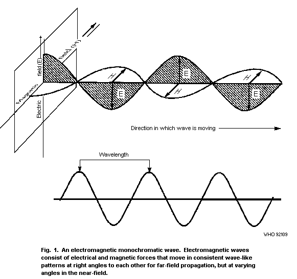 Two idealizations of wave propagation are commonly used:
spherical waves and plane waves (Stuchly, 1983; Grandolfo & Vecchia,
1988). A spherical wave is a good approximation to some
electromagnetic waves that occur. Their wavefronts have spherical
surfaces and each crest and trough has a spherical surface. On every
spherical surface, the E and H fields are constant. The wavefronts
propagate radially outwards from the source and E and H are both
tangential to the spherical surfaces.
A plane wave is another model that approximately represents some
electromagnetic waves. Plane waves have characteristics similar to
spherical waves because, at points far from the source, the curvature
of the spherical wavefronts is so small that they appear to be almost
planar.
The defining characteristics of a plane wave are:
(a) E, H, and the direction of propagation are all mutually
perpendicular.
(b) The quotient E/H is constant and is called the wave
impedance. For free space E/H = 377 OMEGA. For other media and
for sinusoidal steady-state fields, the wave impedance includes
losses in the medium in which the wave is travelling.
(c) Both E and H vary as 1/r, where r is the distance from the
source.
In RF plane wave propagation (far-field), the power crossing a
unit area normal to the direction of wave propagation is usually
designated by the symbol S. When the electric and magnetic field
strengths are expressed in V/m and A/m, respectively, S represents
their product, which yields VA/m2, i.e., W/m2 (watts per square
metre).
In free space, electromagnetic waves spread uniformly in all
directions from a theoretical point (isotropic) source. As the
distance from the point source increases, the area of the wavefront
surface increases as a square of the distance, so that the source
power is spread over a larger area.
As power density S corresponds also to the quotient of the total
radiated power and the spherical surface area enclosing the source, it
is inversely proportional to the square of the distance from the
source, and can be expressed as:
S = P/4pi r2 (Equation 2.6)
where P is the total radiated power and r is the distance from the
source.
In the case of plane waves, frequently called far-field
conditions, the power density can be derived from E2/377 or from 377
H (see Table 1). Therefore, in many practical applications only the
E field or the H field needs to be measured when the point of
measurement is at least one wavelength from the source. In this case,
measurement of E makes possible the determination of H and vice-versa.
Table 1. Comparison of power densities in the more commonly used
units for free-space, far-field conditions (Note: values have been
rounded to one or two significant figures, based on the relationships
above)
W/m2 mW/cm2 µW/cm2 V/m A/m
10-2 10-3 1 2 5 10-3
10-1 10-2 10 6 1.5 10-2
1 10-1 102 20 5 10-2
10 1 103 60 1.5 10-1
102 10 104 2 102 5 10-1
103 102 105 6 102 1.5
104 103 106 2 103 5
The region close to a source is called the near-field. In the
near-field, the E and H fields are not necessarily perpendicular;
in fact, they are not always conveniently characterized by waves. They
are often nonpropagating in nature and are sometimes referred to as
fringing fields, reactive near-fields, or evanescent modes.
Near-fields often vary rapidly with distance; the inverse square law
of the dependence with distance does not apply, and the impedance
(E/H) may differ from 377 OMEGA. Objects located near sources may
strongly affect the nature of the fields. For example, placing a probe
near a source to measure the fields may change the characteristics of
the fields considerably (Dumansky et al., 1986).
When RF fields are incident on a conductive object, RF currents
are induced in the object. These currents produce surface fields that
are highly localized to the object and are often referred to as RF hot
spots. RF hot spots are better characterized as electric and magnetic
fields rather than radiation, since, for many conditions, the fields
leading to the hot spot never propagate away from the object. At
higher frequencies, the electric and magnetic fields maintain an
approximately constant relationship in propagating waves. In general,
the lower the frequency, the less coupled the fields become. This is
particularly so when the wavelength is very large with respect to the
physical size of the source. In practice, the fields of concern from
a hazard perspective will be near-fields at frequencies below about 1
MHz.
3. NATURAL BACKGROUND AND HUMAN-MADE FIELDS
3.1 General
In the last few decades, the use of devices that emit
electromagnetic fields has increased considerably. This proliferation
has been accompanied by an increased concern about possible health
effects of exposure to these fields (Grandolfo et al., 1983;
Repacholi, 1988; Shandala & Zvinyatskovski, 1988, Franceschetti et
al., 1989). As a result, throughout the world, many organizations,
both governmental and nongovernmental, have established safety
standards or guidelines for exposure (see section 10).
Electromagnetic devices already in use and the continuous
addition of new sources result in the expansion to new frequencies in
the spectrum and the increasing presence of RF fields. Comprehensive
data on existing emission systems, and evaluation of present levels of
exposure, are essential for the assessment of potential radiation
hazards (Repacholi, 1983a; Shandala et al., 1983; Savin, 1986; Stuchly
& Mild, 1987).
In this section, sources of electromagnetic fields, both natural
and human-made, in the 300 Hz-300 GHz frequency range are surveyed.
The human-made electromagnetic environment consists of fields that are
produced either intentionally or as by-products of the use of other
devices.
Human-made sources in the spectrum considered here, however,
produce local field levels many orders of magnitude above the natural
background. Therefore, for the practical purposes of hazard
assessment, the electromagnetic fields on the earth's surface arise
from human-made sources. According to the treaty of the International
Telecommunications Union (ITU, 1981), the electromagnetic spectrum up
to 3 THz is subdivided into 12 frequency bands. These bands are
designated by numbers as shown in Table 2; only the bands referred to
in this publication are given.
3.2 Natural background
The natural electromagnetic environment originates from processes
such as discharges in the earth's atmosphere (terrestrial sources) or
in the sun and deep space (extra-terrestrial sources).
Table 2. Frequency bands of the electromagnetic spectrum in the
frequency range 300 Hz-300 GHz a
Band Frequency range Metric Description and symbol
number subdivision
3 0.3 to 3 kHz - voice frequency [VF]
4 3 to 30 kHz myriametric very low frequency [VLF]
Table 2 (continued)
Band Frequency range Metric Description and symbol
number subdivision
5 30 to 300 kHz kilometric low frequency [LF]
6 0.3 to 3 MHz hectometric medium frequency [MF]
7 3 to 30 MHz decametric high frequency [HF]
8 30 to 300 MHz metric very high frequency [VHF]
9 0.3 to 3 GHz decimetric ultra high frequency [UHF]
10 3 to 30 GHz centimetric super high frequency [SHF]
11 30 to 300 GHz millimetric extremely high frequency [EHF]
a From: ITU (1981).
3.2.1 Atmospheric fields
Atmospheric fields of frequencies of less than 30 MHz originate
predominantly from thunderstorms. Their strengths and range of
frequencies vary widely with geographical location, time of day, and
season. Some of these variations are systematic and some are random.
Overall, atmospheric fields have an emission spectrum with the largest
amplitude components having frequencies of between 2 and 30 kHz.
Generally, the atmospheric field level decreases with increasing
frequency. The geographical dependence is such that the highest levels
are observed in equatorial areas and the lowest in polar areas.
3.2.2 Terrestrial emissions
The earth emits electromagnetic radiation (black-body radiation),
as do all media, at a temperature T that is different from that at
absolute zero. In the RF range, the black-body radiation follows the
Rayleigh-Jeans law and the thermal noise from the earth (T about 300
K) is 0.003 W/m2 (0.3 µW/cm2), when integrated up to 300 GHz
(Repacholi 1983).
The human body also emits electromagnetic fields at frequencies of up
to 300 GHz at a power density of approximately 0.003 W/m2. For a
total body surface area of about 1.8 m2, the total radiated power is
approximately 0.0054 W.
3.2.3 Extraterrestrial fields
The atmosphere, ionosphere, and magnetosphere of the earth shield
it from extra-terrestrial sources of nonionizing electromagnetic
energy. Electromagnetic waves that are able to penetrate this shield
are limited to two frequency windows, one optical and the other
encompassing radiowaves of frequencies from about 10 MHz to 37.5 GHz.
The short-wave boundary of the RF-window is due to energy absorption
by molecules contained in the atmosphere (primarily O2 and H2O),
whereas the long-wave boundary is related to the shielding action of
the ionosphere.
RF radiation of cosmic origin observed with earth satellites
ranges in magnitude from 1.8 × 10⁻²⁰ W/m2/Hz at 200 kHz to 8 ×
10-20 W/m2/Hz at 10 MHz (Struzak, 1982).
There are three main types of solar emission. The first is the
so-called background, which is the constant component of the emission
observed during periods of low solar activity. The second is the
component that displays long-term changes, associated with variations
in the number of sunspots. Its main contribution is in the frequency
range from 500 MHz to 10 GHz. The third type of emission arises from
isolated radio flares or radio emission bursts. The intensity of such
emission can exceed the average intensity of the quiet radiation by a
factor of one thousand or more; its duration varies from seconds to
hours.
Natural sources of lesser intensity also exist and include the
moon, Jupiter, Cassiopeia-A, the universal thermal background
radiation at 3 K, hydrogen emissions from ionized clouds, line
emissions from neutral hydrogen, the OH radical and, most recently
observed, from ammonia.
3.3 Human-made sources
3.3.1 General
Radio and television transmitters are examples of human-made RF
sources that intentionally produce electromagnetic emissions for
telecommunication purposes. At frequencies of 3 kHz-3 MHz, normal
service coverage is provided by ground-wave propagation. At VLF,
propagation over distances of thousands of km is possible using this
method. At LF and MF, during night-time, reflections from the
ionosphere make propagation up to 2000 km possible with little
attenuation. At HF, other propagation modes are also possible. At
frequencies of 30 MHz-30 GHz, service coverage is provided by
line-of-sight (short paths), diffraction (intermediate paths), or by
forward scattering (long paths) propagation.
Broadcasting systems vary greatly in terms of their design. This
diversity results in somewhat different approaches in evaluating human
exposure and potential problems. The situations are significantly
different for workers and for the general population. In the case of
some workers, such as those maintaining equipment on broadcasting
towers, there is a potential for exposure to strong RF fields. Workers
may also be exposed to strong fields in the close vicinity of towers
and particularly broadcasting antennas in the VLF, LF, and MF. In
contrast, it is rare for the general population to be exposed to
strong RF fields from broadcasting. However, there is simultaneous
exposure to more than one source.
Some insight on the levels of exposure of the general population
may be gained from data collected in the USA, indicating that, in
large cities, the median exposure level is about 50 µW/m2 (Tell &
Mantiply, 1980). SAR values ranging from 0.05 to 0.3 µW/kg are
expected in the frequency range 170-800 MHz.
There are also human-made sources of electromagnetic fields used
for non-communication purposes, in industry (I), science (S), and
medicine (M). ISM applications are intended to transport and
concentrate electromagnetic energy in a restricted working area to
produce physical, chemical, and/or biological effects.
The frequency bands for ISM applications designated by the ITU
are shown in Table 3. However, in individual countries, different
and/or additional frequencies may be designated for use by ISM
equipment (ITU, 1981; Metaxas & Meredith, 1983).
Table 3. Centre frequencies and frequency bands agreed
internationally and assigned for ISM applications a
Centre frequency Frequency bands Area permitted
70 kHz 60-80 kHz USSR
6.780 MHz 6.765-6.795 MHz subject to agreement
13.560 MHz 13.553-13.567 MHz worldwide
27.120 MHz 26.957-27.283 MHz worldwide
40.68 MHz 40.66-40.70 MHz worldwide
42;49;56;61;66 MHz approx. 0.2% United Kingdom
84;168 MHz approx. 0.005% United Kingdom, Austria,
433.92 MHz 433.05-434.79 MHz Liechtenstein,
The Netherlands, Portugal,
Switzerland W. Germany
Yugoslavia
896 MHz 886-906 MHz United Kingdom
915 MHz 902-928 MHz North and South
America
2.375 GHz 2.325-2.425 GHz Albania, Bulgaria,
Czechoslovakia,
Hungary, Romania,
and USSR
Table 3 (continued)
Centre frequency Frequency bands Area permitted
2.45 GHz 2.4-2.5 GHz worldwide, except
where 2.375 GHz is
used
3.39 GHz 3.37-3.41 GHz The Netherlands
5.8 GHz 5.724-5.875 GHz worldwide
6.78 GHz 6.74-6.82 GHz The Netherlands
24.125 GHz 24.0-24.05 GHz worldwide
40.68 GHz 40.43-40.92 GHz United Kingdom
61.25 GHz 61.0-61.5 GHz subject to agreement
122.5 GHz 122-123 GHz subject to agreement
245 GHz 244-246 GHz subject to agreement
a Adapted from: ITU (1981) and Metaxus & Meredith (1983).
Because of unavoidable imperfections in the construction,
production, and use of ISM equipment, and of fundamental physical
laws, there is always unintentional leakage of electromagnetic energy
from such equipment. As a result, each ISM generator acts as an
unintentional source producing signals capable of causing harmful
effects, depending upon the amount of leakage.
To date, the total number of ISM installations in the world is
estimated at 120 million (Struzak, 1985). The number of ISM generators
constantly increases at a rate of about 3-7% per year. With such
growth, the number of ISM generators expected by the year 2000 will be
2-4 times greater than it is now.
ISM equipment is usually designed at minimum cost, and,
typically, is reduced to the essentials necessary for operation.
Frequency stability and spectral purity of the power delivered to the
work piece are not normally major objectives. In almost every case,
the work piece is strongly coupled to the oscillator/amplifier, and
since the electromagnetic characteristics of the material change
during the work cycle, the magnitude, phase, and frequency of the
radiation may be affected by these changes.
Electromagnetic energy leaks from ISM equipment mainly from the
applicator and associated leads (e.g., RF heaters and sealers), the
oscillator body/cabinet, and also from surrounding structures in which
RF currents are induced. The amount of energy radiated from the
applicator and associated leads depends on the particular arrangement
of the devices and the work piece, which together act like an antenna
the radiation efficiency of which is usually very low. However, the
radiated power may be considerable if the nominal power is high.
Stray fields are also associated with currents flowing over the
surface of the body/cabinet and over the surrounding structures. The
equipment acts as a complex antenna system consisting of coupled
radiating surface elements resonating at some unspecified frequencies.
Often all the power and control wires are situated close to RF power
circuits with no shielding. As a result, a considerable amount of RF
energy may be fed into these leads and is conducted outwards at a
distance and then reradiated.
Table 4. Typical applications of equipment generating electromagnetic
fields in the range 300 Hz-300 GHz
Frequency Wavelength Typical applications
0.3-3 kHz 1000-100 km Broadcast modulation, medical applications,
electric furnaces, induction heating,
hardening, soldering, melting, refining
3-30 kHz 100-10 km Very long range communications, radio
navigation, broadcast modulation, medical
applications, induction heating, hardening,
soldering, melting, refining, VDUs
30-300 kHz 10-1 km Radionavigation, marine and aeronautical
communications, long-range communications,
radiolocation, VDUs, electro-erosion
treatment, induction heating and melting of
metals, power inverters
0.3-3 MHz 1 km-100 m Communications, radionavigation, marine
radiophone, amateur radio, industrial RF
equipment, AM broadcasting, RF excited arc
welders, sealing for packaging, production
of semiconductor material, medical
applications
3-30 MHz 100-10 m Citizen band, amateur radio broadcasting,
international communications, medical
diathermy, magnetic resonance imaging,
dielectric heating, wood drying and gluing,
plasma heating
30-300 MHz 10-1 m Police, fire, amateur FM, VHF-TV, diathermy,
emergency medical radio, air traffic
control, magnetic resonance imaging,
dielectric heating, plastic welding, food
processing, plasma heating, particle
separation
Table 4 (continued)
Frequency Wavelength Typical applications
0.3-3 GHz 100-10 cm Microwave point to point, amateur, taxi,
police, fire, radar, citizen band,
radionavigation, UHF-TV, microwave ovens,
medical diathermy, food processing, material
manufacture, insecticide, plasma heating,
particle acceleration
3-30 GHz 10-1 cm Radar, satellite communications, amateur,
fire, taxi, airborne weather radar, police,
microwave relay, anti-intruder alarms,
plasma heating, thermonuclear fusion
experiments
30-300 GHz 10-1 mm Radar, satellite communications, microwave
relay, radionavigation, amateur radio
Typical uses of equipment generating electromagnetic fields in
the frequency range 300 Hz-300 GHz are shown in Table 4.
3.3.2 Environment, home, and public premises
A comprehensive evaluation of general population exposure to RF
has been performed by the USA Environmental Protection Agency (Tell&
Mantiply, 1980). Broadcasting services, particularly those usingthe
VHF and UHF bands, have been identified as the main sources of ambient
RF fields (Karachev & Bitkin, 1985). Measurements performed in 15
large cities in the USA led to the conclusions that the median
exposure level was 50 µW/m2 and that approximately 1% of the
population studied was potentially exposed to levels greater than 10
mW/m2.
The presence of conducting objects can give rise to field
strengths higher than those expected from theoretical considerations,
since they act as diffracting elements for the electromagnetic fields.
Consequently, the presence of such objects in the near-field zone of
radio stations makes the area between the radiator and the object
potentially more hazardous and indicates that problems of safety
should be considered carefully (Bernardi et al., 1981).
Although measurements as well as theory indicate that there is no
high-level exposure from broadcasting stations, the existence of
limited areas of relatively high irradiation close to the sources
should be checked (Dumansky et al., 1985a). Such situations can exist
in proximity to very powerful, ground-level transmitters. In several
cases, urban areas are served locally by low-power, in-town repeaters.
These are placed, for convenience, on the top of tall buildings;
unless properly designed, this creates the possibility of stray fields
in a densely populated area directly below the RF source. A typical,
high-power, MF transmitter can have a carrier power of 100 kW, plus up
to 50 kW in the sidebands of the propagated field. This is an example
of how high field strengths can occur in a space open to the public.
Although a broadcasting station's property is usually fenced to
keep out unauthorized individuals, the fence may be close to the tower
base and people may be able to get as close as a few tens of metres or
less from the antenna. Because the wavelengths involved are so long,
a near-field exposure situation may exist and a field strength
considerably greater than the theoretical ground-wave field strength
is to be expected (Bini et al., 1980).
Local MF transmitters find widespread use in cities, where they
provide coverage on "blind spots" or other low-signal receiving areas.
Powers range from 100 to 1000 W at the amplifier output and much less
than that can be expected to be radiated into space. In a typical
arrangement, the transmitting module is located at the top of a
stucture. The radiating system is fed via a coaxial cable. It consists
of a dipole over a ground plane or counterpoise laid directly on the
roof. More than one transmitter can serve the same radiator. Currents
can set up fields in a complicated pattern inside the building (Bini
et al., 1980).
When RF fields are incident on conductive objects, RF currents
are induced in the objects. Because of these currents, the objects
become sources of additional fields that are highly localized and in
some situations can constructively add to original fields.
Among the general population, the most popular application of
microwave power is in the cooking of food. Power levels range from 300
W to 1 kW in consumer microwave models, at a frequency of 2.45 GHz. In
a properly designed microwave oven, a very small fraction of this
power escapes from the oven housing through various leakage paths.
When leakage occurs, it is most frequently through the door seal. It
may increase with use or mechanical abuse of the oven. Small amounts
of leakage can also occur through the viewing screen (Osepchuk, 1979).
Personal exposure from microwave ovens is extremely small because
of the rapid decrease of the power flux density with increasing
distance from the oven. For worst case leakage from the microwave oven
of 5 mW/cm2, the power density at a distance of 0.3 m is less than
0.15 mW/cm2 and, at 1 m it is about 10 µW/cm2. Typical leakage
values, therefore, imply exposure values well below the most
conservative RF exposure standards in the world (Stuchly, 1977;
Dumansky et al., 1980).
Recently, the induction heating stove, a new appliance for
domestic use, has been introduced on the market. This appliance
operates in the kilohertz range. Exposure levels at distances greater
than 0.5 m are low compared with existing exposure limits, being less
than 5 V/m and 0.5-10 A/m, respectively, at a distance of 0.3 m
(Stuchly & Lecuyer, 1987).
Microwave anti-intrusion alarms are typical of low-power devices.
These operate continuously to avoid thermal drift or switching
problems, thus exposing people in the protected area. With a typical
power of 10 mW, power densities of the order of 10 µW/cm2 are
measured at a distance of about 0.5 m. Population exposure to RF
fields from commonly encountered sources, such as airport, marine, and
police radar, is similarly very low (Stuchly, 1977; Dumansky et al.,
1980, 1985b, 1988).
3.3.3 Workplace
Levels of occupational exposure vary considerably, and are
strongly dependent upon the particular application. While most
communication and radar workers are exposed to fields of only
relatively low intensity, some can be exposed to high levels of RF.
Workers climbing FM radio or TV broadcasting towers may be exposed to
E fields up to 1 kV/m and H fields up to 5 A/m (Repacholi 1983a; Mild
& Lovstrand, 1990).
Radar systems produce strong RF fields along the axis of the
antenna. However, in most systems, average field strengths are reduced
typically by a factor of 100-1000, because of antenna rotation and
because the field is pulsed. With stationary antennas, which represent
the worst case, peak power flux densities of 10 MW/m2 may occur on
the antenna axis up to a few metres away from the source.
In areas surrounding air traffic control radars (ATCRs), workers
can be exposed to power flux densities of up to tens of W/m2, but
are normally exposed to fields in the range 0.03-0.8 W/m2. In an
exposure survey of civilian airport radar workers in Australia, it was
found that, unless working on open waveguide slots, or within
transmitter cabinets when high voltage arcing was occurring, personnel
were, in general, not exposed to levels of radiation exceeding the
specified limits in the Australian and IRPA radiofrequency exposure
standards (Joyner & Bangay, 1986a).
Dielectric or RF heaters are widespread in many industries. RF
energy produces heat directly within the processed material. This
unique characteristic is commonly used for such purposes as sealing
plastics or drying glue for joining wood. All RF heaters have a higher
efficiency in comparison with conventional ovens. According to several
surveys (Conover et al., 1980; Stuchly et al., 1980; Grandolfo et al.,
1982; Bini et al., 1986; Joyner & Bangay, 1986b; Stuchly & Mild,
1987), the sealing or welding of polyvinyl chloride (PVC) is the most
common use for RF dielectric heaters. Two pieces of plastic are
compressed between electrodes and RF power is applied. The plastic
material heats, partially fuses, and forms a bond. Plastic heaters
frequently operate at the ISM frequency of 27.12 MHz. However, during
the operating cycle, this value may vary by several megahertz. The RF
output power ranges from fractions of a kilowatt to about 100 kW.
Since the exposure of heater sealer operators and other personnel
working in the same area takes place in the near-field, both E and H
field strengths must be measured to evaluate exposure levels. However,
to demonstrate compliance with basic limits of RF exposure, the
development of body current measurement techniques should prove to be
useful (Allen et al., 1986). In the vicinity of RF sources,
measurements of fields must be made with the operators absent from the
positions that they normally occupy. The stray fields are localized in
the immediate vicinity of the sealers, so that exposure of the body is
highly inhomogeneous.
RF industrial heaters (plastic sealers and other devices) have
been found to produce exposure fields exceeding the limits recommended
in various countries and by the IRPA. Furthermore, direct current
measurements have confirmed coupling of the RF energy from the device
to its operator. Various methods have been developed to ameliorate the
situation, such as shields, grounding strips, and others. Potential
overexposure to RF radiation is probably one of the most common
occurrences in the case of RF heaters, unless protective measures are
employed.
Magnetic fields below a few tens of megahertz are used in
industry for the induction heating of metals and semiconductors and in
arc welding. Surveys of the magnetic field strength to which the
operators are exposed have shown that these exposures are, in many
instances, high compared with recommended exposure limits (Stuchly &
Lecuyer, 1985; Conover et al., 1986; Stuchly, 1986; Andreuccetti et
al., 1988; Stuchly & Lecuyer, 1989).
In many practical situations, exposure can be reduced either by
administrative measures (Eriksson & Mild, 1985) or by the use of
protective screening. Screening may be intentional (wire fences) or
incidental (walls of buildings) and may function by reflection or by
absorption. In general, both contribute to the total attenuation
provided.
Thin metal sheets are adequate for the attenuation of RF electric
fields. However, in many cases, it is usual to use wire screens or
perforated sheets, since these have the advantages of transparency,
ventilation, light weight, etc. In all cases, surveys are desirable to
verify the integrity of such screens or shields. Faults in screens
could, in some circumstances, be secondary sources of significant
radiation or reactive fields (White, 1980).
The applications of video display units (VDUs) are numerous and
their use widespread. Even more applications are anticipated in the
future. In the RF region, they emit electric and magnetic fields from
the cathode ray tubes (CRTs). The dominant sources are the horizontal
and vertical sweep generators (fly-back transformers) operating at
frequencies of some 15-35 kHz. VDUs produce fields that have complex
waveforms. Typical electric field stengths at the operator position
(0.5 m from the screen) range from less than 1 to 10 V/m (RMS).
Magnetic flux densities range typically from less than 0.01 µT to 0.1
µT (RMS). In most VDUs, both fields are produced at the lower end of
these ranges. VDUs also produce weak, electric and magnetic fields at
the power line frequency (50 or 60 Hz) and its harmonics. All surveys
have concluded that VDUs do not present any hazard for human health
within the context of existing guidelines for exposures to
non-ionizing electromagnetic fields (see section 10) (BRH, 1981;
Stuchly et al., 1983b; Harvey, 1984; Repacholi, 1985; Elliott et al.,
1986).
A statement has been published by the International Non-Ionizing
Radiation Committee of the International Radiation Protection
Association (IRPA, 1988b). Conclusions in this and other documents
(WHO, 1987; ILO, 1991) are that, on the basis of current biomedical
knowledge, there are no health hazards associated with radiation or
fields from VDUs and that there is no scientific basis to justify
radiation shielding or regular monitoring of the various radiations
and fields emitted by VDUs.
3.3.4 Medical practice
Shortwave and microwave diathermy treatments are used to relieve
pain through the non-invasive application of non-ionizing
electromagnetic energy to body tissues. Several surveys have been
published (EHD, 1980; Ruggera, 1980; Grandolfo et al., 1982; Stuchly
et al., 1982; Delpizzo & Joyner, 1987), with the primary purpose of
measuring the field strengths to which diathermy operators are exposed
during typical treatments. Measurements of the magnitude of fields
near diathermy electrodes (applicators) were made from shortwave
diathermy units operating at 27.12 MHz, and from microwave diathermy
units operating at 434 MHz and 2.45 GHz. They indicate emissions of
high field and radiation levels in directions other than those
intended for treatment. Operators, physiotherapists, and personnel
performing service and maintenance tasks are exposed to stray fields
and radiations. Reduction of unnecessary exposure of both operator and
patient during microwave and shortwave diathermy treatments is
technically achievable through adequate shielding of existing units,
careful design of new equipment, and judicious planning of the
treatment area (Bonkowski & Makiewicz, 1986).
Hyperthermia devices are used in cancer adjuvant therapies (Storm
et al, 1981; Stuchly et al., 1983a; Hagmann et al., 1985). Treatments
have been based on biological studies that suggest hyperthermia
effectiveness in conjunction with radiotherapy and with chemotherapy.
The evaluation of hyperthermia efficacy is proceeding through the
development of therapeutic trials for specific tumours (Arcangeli et
al., 1985; Perez et al., 1991). A few international and national
organizations have independently determined and developed randomized
trials (Lovisolo et al., 1988). For the purposes of safety evaluation,
hyperthemia devices can be classified as: (a) those irradiating
external to the body and intended for superficial and deep
hyperthermia, and (b) those irradiating from inside the body and
intended for interstitial and endocavitary hyperthermia.
All devices present, to a greater or lesser extent, problems of
patient health protection. Adverse effects on patients have included
pain, discomfort, burn, ulceration, and, for deep hyperthemia,
tachycardia and faintness. These are due to an overheating of
superficial tissues, tissues surrounding the tumour, and, in deep
hyperthermia, other tissues far from the tumour and irradiated region
(Myerson et al., 1989; Petrovich et al., 1989). The magnitude of the
electromagnetic field around superficial, interstitial, and
endocavitary applicators is relatively low and does not cause any
health risk to the operators, though the possibility of leakage of RF
energy from generators and connecting cables has to be considered in
some models. Capacitive and phase array devices, however, may leak RF
energy (Storm et al., 1981).
Hagmann et al. (1985) measured the stray electric and magnetic
fields of angular phased array and helical coil applicators for limb
and torso hyperthermia at 70.93 MHz. Field strengths were measured in
excess of 300 V/m and 1A/m, respectively, at a distance of about 10 cm
from the applicator. At 0.5 m, these values were reduced to 14 V/m and
0.1 A/m, respectively. In general, manufacturers of hyperthermia
devices pay too little attention to minimizing the leakage of RF
fields from generators, cables, and applicators, and each new model
generator should be tested for RF emissions.
Magnetic resonance imaging (MRI) is now an established diagnostic
technique while in vivo spectroscopy is undergoing rapid
development. The complexity of exposure associated with MRI requires
the safety consideration of three different fields (Tenforde &
Budinger, 1986; Budinger, 1988). During clinical imaging, patients or
volunteers, and operators are exposed to static magnetic fields,
time-varying magnetic fields, and radiofrequency electromagnetic
fields. RF fields in the frequency range 1-100 MHz, are deposited in
patients, principally as heating associated with eddy currents induced
by the RF magnetic field (Grandolfo et al, 1990). For MRI systems with
static magnetic flux densities below 2 T, power deposition from
electric fields associated with RF transmitter coils is relatively
low, when efficient transmitter coil designs are employed. The power
deposited by transient magnetic field gradients is similarly low
(Bottomley et al., 1985). Staff operating the equipment are
intermittently exposed to weaker fields that are present in the
vicinity of the imaging equipment. Guidelines on "Protection of the
patient undergoing magnetic resonance examinations" have been
published by the International Non-Ionizing Radiation Committee of the
International Radiation Protection Association (IRPA, 1991).
4. EXPOSURE EVALUATION - CALCULATION AND MEASUREMENT
4.1 Introduction
Exposure evaluation provides information necessary to perform
risk assessments. Two methods are available: (i) a theoretical
estimation; and (ii) measurements of the fields or related parameters,
such as energy absorption rates and currents.
Estimates of exposures are necessary before an installation is
constructed. Whenever possible, estimates of radiation fields should
be made before detailed surveys of potentially hazardous exposures are
carried out. This procedure is needed to select suitable survey
instruments, and to determine whether potentially hazardous exposure
of the surveyor could occur, if the choice of the instrument were
inappropriate or if the instrument were faulty.
4.2 Theoretical estimation
Electromagnetic waves may be harmonic, i.e., the electric and
magnetic fields oscillate as sine waves, and power is generated as a
continuous wave (CW) at essentially a single frequency. The waves may
be also modulated, i.e., the amplitude, phase, or frequency may be
changed in a chosen manner, for example, if pulse modulation,
short-duration electromagnetic pulses are emitted at certain time
intervals. The duration of the pulse (pulse length or pulse width),
which may be of the order of small fractions of a second, is
designated by t. Its reciprocal, the pulse repetition frequency (pulse
repetition rate), is expressed in hertz. The product of pulse length
and repetition rate is referred to as the duty cycle, D. In case of
pulsed-wave generation, the emitted power increases rapidly, reaches
a peak pulse power, and rapidly decreases.
This may be averaged for pulse length or per unit time, which
introduces the concept of mean (average) power emitted, according to:
Pp = Pa/tfr or Pa = Ppfr t (Equation 4.1)
where Pp is the peak power, Pa the average power, fr the
repetition frequency, and t the pulse length.
In practice, average power is usually measured, and, for safety
purposes, mean power density is used. The peak pulse power may be many
times higher than the average power output. The average and peak power
flux densities (Sa and Sp) are given by:
Sa = DSp (Equation 4.2)
Universally used sources with moveable antennas and/or beams, such as
scanning or rotating radars, introduce an additional complication from
the safety viewpoint. Electromagnetic power from such installations
arrives intermittently.
The power flux density for a scanning antenna in motion can be
estimated from the power flux density measured with the antenna
stationary using the expression:
Wm = ksWs (Equation 4.3)
where Wm is the power flux density for the antenna in motion, ks
is the antenna rotational reduction factor, and Ws is the power flux
density measured on the axis of the stationary antenna at a given
distance.
In most radar installations, the antenna rotates and therefore an
occupied position is exposed only when the radar beam sweeps it. The
average exposure level is obtained by multiplying the measured or
estimated level from a stationary antenna by the rotational reduction
factor (RRF). In the far-field, RRF equals the ratio of the half power
beam width to the antenna scan angle.
The rotational reduction factor (ks) for the near-field region
is equal to:
a/Rk (Equation 4.4)
where "a" is the dimension of the antenna in the scan (rotation) plane
and Rk is the circumference of the antenna scan sector at the given
distance r, at which the measurements have been made.
The region close to a source antenna is called the near-field. As
shown in Fig.2, the near-field can be divided into two subregions: the
reactive near-field region and the radiating near-field region. The
region of space surrounding the antenna in which the reactive
components predominate is known as the reactive near-field region. In
the radiating near-field region, the radiation pattern varies with the
distance from the antenna. The near-fields often vary rapidly with
distance and mathematical expressions generally contain the terms 1/r,
1/r2, ...., 1/rn, where r is the distance from the source to the
point at which the field is being determined. At greater distances
from the source, the 1/r2, 1/r3, and higher-order terms are
negligible compared with the 1/r term and the fields are called
far-fields. These fields are approximately spherical waves that can,
in turn, be approximated in a limited region of space by plane waves.
Measurements and calculations are usually easier in far-fields than in
near-fields.
When the longest dimension (L) of the source antenna is greater
than the wavelength (lamda), the distance from the source to the
far-field is 2L2/lamda. For L<lamda, this distance is lamda/2pi
(see Fig. 2). In practice, the distance from the source that
represents the boundary between the near-field and far-field regions
is often taken to be the greater of the two quantities, lamda and
2L2/lamda. However, the appropriate empirical relationship depends
on the type of aperture of the source and, for example, for a circular
aperture, such as on a microwave relay tower, the relationship
L2/lamda may be more appropriate. In this case, with a frequency of
2 GHz (lamda = 15 cm), L is approximately 3 m and, consequently, the
quantity L2/lamda = 60 m. Because 60 m is much greater than 15 cm,
this is the distance that can be assumed as a boundary between the
near- and far-field regions.
The boundary between the near-field and far-field regions,
however, is not sharp, because the near-fields gradually become less
as the distance from the source increases.
In free space, electromagnetic waves spread uniformly in all
directions from a theoretical point source. In this case, the
wavefront is spherical. As the distance from the point source
increases, the area of the wavefront surface increases as a square of
the distance, so that the source power is spread over a larger area.
Two idealizations of wave propagation are commonly used:
spherical waves and plane waves (Stuchly, 1983; Grandolfo & Vecchia,
1988). A spherical wave is a good approximation to some
electromagnetic waves that occur. Their wavefronts have spherical
surfaces and each crest and trough has a spherical surface. On every
spherical surface, the E and H fields are constant. The wavefronts
propagate radially outwards from the source and E and H are both
tangential to the spherical surfaces.
A plane wave is another model that approximately represents some
electromagnetic waves. Plane waves have characteristics similar to
spherical waves because, at points far from the source, the curvature
of the spherical wavefronts is so small that they appear to be almost
planar.
The defining characteristics of a plane wave are:
(a) E, H, and the direction of propagation are all mutually
perpendicular.
(b) The quotient E/H is constant and is called the wave
impedance. For free space E/H = 377 OMEGA. For other media and
for sinusoidal steady-state fields, the wave impedance includes
losses in the medium in which the wave is travelling.
(c) Both E and H vary as 1/r, where r is the distance from the
source.
In RF plane wave propagation (far-field), the power crossing a
unit area normal to the direction of wave propagation is usually
designated by the symbol S. When the electric and magnetic field
strengths are expressed in V/m and A/m, respectively, S represents
their product, which yields VA/m2, i.e., W/m2 (watts per square
metre).
In free space, electromagnetic waves spread uniformly in all
directions from a theoretical point (isotropic) source. As the
distance from the point source increases, the area of the wavefront
surface increases as a square of the distance, so that the source
power is spread over a larger area.
As power density S corresponds also to the quotient of the total
radiated power and the spherical surface area enclosing the source, it
is inversely proportional to the square of the distance from the
source, and can be expressed as:
S = P/4pi r2 (Equation 2.6)
where P is the total radiated power and r is the distance from the
source.
In the case of plane waves, frequently called far-field
conditions, the power density can be derived from E2/377 or from 377
H (see Table 1). Therefore, in many practical applications only the
E field or the H field needs to be measured when the point of
measurement is at least one wavelength from the source. In this case,
measurement of E makes possible the determination of H and vice-versa.
Table 1. Comparison of power densities in the more commonly used
units for free-space, far-field conditions (Note: values have been
rounded to one or two significant figures, based on the relationships
above)
W/m2 mW/cm2 µW/cm2 V/m A/m
10-2 10-3 1 2 5 10-3
10-1 10-2 10 6 1.5 10-2
1 10-1 102 20 5 10-2
10 1 103 60 1.5 10-1
102 10 104 2 102 5 10-1
103 102 105 6 102 1.5
104 103 106 2 103 5
The region close to a source is called the near-field. In the
near-field, the E and H fields are not necessarily perpendicular;
in fact, they are not always conveniently characterized by waves. They
are often nonpropagating in nature and are sometimes referred to as
fringing fields, reactive near-fields, or evanescent modes.
Near-fields often vary rapidly with distance; the inverse square law
of the dependence with distance does not apply, and the impedance
(E/H) may differ from 377 OMEGA. Objects located near sources may
strongly affect the nature of the fields. For example, placing a probe
near a source to measure the fields may change the characteristics of
the fields considerably (Dumansky et al., 1986).
When RF fields are incident on a conductive object, RF currents
are induced in the object. These currents produce surface fields that
are highly localized to the object and are often referred to as RF hot
spots. RF hot spots are better characterized as electric and magnetic
fields rather than radiation, since, for many conditions, the fields
leading to the hot spot never propagate away from the object. At
higher frequencies, the electric and magnetic fields maintain an
approximately constant relationship in propagating waves. In general,
the lower the frequency, the less coupled the fields become. This is
particularly so when the wavelength is very large with respect to the
physical size of the source. In practice, the fields of concern from
a hazard perspective will be near-fields at frequencies below about 1
MHz.
3. NATURAL BACKGROUND AND HUMAN-MADE FIELDS
3.1 General
In the last few decades, the use of devices that emit
electromagnetic fields has increased considerably. This proliferation
has been accompanied by an increased concern about possible health
effects of exposure to these fields (Grandolfo et al., 1983;
Repacholi, 1988; Shandala & Zvinyatskovski, 1988, Franceschetti et
al., 1989). As a result, throughout the world, many organizations,
both governmental and nongovernmental, have established safety
standards or guidelines for exposure (see section 10).
Electromagnetic devices already in use and the continuous
addition of new sources result in the expansion to new frequencies in
the spectrum and the increasing presence of RF fields. Comprehensive
data on existing emission systems, and evaluation of present levels of
exposure, are essential for the assessment of potential radiation
hazards (Repacholi, 1983a; Shandala et al., 1983; Savin, 1986; Stuchly
& Mild, 1987).
In this section, sources of electromagnetic fields, both natural
and human-made, in the 300 Hz-300 GHz frequency range are surveyed.
The human-made electromagnetic environment consists of fields that are
produced either intentionally or as by-products of the use of other
devices.
Human-made sources in the spectrum considered here, however,
produce local field levels many orders of magnitude above the natural
background. Therefore, for the practical purposes of hazard
assessment, the electromagnetic fields on the earth's surface arise
from human-made sources. According to the treaty of the International
Telecommunications Union (ITU, 1981), the electromagnetic spectrum up
to 3 THz is subdivided into 12 frequency bands. These bands are
designated by numbers as shown in Table 2; only the bands referred to
in this publication are given.
3.2 Natural background
The natural electromagnetic environment originates from processes
such as discharges in the earth's atmosphere (terrestrial sources) or
in the sun and deep space (extra-terrestrial sources).
Table 2. Frequency bands of the electromagnetic spectrum in the
frequency range 300 Hz-300 GHz a
Band Frequency range Metric Description and symbol
number subdivision
3 0.3 to 3 kHz - voice frequency [VF]
4 3 to 30 kHz myriametric very low frequency [VLF]
Table 2 (continued)
Band Frequency range Metric Description and symbol
number subdivision
5 30 to 300 kHz kilometric low frequency [LF]
6 0.3 to 3 MHz hectometric medium frequency [MF]
7 3 to 30 MHz decametric high frequency [HF]
8 30 to 300 MHz metric very high frequency [VHF]
9 0.3 to 3 GHz decimetric ultra high frequency [UHF]
10 3 to 30 GHz centimetric super high frequency [SHF]
11 30 to 300 GHz millimetric extremely high frequency [EHF]
a From: ITU (1981).
3.2.1 Atmospheric fields
Atmospheric fields of frequencies of less than 30 MHz originate
predominantly from thunderstorms. Their strengths and range of
frequencies vary widely with geographical location, time of day, and
season. Some of these variations are systematic and some are random.
Overall, atmospheric fields have an emission spectrum with the largest
amplitude components having frequencies of between 2 and 30 kHz.
Generally, the atmospheric field level decreases with increasing
frequency. The geographical dependence is such that the highest levels
are observed in equatorial areas and the lowest in polar areas.
3.2.2 Terrestrial emissions
The earth emits electromagnetic radiation (black-body radiation),
as do all media, at a temperature T that is different from that at
absolute zero. In the RF range, the black-body radiation follows the
Rayleigh-Jeans law and the thermal noise from the earth (T about 300
K) is 0.003 W/m2 (0.3 µW/cm2), when integrated up to 300 GHz
(Repacholi 1983).
The human body also emits electromagnetic fields at frequencies of up
to 300 GHz at a power density of approximately 0.003 W/m2. For a
total body surface area of about 1.8 m2, the total radiated power is
approximately 0.0054 W.
3.2.3 Extraterrestrial fields
The atmosphere, ionosphere, and magnetosphere of the earth shield
it from extra-terrestrial sources of nonionizing electromagnetic
energy. Electromagnetic waves that are able to penetrate this shield
are limited to two frequency windows, one optical and the other
encompassing radiowaves of frequencies from about 10 MHz to 37.5 GHz.
The short-wave boundary of the RF-window is due to energy absorption
by molecules contained in the atmosphere (primarily O2 and H2O),
whereas the long-wave boundary is related to the shielding action of
the ionosphere.
RF radiation of cosmic origin observed with earth satellites
ranges in magnitude from 1.8 × 10⁻²⁰ W/m2/Hz at 200 kHz to 8 ×
10-20 W/m2/Hz at 10 MHz (Struzak, 1982).
There are three main types of solar emission. The first is the
so-called background, which is the constant component of the emission
observed during periods of low solar activity. The second is the
component that displays long-term changes, associated with variations
in the number of sunspots. Its main contribution is in the frequency
range from 500 MHz to 10 GHz. The third type of emission arises from
isolated radio flares or radio emission bursts. The intensity of such
emission can exceed the average intensity of the quiet radiation by a
factor of one thousand or more; its duration varies from seconds to
hours.
Natural sources of lesser intensity also exist and include the
moon, Jupiter, Cassiopeia-A, the universal thermal background
radiation at 3 K, hydrogen emissions from ionized clouds, line
emissions from neutral hydrogen, the OH radical and, most recently
observed, from ammonia.
3.3 Human-made sources
3.3.1 General
Radio and television transmitters are examples of human-made RF
sources that intentionally produce electromagnetic emissions for
telecommunication purposes. At frequencies of 3 kHz-3 MHz, normal
service coverage is provided by ground-wave propagation. At VLF,
propagation over distances of thousands of km is possible using this
method. At LF and MF, during night-time, reflections from the
ionosphere make propagation up to 2000 km possible with little
attenuation. At HF, other propagation modes are also possible. At
frequencies of 30 MHz-30 GHz, service coverage is provided by
line-of-sight (short paths), diffraction (intermediate paths), or by
forward scattering (long paths) propagation.
Broadcasting systems vary greatly in terms of their design. This
diversity results in somewhat different approaches in evaluating human
exposure and potential problems. The situations are significantly
different for workers and for the general population. In the case of
some workers, such as those maintaining equipment on broadcasting
towers, there is a potential for exposure to strong RF fields. Workers
may also be exposed to strong fields in the close vicinity of towers
and particularly broadcasting antennas in the VLF, LF, and MF. In
contrast, it is rare for the general population to be exposed to
strong RF fields from broadcasting. However, there is simultaneous
exposure to more than one source.
Some insight on the levels of exposure of the general population
may be gained from data collected in the USA, indicating that, in
large cities, the median exposure level is about 50 µW/m2 (Tell &
Mantiply, 1980). SAR values ranging from 0.05 to 0.3 µW/kg are
expected in the frequency range 170-800 MHz.
There are also human-made sources of electromagnetic fields used
for non-communication purposes, in industry (I), science (S), and
medicine (M). ISM applications are intended to transport and
concentrate electromagnetic energy in a restricted working area to
produce physical, chemical, and/or biological effects.
The frequency bands for ISM applications designated by the ITU
are shown in Table 3. However, in individual countries, different
and/or additional frequencies may be designated for use by ISM
equipment (ITU, 1981; Metaxas & Meredith, 1983).
Table 3. Centre frequencies and frequency bands agreed
internationally and assigned for ISM applications a
Centre frequency Frequency bands Area permitted
70 kHz 60-80 kHz USSR
6.780 MHz 6.765-6.795 MHz subject to agreement
13.560 MHz 13.553-13.567 MHz worldwide
27.120 MHz 26.957-27.283 MHz worldwide
40.68 MHz 40.66-40.70 MHz worldwide
42;49;56;61;66 MHz approx. 0.2% United Kingdom
84;168 MHz approx. 0.005% United Kingdom, Austria,
433.92 MHz 433.05-434.79 MHz Liechtenstein,
The Netherlands, Portugal,
Switzerland W. Germany
Yugoslavia
896 MHz 886-906 MHz United Kingdom
915 MHz 902-928 MHz North and South
America
2.375 GHz 2.325-2.425 GHz Albania, Bulgaria,
Czechoslovakia,
Hungary, Romania,
and USSR
Table 3 (continued)
Centre frequency Frequency bands Area permitted
2.45 GHz 2.4-2.5 GHz worldwide, except
where 2.375 GHz is
used
3.39 GHz 3.37-3.41 GHz The Netherlands
5.8 GHz 5.724-5.875 GHz worldwide
6.78 GHz 6.74-6.82 GHz The Netherlands
24.125 GHz 24.0-24.05 GHz worldwide
40.68 GHz 40.43-40.92 GHz United Kingdom
61.25 GHz 61.0-61.5 GHz subject to agreement
122.5 GHz 122-123 GHz subject to agreement
245 GHz 244-246 GHz subject to agreement
a Adapted from: ITU (1981) and Metaxus & Meredith (1983).
Because of unavoidable imperfections in the construction,
production, and use of ISM equipment, and of fundamental physical
laws, there is always unintentional leakage of electromagnetic energy
from such equipment. As a result, each ISM generator acts as an
unintentional source producing signals capable of causing harmful
effects, depending upon the amount of leakage.
To date, the total number of ISM installations in the world is
estimated at 120 million (Struzak, 1985). The number of ISM generators
constantly increases at a rate of about 3-7% per year. With such
growth, the number of ISM generators expected by the year 2000 will be
2-4 times greater than it is now.
ISM equipment is usually designed at minimum cost, and,
typically, is reduced to the essentials necessary for operation.
Frequency stability and spectral purity of the power delivered to the
work piece are not normally major objectives. In almost every case,
the work piece is strongly coupled to the oscillator/amplifier, and
since the electromagnetic characteristics of the material change
during the work cycle, the magnitude, phase, and frequency of the
radiation may be affected by these changes.
Electromagnetic energy leaks from ISM equipment mainly from the
applicator and associated leads (e.g., RF heaters and sealers), the
oscillator body/cabinet, and also from surrounding structures in which
RF currents are induced. The amount of energy radiated from the
applicator and associated leads depends on the particular arrangement
of the devices and the work piece, which together act like an antenna
the radiation efficiency of which is usually very low. However, the
radiated power may be considerable if the nominal power is high.
Stray fields are also associated with currents flowing over the
surface of the body/cabinet and over the surrounding structures. The
equipment acts as a complex antenna system consisting of coupled
radiating surface elements resonating at some unspecified frequencies.
Often all the power and control wires are situated close to RF power
circuits with no shielding. As a result, a considerable amount of RF
energy may be fed into these leads and is conducted outwards at a
distance and then reradiated.
Table 4. Typical applications of equipment generating electromagnetic
fields in the range 300 Hz-300 GHz
Frequency Wavelength Typical applications
0.3-3 kHz 1000-100 km Broadcast modulation, medical applications,
electric furnaces, induction heating,
hardening, soldering, melting, refining
3-30 kHz 100-10 km Very long range communications, radio
navigation, broadcast modulation, medical
applications, induction heating, hardening,
soldering, melting, refining, VDUs
30-300 kHz 10-1 km Radionavigation, marine and aeronautical
communications, long-range communications,
radiolocation, VDUs, electro-erosion
treatment, induction heating and melting of
metals, power inverters
0.3-3 MHz 1 km-100 m Communications, radionavigation, marine
radiophone, amateur radio, industrial RF
equipment, AM broadcasting, RF excited arc
welders, sealing for packaging, production
of semiconductor material, medical
applications
3-30 MHz 100-10 m Citizen band, amateur radio broadcasting,
international communications, medical
diathermy, magnetic resonance imaging,
dielectric heating, wood drying and gluing,
plasma heating
30-300 MHz 10-1 m Police, fire, amateur FM, VHF-TV, diathermy,
emergency medical radio, air traffic
control, magnetic resonance imaging,
dielectric heating, plastic welding, food
processing, plasma heating, particle
separation
Table 4 (continued)
Frequency Wavelength Typical applications
0.3-3 GHz 100-10 cm Microwave point to point, amateur, taxi,
police, fire, radar, citizen band,
radionavigation, UHF-TV, microwave ovens,
medical diathermy, food processing, material
manufacture, insecticide, plasma heating,
particle acceleration
3-30 GHz 10-1 cm Radar, satellite communications, amateur,
fire, taxi, airborne weather radar, police,
microwave relay, anti-intruder alarms,
plasma heating, thermonuclear fusion
experiments
30-300 GHz 10-1 mm Radar, satellite communications, microwave
relay, radionavigation, amateur radio
Typical uses of equipment generating electromagnetic fields in
the frequency range 300 Hz-300 GHz are shown in Table 4.
3.3.2 Environment, home, and public premises
A comprehensive evaluation of general population exposure to RF
has been performed by the USA Environmental Protection Agency (Tell&
Mantiply, 1980). Broadcasting services, particularly those usingthe
VHF and UHF bands, have been identified as the main sources of ambient
RF fields (Karachev & Bitkin, 1985). Measurements performed in 15
large cities in the USA led to the conclusions that the median
exposure level was 50 µW/m2 and that approximately 1% of the
population studied was potentially exposed to levels greater than 10
mW/m2.
The presence of conducting objects can give rise to field
strengths higher than those expected from theoretical considerations,
since they act as diffracting elements for the electromagnetic fields.
Consequently, the presence of such objects in the near-field zone of
radio stations makes the area between the radiator and the object
potentially more hazardous and indicates that problems of safety
should be considered carefully (Bernardi et al., 1981).
Although measurements as well as theory indicate that there is no
high-level exposure from broadcasting stations, the existence of
limited areas of relatively high irradiation close to the sources
should be checked (Dumansky et al., 1985a). Such situations can exist
in proximity to very powerful, ground-level transmitters. In several
cases, urban areas are served locally by low-power, in-town repeaters.
These are placed, for convenience, on the top of tall buildings;
unless properly designed, this creates the possibility of stray fields
in a densely populated area directly below the RF source. A typical,
high-power, MF transmitter can have a carrier power of 100 kW, plus up
to 50 kW in the sidebands of the propagated field. This is an example
of how high field strengths can occur in a space open to the public.
Although a broadcasting station's property is usually fenced to
keep out unauthorized individuals, the fence may be close to the tower
base and people may be able to get as close as a few tens of metres or
less from the antenna. Because the wavelengths involved are so long,
a near-field exposure situation may exist and a field strength
considerably greater than the theoretical ground-wave field strength
is to be expected (Bini et al., 1980).
Local MF transmitters find widespread use in cities, where they
provide coverage on "blind spots" or other low-signal receiving areas.
Powers range from 100 to 1000 W at the amplifier output and much less
than that can be expected to be radiated into space. In a typical
arrangement, the transmitting module is located at the top of a
stucture. The radiating system is fed via a coaxial cable. It consists
of a dipole over a ground plane or counterpoise laid directly on the
roof. More than one transmitter can serve the same radiator. Currents
can set up fields in a complicated pattern inside the building (Bini
et al., 1980).
When RF fields are incident on conductive objects, RF currents
are induced in the objects. Because of these currents, the objects
become sources of additional fields that are highly localized and in
some situations can constructively add to original fields.
Among the general population, the most popular application of
microwave power is in the cooking of food. Power levels range from 300
W to 1 kW in consumer microwave models, at a frequency of 2.45 GHz. In
a properly designed microwave oven, a very small fraction of this
power escapes from the oven housing through various leakage paths.
When leakage occurs, it is most frequently through the door seal. It
may increase with use or mechanical abuse of the oven. Small amounts
of leakage can also occur through the viewing screen (Osepchuk, 1979).
Personal exposure from microwave ovens is extremely small because
of the rapid decrease of the power flux density with increasing
distance from the oven. For worst case leakage from the microwave oven
of 5 mW/cm2, the power density at a distance of 0.3 m is less than
0.15 mW/cm2 and, at 1 m it is about 10 µW/cm2. Typical leakage
values, therefore, imply exposure values well below the most
conservative RF exposure standards in the world (Stuchly, 1977;
Dumansky et al., 1980).
Recently, the induction heating stove, a new appliance for
domestic use, has been introduced on the market. This appliance
operates in the kilohertz range. Exposure levels at distances greater
than 0.5 m are low compared with existing exposure limits, being less
than 5 V/m and 0.5-10 A/m, respectively, at a distance of 0.3 m
(Stuchly & Lecuyer, 1987).
Microwave anti-intrusion alarms are typical of low-power devices.
These operate continuously to avoid thermal drift or switching
problems, thus exposing people in the protected area. With a typical
power of 10 mW, power densities of the order of 10 µW/cm2 are
measured at a distance of about 0.5 m. Population exposure to RF
fields from commonly encountered sources, such as airport, marine, and
police radar, is similarly very low (Stuchly, 1977; Dumansky et al.,
1980, 1985b, 1988).
3.3.3 Workplace
Levels of occupational exposure vary considerably, and are
strongly dependent upon the particular application. While most
communication and radar workers are exposed to fields of only
relatively low intensity, some can be exposed to high levels of RF.
Workers climbing FM radio or TV broadcasting towers may be exposed to
E fields up to 1 kV/m and H fields up to 5 A/m (Repacholi 1983a; Mild
& Lovstrand, 1990).
Radar systems produce strong RF fields along the axis of the
antenna. However, in most systems, average field strengths are reduced
typically by a factor of 100-1000, because of antenna rotation and
because the field is pulsed. With stationary antennas, which represent
the worst case, peak power flux densities of 10 MW/m2 may occur on
the antenna axis up to a few metres away from the source.
In areas surrounding air traffic control radars (ATCRs), workers
can be exposed to power flux densities of up to tens of W/m2, but
are normally exposed to fields in the range 0.03-0.8 W/m2. In an
exposure survey of civilian airport radar workers in Australia, it was
found that, unless working on open waveguide slots, or within
transmitter cabinets when high voltage arcing was occurring, personnel
were, in general, not exposed to levels of radiation exceeding the
specified limits in the Australian and IRPA radiofrequency exposure
standards (Joyner & Bangay, 1986a).
Dielectric or RF heaters are widespread in many industries. RF
energy produces heat directly within the processed material. This
unique characteristic is commonly used for such purposes as sealing
plastics or drying glue for joining wood. All RF heaters have a higher
efficiency in comparison with conventional ovens. According to several
surveys (Conover et al., 1980; Stuchly et al., 1980; Grandolfo et al.,
1982; Bini et al., 1986; Joyner & Bangay, 1986b; Stuchly & Mild,
1987), the sealing or welding of polyvinyl chloride (PVC) is the most
common use for RF dielectric heaters. Two pieces of plastic are
compressed between electrodes and RF power is applied. The plastic
material heats, partially fuses, and forms a bond. Plastic heaters
frequently operate at the ISM frequency of 27.12 MHz. However, during
the operating cycle, this value may vary by several megahertz. The RF
output power ranges from fractions of a kilowatt to about 100 kW.
Since the exposure of heater sealer operators and other personnel
working in the same area takes place in the near-field, both E and H
field strengths must be measured to evaluate exposure levels. However,
to demonstrate compliance with basic limits of RF exposure, the
development of body current measurement techniques should prove to be
useful (Allen et al., 1986). In the vicinity of RF sources,
measurements of fields must be made with the operators absent from the
positions that they normally occupy. The stray fields are localized in
the immediate vicinity of the sealers, so that exposure of the body is
highly inhomogeneous.
RF industrial heaters (plastic sealers and other devices) have
been found to produce exposure fields exceeding the limits recommended
in various countries and by the IRPA. Furthermore, direct current
measurements have confirmed coupling of the RF energy from the device
to its operator. Various methods have been developed to ameliorate the
situation, such as shields, grounding strips, and others. Potential
overexposure to RF radiation is probably one of the most common
occurrences in the case of RF heaters, unless protective measures are
employed.
Magnetic fields below a few tens of megahertz are used in
industry for the induction heating of metals and semiconductors and in
arc welding. Surveys of the magnetic field strength to which the
operators are exposed have shown that these exposures are, in many
instances, high compared with recommended exposure limits (Stuchly &
Lecuyer, 1985; Conover et al., 1986; Stuchly, 1986; Andreuccetti et
al., 1988; Stuchly & Lecuyer, 1989).
In many practical situations, exposure can be reduced either by
administrative measures (Eriksson & Mild, 1985) or by the use of
protective screening. Screening may be intentional (wire fences) or
incidental (walls of buildings) and may function by reflection or by
absorption. In general, both contribute to the total attenuation
provided.
Thin metal sheets are adequate for the attenuation of RF electric
fields. However, in many cases, it is usual to use wire screens or
perforated sheets, since these have the advantages of transparency,
ventilation, light weight, etc. In all cases, surveys are desirable to
verify the integrity of such screens or shields. Faults in screens
could, in some circumstances, be secondary sources of significant
radiation or reactive fields (White, 1980).
The applications of video display units (VDUs) are numerous and
their use widespread. Even more applications are anticipated in the
future. In the RF region, they emit electric and magnetic fields from
the cathode ray tubes (CRTs). The dominant sources are the horizontal
and vertical sweep generators (fly-back transformers) operating at
frequencies of some 15-35 kHz. VDUs produce fields that have complex
waveforms. Typical electric field stengths at the operator position
(0.5 m from the screen) range from less than 1 to 10 V/m (RMS).
Magnetic flux densities range typically from less than 0.01 µT to 0.1
µT (RMS). In most VDUs, both fields are produced at the lower end of
these ranges. VDUs also produce weak, electric and magnetic fields at
the power line frequency (50 or 60 Hz) and its harmonics. All surveys
have concluded that VDUs do not present any hazard for human health
within the context of existing guidelines for exposures to
non-ionizing electromagnetic fields (see section 10) (BRH, 1981;
Stuchly et al., 1983b; Harvey, 1984; Repacholi, 1985; Elliott et al.,
1986).
A statement has been published by the International Non-Ionizing
Radiation Committee of the International Radiation Protection
Association (IRPA, 1988b). Conclusions in this and other documents
(WHO, 1987; ILO, 1991) are that, on the basis of current biomedical
knowledge, there are no health hazards associated with radiation or
fields from VDUs and that there is no scientific basis to justify
radiation shielding or regular monitoring of the various radiations
and fields emitted by VDUs.
3.3.4 Medical practice
Shortwave and microwave diathermy treatments are used to relieve
pain through the non-invasive application of non-ionizing
electromagnetic energy to body tissues. Several surveys have been
published (EHD, 1980; Ruggera, 1980; Grandolfo et al., 1982; Stuchly
et al., 1982; Delpizzo & Joyner, 1987), with the primary purpose of
measuring the field strengths to which diathermy operators are exposed
during typical treatments. Measurements of the magnitude of fields
near diathermy electrodes (applicators) were made from shortwave
diathermy units operating at 27.12 MHz, and from microwave diathermy
units operating at 434 MHz and 2.45 GHz. They indicate emissions of
high field and radiation levels in directions other than those
intended for treatment. Operators, physiotherapists, and personnel
performing service and maintenance tasks are exposed to stray fields
and radiations. Reduction of unnecessary exposure of both operator and
patient during microwave and shortwave diathermy treatments is
technically achievable through adequate shielding of existing units,
careful design of new equipment, and judicious planning of the
treatment area (Bonkowski & Makiewicz, 1986).
Hyperthermia devices are used in cancer adjuvant therapies (Storm
et al, 1981; Stuchly et al., 1983a; Hagmann et al., 1985). Treatments
have been based on biological studies that suggest hyperthermia
effectiveness in conjunction with radiotherapy and with chemotherapy.
The evaluation of hyperthermia efficacy is proceeding through the
development of therapeutic trials for specific tumours (Arcangeli et
al., 1985; Perez et al., 1991). A few international and national
organizations have independently determined and developed randomized
trials (Lovisolo et al., 1988). For the purposes of safety evaluation,
hyperthemia devices can be classified as: (a) those irradiating
external to the body and intended for superficial and deep
hyperthermia, and (b) those irradiating from inside the body and
intended for interstitial and endocavitary hyperthermia.
All devices present, to a greater or lesser extent, problems of
patient health protection. Adverse effects on patients have included
pain, discomfort, burn, ulceration, and, for deep hyperthemia,
tachycardia and faintness. These are due to an overheating of
superficial tissues, tissues surrounding the tumour, and, in deep
hyperthermia, other tissues far from the tumour and irradiated region
(Myerson et al., 1989; Petrovich et al., 1989). The magnitude of the
electromagnetic field around superficial, interstitial, and
endocavitary applicators is relatively low and does not cause any
health risk to the operators, though the possibility of leakage of RF
energy from generators and connecting cables has to be considered in
some models. Capacitive and phase array devices, however, may leak RF
energy (Storm et al., 1981).
Hagmann et al. (1985) measured the stray electric and magnetic
fields of angular phased array and helical coil applicators for limb
and torso hyperthermia at 70.93 MHz. Field strengths were measured in
excess of 300 V/m and 1A/m, respectively, at a distance of about 10 cm
from the applicator. At 0.5 m, these values were reduced to 14 V/m and
0.1 A/m, respectively. In general, manufacturers of hyperthermia
devices pay too little attention to minimizing the leakage of RF
fields from generators, cables, and applicators, and each new model
generator should be tested for RF emissions.
Magnetic resonance imaging (MRI) is now an established diagnostic
technique while in vivo spectroscopy is undergoing rapid
development. The complexity of exposure associated with MRI requires
the safety consideration of three different fields (Tenforde &
Budinger, 1986; Budinger, 1988). During clinical imaging, patients or
volunteers, and operators are exposed to static magnetic fields,
time-varying magnetic fields, and radiofrequency electromagnetic
fields. RF fields in the frequency range 1-100 MHz, are deposited in
patients, principally as heating associated with eddy currents induced
by the RF magnetic field (Grandolfo et al, 1990). For MRI systems with
static magnetic flux densities below 2 T, power deposition from
electric fields associated with RF transmitter coils is relatively
low, when efficient transmitter coil designs are employed. The power
deposited by transient magnetic field gradients is similarly low
(Bottomley et al., 1985). Staff operating the equipment are
intermittently exposed to weaker fields that are present in the
vicinity of the imaging equipment. Guidelines on "Protection of the
patient undergoing magnetic resonance examinations" have been
published by the International Non-Ionizing Radiation Committee of the
International Radiation Protection Association (IRPA, 1991).
4. EXPOSURE EVALUATION - CALCULATION AND MEASUREMENT
4.1 Introduction
Exposure evaluation provides information necessary to perform
risk assessments. Two methods are available: (i) a theoretical
estimation; and (ii) measurements of the fields or related parameters,
such as energy absorption rates and currents.
Estimates of exposures are necessary before an installation is
constructed. Whenever possible, estimates of radiation fields should
be made before detailed surveys of potentially hazardous exposures are
carried out. This procedure is needed to select suitable survey
instruments, and to determine whether potentially hazardous exposure
of the surveyor could occur, if the choice of the instrument were
inappropriate or if the instrument were faulty.
4.2 Theoretical estimation
Electromagnetic waves may be harmonic, i.e., the electric and
magnetic fields oscillate as sine waves, and power is generated as a
continuous wave (CW) at essentially a single frequency. The waves may
be also modulated, i.e., the amplitude, phase, or frequency may be
changed in a chosen manner, for example, if pulse modulation,
short-duration electromagnetic pulses are emitted at certain time
intervals. The duration of the pulse (pulse length or pulse width),
which may be of the order of small fractions of a second, is
designated by t. Its reciprocal, the pulse repetition frequency (pulse
repetition rate), is expressed in hertz. The product of pulse length
and repetition rate is referred to as the duty cycle, D. In case of
pulsed-wave generation, the emitted power increases rapidly, reaches
a peak pulse power, and rapidly decreases.
This may be averaged for pulse length or per unit time, which
introduces the concept of mean (average) power emitted, according to:
Pp = Pa/tfr or Pa = Ppfr t (Equation 4.1)
where Pp is the peak power, Pa the average power, fr the
repetition frequency, and t the pulse length.
In practice, average power is usually measured, and, for safety
purposes, mean power density is used. The peak pulse power may be many
times higher than the average power output. The average and peak power
flux densities (Sa and Sp) are given by:
Sa = DSp (Equation 4.2)
Universally used sources with moveable antennas and/or beams, such as
scanning or rotating radars, introduce an additional complication from
the safety viewpoint. Electromagnetic power from such installations
arrives intermittently.
The power flux density for a scanning antenna in motion can be
estimated from the power flux density measured with the antenna
stationary using the expression:
Wm = ksWs (Equation 4.3)
where Wm is the power flux density for the antenna in motion, ks
is the antenna rotational reduction factor, and Ws is the power flux
density measured on the axis of the stationary antenna at a given
distance.
In most radar installations, the antenna rotates and therefore an
occupied position is exposed only when the radar beam sweeps it. The
average exposure level is obtained by multiplying the measured or
estimated level from a stationary antenna by the rotational reduction
factor (RRF). In the far-field, RRF equals the ratio of the half power
beam width to the antenna scan angle.
The rotational reduction factor (ks) for the near-field region
is equal to:
a/Rk (Equation 4.4)
where "a" is the dimension of the antenna in the scan (rotation) plane
and Rk is the circumference of the antenna scan sector at the given
distance r, at which the measurements have been made.
The region close to a source antenna is called the near-field. As
shown in Fig.2, the near-field can be divided into two subregions: the
reactive near-field region and the radiating near-field region. The
region of space surrounding the antenna in which the reactive
components predominate is known as the reactive near-field region. In
the radiating near-field region, the radiation pattern varies with the
distance from the antenna. The near-fields often vary rapidly with
distance and mathematical expressions generally contain the terms 1/r,
1/r2, ...., 1/rn, where r is the distance from the source to the
point at which the field is being determined. At greater distances
from the source, the 1/r2, 1/r3, and higher-order terms are
negligible compared with the 1/r term and the fields are called
far-fields. These fields are approximately spherical waves that can,
in turn, be approximated in a limited region of space by plane waves.
Measurements and calculations are usually easier in far-fields than in
near-fields.
When the longest dimension (L) of the source antenna is greater
than the wavelength (lamda), the distance from the source to the
far-field is 2L2/lamda. For L<lamda, this distance is lamda/2pi
(see Fig. 2). In practice, the distance from the source that
represents the boundary between the near-field and far-field regions
is often taken to be the greater of the two quantities, lamda and
2L2/lamda. However, the appropriate empirical relationship depends
on the type of aperture of the source and, for example, for a circular
aperture, such as on a microwave relay tower, the relationship
L2/lamda may be more appropriate. In this case, with a frequency of
2 GHz (lamda = 15 cm), L is approximately 3 m and, consequently, the
quantity L2/lamda = 60 m. Because 60 m is much greater than 15 cm,
this is the distance that can be assumed as a boundary between the
near- and far-field regions.
The boundary between the near-field and far-field regions,
however, is not sharp, because the near-fields gradually become less
as the distance from the source increases.
In free space, electromagnetic waves spread uniformly in all
directions from a theoretical point source. In this case, the
wavefront is spherical. As the distance from the point source
increases, the area of the wavefront surface increases as a square of
the distance, so that the source power is spread over a larger area.
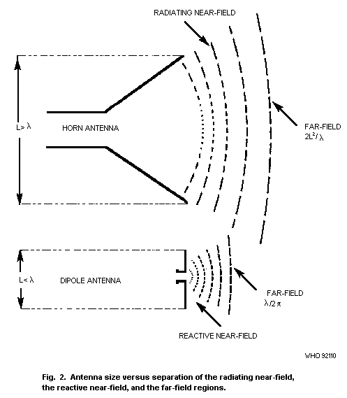 If the exposure takes place in the far-field of a well
characterized antenna in free space, then S is calculated by the
formula:
S = GPt /4pi r2 (W/m2) (Equation 4.5)
where G is the far-field power gain, Pt is the power transmitted (W)
and r is the distance (m) from the antenna.
For a horn or reflector type antenna:
G = 4pi Ae/lamda2 (Equation 4.6)
where Ae is the effective area of the antenna.
If G is not known, a useful approximation of S can be obtained by
substituting the physical area A for Ae in equation 4.6. This gives
a somewhat larger value for S, since A is generally larger than Ae.
Although the equations are far-field relationships, i.e., correct
for distances greater than approximately 2L2/lamda (L > lamda),
they can be used with an acceptable error for distances greater than
0.5 L2/lamda. The error is on the safe side, since the equations
predict greater values of S. However, at distances closer than 0.5
L2/lamda, the values of S predicted by the equations become
unrealistically large and radiating near-field estimates must be used.
For commonly encountered horn and reflector type antennas, the maximum
expected radiating, near-field, power flux density Sm can be
estimated (Hankin, 1974) from:
Sm = 4Pt/A (Equation 4.7)
Unfortunately, there are no equivalent reactive near-field
formulae for small radiators. The radiating near-field behaviour of
horn and reflector type antennas is discussed in detail elsewhere
(Bickmore & Hansen,1959; SAA, 1988). A detailed discussion of the
reactive near-field of small radiators can be found in Jordan &
Balmain (1968).
In the near-field, the situation is somewhat complicated, because
the maxima and minima of E and H fields do not occur at the same
points along the direction of propagation as they do in the the case
of the far-field. In this region, the electromagnetic field structure
may be highly inhomogeneous and typically, there may be substantial
variations from the plane wave impedance of 377 OMEGA; i.e., in some
regions, almost pure E-fields may exist and, in other regions, almost
pure H-fields. Field strengths in the near-field are more difficult to
specify, because both the E and H fields must be measured and because
the field patterns are more complicated; the power density tends to
vary inversely with r instead of r2 (as in the far-field), and may
display interference patterns. Near-field exposures become
particularly important when considering fields from microwave
diathermy equipment, RF sealers, broadcasting antennas, and microwave
oscillators under test.
4.3 Measurements
4.3.1 Preliminary considerations
Several steps are necessary for the accurate assessment of RF
exposure. The source and exposure situation must be characterized, so
that the most appropriate measurement technique and instrumentation
can be selected (ANSI, 1990; Tell, 1983). The correct use of this
instrumentation requires knowledge of the quantity to be measured and
the limitations of the instrument used. A knowledge of relevant
exposure standards is essential.
In the following sections, information is given concerning
preliminary RF survey considerations, measurement procedures, and
calibration facilities.
Prior to the commencement of a survey, it is important to obtain
as much information as possible about the characteristics of the RF
source and the exposure situation. This information is required for
the estimation of the expected field strengths and the selection of
the most appropriate survey instrumentation.
Information about the RF source should include:
- frequencies present, including harmonics;
- power transmitted;
- polarization (orientation of E field);
- modulation characteristics (peak and average values);
- duty cycle, pulse width, and pulse repetition frequency;
- antenna characteristics, such as type, gain, beam width and scan
rate.
Information about the exposure situation must include:
- distance from the source;
- existence of any scattering objects. Scattering by plane surfaces
can enhance the E field by a factor of 2, hence, S, by a factor
of 4. Even greater enhancement may result from curved surfaces,
e.g., corner reflectors.
4.3.2 Near-field versus far-field
For the practical purposes of measurement, the reactive
near-field exists within 0.5 lamda from the source with a practical
outer limit of several wavelengths (Jordan & Balmain, 1968). Both E
and H field components must be measured within the reactive
near-field. At present, no instruments are available commercially for
the measurement of H-fields above 300 MHz, which imposes a de facto
frequency limit on the measurements.
4.3.3 Instrumentation
An electric or magnetic field-measuring instrument consists of
three basic parts; the probe, the leads, and the monitor. To ensure
appropriate measurements, the following instrumentation
characteristics are required or are desirable:
- The probe must respond to only the E field or the H field and not
to both simultaneously.
- The probe must not produce significant perturbation of the field.
- The leads from the probe to the monitor must not disturb the
field at the probe significantly, or couple energy from the
field.
- The frequency response of the probe must cover the range of
frequencies required to be measured.
- If used in the reactive near-field, the dimensions of the probe
sensor should preferably be less than a quarter of a wavelength
at the highest frequency present (see next section).
- The instrument should indicate the root mean square (rms) value
of the measured field parameter.
- The response time of the instrument should be known. It is
desirable to have a response time of about 1 second or less, so
that intermittent fields are easily detected.
- The probe should be responsive to all polarization components of
the field. This may be accomplished, either by inherent isotropic
response, or by physical rotation of the probe through three
orthogonal directions.
- Good overload protection, battery operation, portability, and
rugged construction are other desirable characteristics.
- Instruments provide an indication of one or more of the following
parameters:
(a) Average power density (W/m2, mW/cm2);
(b) Average E field (V/m) or mean square E field (V2/m2);
(c) Average H field (A/m) or mean square H field (A2/m2).
However, no instrument actually measures average power density
and this quantity is not useful in the near-field of antennas. Power
density is measured in the far-field by E-field or H-field probes. The
surveyor should be aware of the field parameter (E or H) to which the
instrument responds, and that exposure standards generally stipulate
limits corresponding to both field parameters. Equivalent plane wave
power density is certainly a convenient unit, but in the reactive
near-field, E and H field components must be measured and compared
with the corresponding exposure limits.
Some factors that can influence the signal levels of the
instruments (e.g.,influence of multiple signals, pulse modulation,
lead pick-up, coupling into probes) are discussed in detail in ANSI
(1981) and Joyner (1988).
4.3.4 Measurement procedures
If information on the RF source and exposure situation is well
defined, then a surveyor, after making estimates of the expected field
strengths and selecting appropriate instruments, may proceed with the
survey using a high-range probe to avoid inadvertent probe burnout and
a high-range scale to avoid possible over-exposure.
In the reactive near-field of radiators operating at frequencies
of less than 300 MHz, an electrically small (largest dimension <0.25
lamda) probe sensor is required since large gradients in field
components exist. Spatial resolution is critical (large probes will
yield spatially averaged values) and the use of an isotropic probe is
strongly recommended. E and H field measurements should not be made
closer than a distance of 20 cm from metallic objects. In some such
cases, it may be possible to assess compliance with exposure standards
by making contact current measurements.
Non-uniform field distributions result from reflections from
various structures. Peaks in the field distribution are separated by
at least one-half wavelength with the maximum levels of E and H fields
occurring in different locations. Temporal variations occur also as a
result of scanning antennas, scanning radiation beams, and changes in
frequency. Therefore, it is imperative that any survey include a
sufficiently large sample of data to preclude omission of hazardous
combinations of conditions. When surveying sources of leakage
radiation, such as waveguide flanges, equipment cabinet doors, and
viewing or shielding screens, a "sniffing" procedure in the immediate
vicinity of the equipment is required. A low-power probe and
high-range setting should first be used to determine leakage sources
from a distance, and lower-range settings used as a closer approach is
made. Usually, leakage power varies as the inverse square of the
distance.
When surveying radar antennas, it is necessary to have the
antenna or the beam stationary, because the response time of the
instruments is generally not short enough to indicate the maximum
levels for common beam sweep and scan speed. It is important to
estimate the peak exposure level, in order to ensure that the probe
chosen can withstand such a peak level. Also, instruments that
time-sample the field at insufficiently low sample rates should not be
used for radar applications (Tell, 1983). Appropriate equations are
then used to convert back to time-averaged levels for a rotating
antenna.
All occupied and accessible locations should be surveyed. The
operator of the equipment under test and the surveyor should be as far
away as practicable from the test area. All objects normally present,
which may reflect or absorb energy, must be in position. The surveyor
should take precautions against RF burns and shock, particularly near
high-power, low-frequency systems.
With careful measurement techniques and the correct choice of
instrument, overall measurement uncertainties that are acceptable can
be achieved. Direct field measurements frequently do not provide
reliable means for exposure evaluation at distances from the field
source (an antenna, or a re-radiating surface) of less than about 0.2
m or lamda/2, whichever is smaller. In such a case, it may be
necessary to evaluate the specific absorption rates (SARs) in a model
of the human body using one of the dosimetric measures (Stuchly &
Stuchly, 1986), or to measure directly the RF current flowing through
the person (Blackwell, 1990; Tell, 1990a).
5. DOSIMETRY
5.1 General
Time-varying electric and magnetic fields induce electric fields
and corresponding electric currents in biological systems exposed to
these fields. The intensities and spatial distribution of induced
currents and fields are dependent on various characteristics of the
exposure field, the exposure geometry, and the exposed biological
system. The exposure field characteristics that play a role include
the type of field (electric, magnetic, or electromagnetic radiation),
frequency, polarization, direction, and strength. Important
characteristics of the exposed biological body system include its
size, geometry, and electrical properties. The electrical properties
of biological systems described by the complex permittivity and
electrical conductivity differ for various tissues.
The biological responses and effects due to exposure to
electromagnetic fields generally depend on the strength of induced
currents and fields. However, only the external fields can be measured
easily and dosimetry has been developed to correlate the induced
currents and fields with the exposure conditions. Induced currents, as
a measure of dose, have been used in the quantification of
experimentally induced effects in animals and the results have been
extrapolated to humans.
In the broad range of frequencies considered in this publication,
i.e., 300 Hz-300 GHz, two different, but interrelated, quantities are
commonly used in dosimetry. At lower frequencies (below approximately
100 kHz), many biological effects can be quantified in terms of the
current density in tissue. Therefore, this parameter is most often
used as a dosimetric quantity. At higher frequencies, where many (but
not all) interactions are due to the rate of energy deposition per
unit mass, the parameter specific absorption rate (SAR) is used. The
SAR is defined as "the time derivative of the incremental energy, dW,
absorbed by, or dissipated in, an incremental mass, dm, contained in
a volume element, dV, of a given density, rho" (NCRP 1981). The SAR is
most often expressed in units of watts per kilogram (W/kg).
5.2 Low frequency range
At frequencies below approximately 0.1-1 MHz, interactions of
electromagnetic fields with biological systems can be considered in
terms of induced currents and their density. This approach is
particularly well suited for calculations at frequencies for which the
dimensions of the object are small compared with the wavelength. Under
these circumstances, quasi-static approximations are valid, i.e., the
effects of the electric and the magnetic field can be considered
separately. The advantages of considering induced currents are
twofold. First, the current densities induced in humans can be
compared with those known to produce physiological responses, e.g.,
nerve or muscle stimulation, or they can be compared with endogenous
body currents. Second, consideration of induced currents in ungrounded
metallic objects can be used to assess thresholds for shocks and burns
for people, who are fully or partially grounded and come in contact
with such objects. Maximum current densities and the resulting maximum
SARs, in some parts of the human body under certain exposure
conditions, can be conveniently evaluated using the induced current
approach. The direct evaluation of the internal electric fields would
be much more complex and difficult. Under these conditions, limits of
exposure may be expressed more appropriately in terms of induced
currents rather than external field strengths.
The use of induced currents or current densities is appropriate
for the assessment of acute, immediate, safety hazards, while it may
have limitations for the complete evaluation of long-term effects.
This has yet to be determined.
5.2.1 Magnetic fields
In accordance with Faraday's law, magnetic fields that vary in
time induce the movement of electrical charge and cause potentials and
circulating (eddy) currents in biological systems. These currents can
be estimated using the following equation, provided that the current
paths are circular:
J = sigma E = 0.5 r sigma dB/dt (Equation 5.1)
where:
J = current density (A/m2)
E = induced electric field strength (V/m)
r = radius of the loop (m) (usually several cm up to 20 cm)
sigma = tissue conductivity (S/m)
dB/dt = rate of change of magnetic flux density B (T/s).
For sinusoidal fields of frequency f, equation 5.1 reduces to:
J = pi r sigma fB0 (Equation 5.2)
where B0 is the magnetic flux density peak amplitude.
The current density, internal electric field, and SAR, at any
location in an exposed biological body, are inter-related as follows:
SAR = sigma E2/rho (Equation 5.3)
where rho is the physical density (kg/m3) and
SAR = J2/sigma rho (Equation 5.4)
Because of the paucity of experimental data on the biological
effects of electromagnetic fields at frequencies below a few tens of
megahertz, consideration of the following effects of induced current
densities provides a useful alternative.
The magnitude of the magnetically induced electric fields and
current densities is proportional to the radius of the induction loop
in the body, to the tissue conductivity, and to the rate of change of
magnetic flux density. The dependence of the induced field and current
on the radius of the loop through which magnetic flux linkage occurs
is an important consideration for biological systems. The induced
current density is greatest at the periphery of the body, where the
conducting paths are longest, whereas microscopic current loops
anywhere within the body would have proportionally smaller current
densities dependent on the loop size. The magnitude of the current
density is influenced also by tissue electrical conductivity. In
biological bodies, the exact paths of the current flow depend in a
complicated way on the electrical conducting properties of the various
tissues.
It is difficult to calculate the complex current distributions in
biological bodies. Therefore, the treatment of this problem is
restricted, at present, to relatively simplified situations.
Typical values for the low-frequency electrical conductivity are
0.1-0.35 S/m for cardiac muscle and 0.1-0.3 S/m for nerve tissue.
Additionally, high ratios of transverse to longitudinal impedance up
to 7:1 have been observed (Epstein & Foster, 1983).
There is very little experimental or theoretical work dealing
with the coupling of magnetic fields to models of living organisms
(e.g., Spiegel (1976) described magnetic field coupling with spherical
models, Gandhi et al. (1984) calculated induced current densities in
the torso of a human using a multidimensional lattice of impedance
elements). Bernhardt (1979, 1985, 1988) performed calculations, using
"worst case" assumptions, to estimate the order of magnitude for
"safe" and "dangerous" values of magnetic field strengths and their
frequency dependence. Considering the cardiac region and the brain as
"critical" organs, approximate "worst case" calculations can be made
(Bernhardt, 1979, 1985). For the purpose of these calculations, both
regions can be considered as homogeneous spheres of different radii.
Differences in electrical conductivity of the white and grey cerebral
matter, and the anisotropic nature of conductivity at frequencies
below approximately 10 kHz are not considered. A value of sigma =
0.2S/m is used for the specific electrical conductivity of the
cerebral substance, and a value of 0.25 S/m is used for the myocardial
tissue. When a radius r of 7.5 cm of the induction loop is assumed for
the head, and 6 cm for the heart, the product sigma r is the same for
both the heart and head.
Therefore, approximately the same current densities are
calculated to result in the peripheral regions of the heart and brain
for a vertical magnetic field. Because of the uncertainties of the
current loops and of the values for the electrical conductivities, the
accuracies of these calculations are limited to about one order of
magnitude. For larger effective current loops and electrical
conductivities, smaller values of magnetic flux density may induce the
same current densities.
The waveform is an important factor to be considered in the
response of biological systems to a time-varying magnetic field. Many
different waveforms of magnetic field are used in medicine and
industry, including sinusoidal, square-wave, saw-tooth, and pulsed.
For these fields, the parameters of key importance are the rise and
decay signal times. These determine the maximum rates of change of the
field (dB/dt) and the maximum instantaneous current densities induced
in tissues. In order to provide an "effective" value for a variety of
waveforms, root-mean-square (rms) values are often used. However, peak
instantaneous field strengths appear to be important in considering
nerve and muscle cell stimulation, and for perturbing cell functions.
The effects depend strongly on frequency.
5.2.2 Electric fields
Exposure of a living organism to electric fields is normally
specified by the unperturbed electric field strength. The fields that
actually act on an exposed organism include electric fields acting on
the outer surface of the body and electric fields and current
densities induced inside the body. These fields can be different from
the exposure, because of perturbations caused by placing the body in
the external field. They must, however, be determined in order to
specify exposure at the level of living tissues or to relate exposure
levels and conditions from one species to another.
The electric fields that act directly on an exposed subject can
be categorized as follows:
(a) Electric fields acting on the outer surface of the body.
These fields can cause hair to vibrate and thereby can be perceived;
they may also be able to stimulate other sensory receptors in the
skin.
(b) Electric fields induced inside the body.
These fields act at the cellular level, and their presence is
accompanied by electric currents because of the electrically
conductive nature of living tissues.
Secondary short-term effects must also be considered when evaluating
health risks resulting from electric field exposure. Hazardous
thresholds for some indirect effects are lower than the thresholds for
biological effects due to the direct influence of electric fields. In
this case, the following points are important:
* Contact currents enter a person through electrical conductors in
contact with the skin.
* For static and low-frequency fields, spark discharges introduce
transient currents into the body via an arc gap, when the
electrical breakdown potential of air is exceeded.
* Electric or magnetic fields may interfere with the performance of
unipolar cardiac pacemakers.
Therefore, a clear distinction is necessary between effects
caused by the direct influence of electric fields and indirect effects
caused by approaching or touching electrically charged objects, or by
electromagnetic interference with implanted electromedical devices.
Within the body, the current and the current density are the two
main quantities of interest. The total current is more easily measured
or calculated, but the current density is more directly relevant to
electric field effects in a particular tissue or organ. The electrical
complexity of the interior of the human body, due to the presence of
insulating membranes and tissues of various impedances, has so far
frustrated confident analysis of precise interior current densities
(Kaune & Phillips, 1980; Spiegel, 1981).
Electric field coupling occurs through capacitive and conductive
mechanisms. A body is coupled to an electric field in proportion to
its capacitance to the ground as one equipotential surface, such that
the greater the capacitance the greater the current flow in the body.
By definition, in capacitive coupling, the body, according to its
capacitance C, "acquires" a certain amount of surface charge Q and
attains a potential V = Q/C. The capacitance, and, thus, the induced
current, decreases for a body separated from the ground and not close
to an energized electrode. The capacitance is dependent on the size
(especially on the surface area), the shape, and the orientation of
the body. Internal currents will differ between fat and thin persons,
between persons standing and reclining, and between persons walking
barefoot and those wearing shoes or standing on a non-conductive
platform. In all cases, it is necessary, to define the conditions
under which the capacitance has been measured.
A short-circuit current, Isc, flows in a body placed in an
electric field and connected to the ground through a low resistance
path (paws, bare feet, a hand grasping an earthed pole). This current
is the sum of all the displacement currents collected over the surface
of the body. The only place on the body where a current of the
magnitude of the short-circuit current flows is where there is
connection with the ground. The total current induced in the body is
simply the Maxwell's displacement current density multiplied by the
effective area of the body. Since the body is highly conducting, this
current is completely independent of the body's dielectric parameters.
Deno (1977) determined this effective area by measuring the surface
currents induced in hollow metal mannequins exposed to 60 Hz electric
fields. He characterized the complete current distribution and
determined the total short-circuit current to ground.
The equivalent area for an adult human corresponds to an
effective surface area of 5.08 m2 for a 1.77 m-tall subject. This
results in a total short circuit to ground current Isc (mA) for a
grounded person given by:
Isc = 0.09 h2Ef (Equation 5.5)
where h(in m) is the height of the person, E (in kV/m) is the electric
field strength, and f (in kHz) is the frequency.
From measurements by Guy & Chou (1982) and Tell et al. (1982),
the values of short-circuit current obtained by Deno for the metal
foil models were confirmed to be the same for humans at frequencies of
up to 1 MHz.
The results are shown in Fig. 3, normalized to an exposure level
of 614 V/m. Since the threshold for RF burns was found by Rogers
(1981) to be 200 mA, it is clear that an exposure level of 614 V/m
does not protect humans against RF burns resulting from contact with
grounded objects.
Deno's current distributions can be used to calculate spatial
distributions of SAR as well as average SAR for real human bodies
exposed to electric fields of wavelengths that are large compared with
the size of the body.
To make accurate calculations of the SAR distributions from the
body current distributions for various exposure conditions, it is
necessary to determine the electrical conductivity and resistance per
unit length along the axis of the body and limbs. At frequencies
between approximately 60 kHz and 3 MHz, this can be simply achieved by
passing a known very low-level (VLF-MF) current through the whole body
and measuring the potential at various points.
If the exposure takes place in the far-field of a well
characterized antenna in free space, then S is calculated by the
formula:
S = GPt /4pi r2 (W/m2) (Equation 4.5)
where G is the far-field power gain, Pt is the power transmitted (W)
and r is the distance (m) from the antenna.
For a horn or reflector type antenna:
G = 4pi Ae/lamda2 (Equation 4.6)
where Ae is the effective area of the antenna.
If G is not known, a useful approximation of S can be obtained by
substituting the physical area A for Ae in equation 4.6. This gives
a somewhat larger value for S, since A is generally larger than Ae.
Although the equations are far-field relationships, i.e., correct
for distances greater than approximately 2L2/lamda (L > lamda),
they can be used with an acceptable error for distances greater than
0.5 L2/lamda. The error is on the safe side, since the equations
predict greater values of S. However, at distances closer than 0.5
L2/lamda, the values of S predicted by the equations become
unrealistically large and radiating near-field estimates must be used.
For commonly encountered horn and reflector type antennas, the maximum
expected radiating, near-field, power flux density Sm can be
estimated (Hankin, 1974) from:
Sm = 4Pt/A (Equation 4.7)
Unfortunately, there are no equivalent reactive near-field
formulae for small radiators. The radiating near-field behaviour of
horn and reflector type antennas is discussed in detail elsewhere
(Bickmore & Hansen,1959; SAA, 1988). A detailed discussion of the
reactive near-field of small radiators can be found in Jordan &
Balmain (1968).
In the near-field, the situation is somewhat complicated, because
the maxima and minima of E and H fields do not occur at the same
points along the direction of propagation as they do in the the case
of the far-field. In this region, the electromagnetic field structure
may be highly inhomogeneous and typically, there may be substantial
variations from the plane wave impedance of 377 OMEGA; i.e., in some
regions, almost pure E-fields may exist and, in other regions, almost
pure H-fields. Field strengths in the near-field are more difficult to
specify, because both the E and H fields must be measured and because
the field patterns are more complicated; the power density tends to
vary inversely with r instead of r2 (as in the far-field), and may
display interference patterns. Near-field exposures become
particularly important when considering fields from microwave
diathermy equipment, RF sealers, broadcasting antennas, and microwave
oscillators under test.
4.3 Measurements
4.3.1 Preliminary considerations
Several steps are necessary for the accurate assessment of RF
exposure. The source and exposure situation must be characterized, so
that the most appropriate measurement technique and instrumentation
can be selected (ANSI, 1990; Tell, 1983). The correct use of this
instrumentation requires knowledge of the quantity to be measured and
the limitations of the instrument used. A knowledge of relevant
exposure standards is essential.
In the following sections, information is given concerning
preliminary RF survey considerations, measurement procedures, and
calibration facilities.
Prior to the commencement of a survey, it is important to obtain
as much information as possible about the characteristics of the RF
source and the exposure situation. This information is required for
the estimation of the expected field strengths and the selection of
the most appropriate survey instrumentation.
Information about the RF source should include:
- frequencies present, including harmonics;
- power transmitted;
- polarization (orientation of E field);
- modulation characteristics (peak and average values);
- duty cycle, pulse width, and pulse repetition frequency;
- antenna characteristics, such as type, gain, beam width and scan
rate.
Information about the exposure situation must include:
- distance from the source;
- existence of any scattering objects. Scattering by plane surfaces
can enhance the E field by a factor of 2, hence, S, by a factor
of 4. Even greater enhancement may result from curved surfaces,
e.g., corner reflectors.
4.3.2 Near-field versus far-field
For the practical purposes of measurement, the reactive
near-field exists within 0.5 lamda from the source with a practical
outer limit of several wavelengths (Jordan & Balmain, 1968). Both E
and H field components must be measured within the reactive
near-field. At present, no instruments are available commercially for
the measurement of H-fields above 300 MHz, which imposes a de facto
frequency limit on the measurements.
4.3.3 Instrumentation
An electric or magnetic field-measuring instrument consists of
three basic parts; the probe, the leads, and the monitor. To ensure
appropriate measurements, the following instrumentation
characteristics are required or are desirable:
- The probe must respond to only the E field or the H field and not
to both simultaneously.
- The probe must not produce significant perturbation of the field.
- The leads from the probe to the monitor must not disturb the
field at the probe significantly, or couple energy from the
field.
- The frequency response of the probe must cover the range of
frequencies required to be measured.
- If used in the reactive near-field, the dimensions of the probe
sensor should preferably be less than a quarter of a wavelength
at the highest frequency present (see next section).
- The instrument should indicate the root mean square (rms) value
of the measured field parameter.
- The response time of the instrument should be known. It is
desirable to have a response time of about 1 second or less, so
that intermittent fields are easily detected.
- The probe should be responsive to all polarization components of
the field. This may be accomplished, either by inherent isotropic
response, or by physical rotation of the probe through three
orthogonal directions.
- Good overload protection, battery operation, portability, and
rugged construction are other desirable characteristics.
- Instruments provide an indication of one or more of the following
parameters:
(a) Average power density (W/m2, mW/cm2);
(b) Average E field (V/m) or mean square E field (V2/m2);
(c) Average H field (A/m) or mean square H field (A2/m2).
However, no instrument actually measures average power density
and this quantity is not useful in the near-field of antennas. Power
density is measured in the far-field by E-field or H-field probes. The
surveyor should be aware of the field parameter (E or H) to which the
instrument responds, and that exposure standards generally stipulate
limits corresponding to both field parameters. Equivalent plane wave
power density is certainly a convenient unit, but in the reactive
near-field, E and H field components must be measured and compared
with the corresponding exposure limits.
Some factors that can influence the signal levels of the
instruments (e.g.,influence of multiple signals, pulse modulation,
lead pick-up, coupling into probes) are discussed in detail in ANSI
(1981) and Joyner (1988).
4.3.4 Measurement procedures
If information on the RF source and exposure situation is well
defined, then a surveyor, after making estimates of the expected field
strengths and selecting appropriate instruments, may proceed with the
survey using a high-range probe to avoid inadvertent probe burnout and
a high-range scale to avoid possible over-exposure.
In the reactive near-field of radiators operating at frequencies
of less than 300 MHz, an electrically small (largest dimension <0.25
lamda) probe sensor is required since large gradients in field
components exist. Spatial resolution is critical (large probes will
yield spatially averaged values) and the use of an isotropic probe is
strongly recommended. E and H field measurements should not be made
closer than a distance of 20 cm from metallic objects. In some such
cases, it may be possible to assess compliance with exposure standards
by making contact current measurements.
Non-uniform field distributions result from reflections from
various structures. Peaks in the field distribution are separated by
at least one-half wavelength with the maximum levels of E and H fields
occurring in different locations. Temporal variations occur also as a
result of scanning antennas, scanning radiation beams, and changes in
frequency. Therefore, it is imperative that any survey include a
sufficiently large sample of data to preclude omission of hazardous
combinations of conditions. When surveying sources of leakage
radiation, such as waveguide flanges, equipment cabinet doors, and
viewing or shielding screens, a "sniffing" procedure in the immediate
vicinity of the equipment is required. A low-power probe and
high-range setting should first be used to determine leakage sources
from a distance, and lower-range settings used as a closer approach is
made. Usually, leakage power varies as the inverse square of the
distance.
When surveying radar antennas, it is necessary to have the
antenna or the beam stationary, because the response time of the
instruments is generally not short enough to indicate the maximum
levels for common beam sweep and scan speed. It is important to
estimate the peak exposure level, in order to ensure that the probe
chosen can withstand such a peak level. Also, instruments that
time-sample the field at insufficiently low sample rates should not be
used for radar applications (Tell, 1983). Appropriate equations are
then used to convert back to time-averaged levels for a rotating
antenna.
All occupied and accessible locations should be surveyed. The
operator of the equipment under test and the surveyor should be as far
away as practicable from the test area. All objects normally present,
which may reflect or absorb energy, must be in position. The surveyor
should take precautions against RF burns and shock, particularly near
high-power, low-frequency systems.
With careful measurement techniques and the correct choice of
instrument, overall measurement uncertainties that are acceptable can
be achieved. Direct field measurements frequently do not provide
reliable means for exposure evaluation at distances from the field
source (an antenna, or a re-radiating surface) of less than about 0.2
m or lamda/2, whichever is smaller. In such a case, it may be
necessary to evaluate the specific absorption rates (SARs) in a model
of the human body using one of the dosimetric measures (Stuchly &
Stuchly, 1986), or to measure directly the RF current flowing through
the person (Blackwell, 1990; Tell, 1990a).
5. DOSIMETRY
5.1 General
Time-varying electric and magnetic fields induce electric fields
and corresponding electric currents in biological systems exposed to
these fields. The intensities and spatial distribution of induced
currents and fields are dependent on various characteristics of the
exposure field, the exposure geometry, and the exposed biological
system. The exposure field characteristics that play a role include
the type of field (electric, magnetic, or electromagnetic radiation),
frequency, polarization, direction, and strength. Important
characteristics of the exposed biological body system include its
size, geometry, and electrical properties. The electrical properties
of biological systems described by the complex permittivity and
electrical conductivity differ for various tissues.
The biological responses and effects due to exposure to
electromagnetic fields generally depend on the strength of induced
currents and fields. However, only the external fields can be measured
easily and dosimetry has been developed to correlate the induced
currents and fields with the exposure conditions. Induced currents, as
a measure of dose, have been used in the quantification of
experimentally induced effects in animals and the results have been
extrapolated to humans.
In the broad range of frequencies considered in this publication,
i.e., 300 Hz-300 GHz, two different, but interrelated, quantities are
commonly used in dosimetry. At lower frequencies (below approximately
100 kHz), many biological effects can be quantified in terms of the
current density in tissue. Therefore, this parameter is most often
used as a dosimetric quantity. At higher frequencies, where many (but
not all) interactions are due to the rate of energy deposition per
unit mass, the parameter specific absorption rate (SAR) is used. The
SAR is defined as "the time derivative of the incremental energy, dW,
absorbed by, or dissipated in, an incremental mass, dm, contained in
a volume element, dV, of a given density, rho" (NCRP 1981). The SAR is
most often expressed in units of watts per kilogram (W/kg).
5.2 Low frequency range
At frequencies below approximately 0.1-1 MHz, interactions of
electromagnetic fields with biological systems can be considered in
terms of induced currents and their density. This approach is
particularly well suited for calculations at frequencies for which the
dimensions of the object are small compared with the wavelength. Under
these circumstances, quasi-static approximations are valid, i.e., the
effects of the electric and the magnetic field can be considered
separately. The advantages of considering induced currents are
twofold. First, the current densities induced in humans can be
compared with those known to produce physiological responses, e.g.,
nerve or muscle stimulation, or they can be compared with endogenous
body currents. Second, consideration of induced currents in ungrounded
metallic objects can be used to assess thresholds for shocks and burns
for people, who are fully or partially grounded and come in contact
with such objects. Maximum current densities and the resulting maximum
SARs, in some parts of the human body under certain exposure
conditions, can be conveniently evaluated using the induced current
approach. The direct evaluation of the internal electric fields would
be much more complex and difficult. Under these conditions, limits of
exposure may be expressed more appropriately in terms of induced
currents rather than external field strengths.
The use of induced currents or current densities is appropriate
for the assessment of acute, immediate, safety hazards, while it may
have limitations for the complete evaluation of long-term effects.
This has yet to be determined.
5.2.1 Magnetic fields
In accordance with Faraday's law, magnetic fields that vary in
time induce the movement of electrical charge and cause potentials and
circulating (eddy) currents in biological systems. These currents can
be estimated using the following equation, provided that the current
paths are circular:
J = sigma E = 0.5 r sigma dB/dt (Equation 5.1)
where:
J = current density (A/m2)
E = induced electric field strength (V/m)
r = radius of the loop (m) (usually several cm up to 20 cm)
sigma = tissue conductivity (S/m)
dB/dt = rate of change of magnetic flux density B (T/s).
For sinusoidal fields of frequency f, equation 5.1 reduces to:
J = pi r sigma fB0 (Equation 5.2)
where B0 is the magnetic flux density peak amplitude.
The current density, internal electric field, and SAR, at any
location in an exposed biological body, are inter-related as follows:
SAR = sigma E2/rho (Equation 5.3)
where rho is the physical density (kg/m3) and
SAR = J2/sigma rho (Equation 5.4)
Because of the paucity of experimental data on the biological
effects of electromagnetic fields at frequencies below a few tens of
megahertz, consideration of the following effects of induced current
densities provides a useful alternative.
The magnitude of the magnetically induced electric fields and
current densities is proportional to the radius of the induction loop
in the body, to the tissue conductivity, and to the rate of change of
magnetic flux density. The dependence of the induced field and current
on the radius of the loop through which magnetic flux linkage occurs
is an important consideration for biological systems. The induced
current density is greatest at the periphery of the body, where the
conducting paths are longest, whereas microscopic current loops
anywhere within the body would have proportionally smaller current
densities dependent on the loop size. The magnitude of the current
density is influenced also by tissue electrical conductivity. In
biological bodies, the exact paths of the current flow depend in a
complicated way on the electrical conducting properties of the various
tissues.
It is difficult to calculate the complex current distributions in
biological bodies. Therefore, the treatment of this problem is
restricted, at present, to relatively simplified situations.
Typical values for the low-frequency electrical conductivity are
0.1-0.35 S/m for cardiac muscle and 0.1-0.3 S/m for nerve tissue.
Additionally, high ratios of transverse to longitudinal impedance up
to 7:1 have been observed (Epstein & Foster, 1983).
There is very little experimental or theoretical work dealing
with the coupling of magnetic fields to models of living organisms
(e.g., Spiegel (1976) described magnetic field coupling with spherical
models, Gandhi et al. (1984) calculated induced current densities in
the torso of a human using a multidimensional lattice of impedance
elements). Bernhardt (1979, 1985, 1988) performed calculations, using
"worst case" assumptions, to estimate the order of magnitude for
"safe" and "dangerous" values of magnetic field strengths and their
frequency dependence. Considering the cardiac region and the brain as
"critical" organs, approximate "worst case" calculations can be made
(Bernhardt, 1979, 1985). For the purpose of these calculations, both
regions can be considered as homogeneous spheres of different radii.
Differences in electrical conductivity of the white and grey cerebral
matter, and the anisotropic nature of conductivity at frequencies
below approximately 10 kHz are not considered. A value of sigma =
0.2S/m is used for the specific electrical conductivity of the
cerebral substance, and a value of 0.25 S/m is used for the myocardial
tissue. When a radius r of 7.5 cm of the induction loop is assumed for
the head, and 6 cm for the heart, the product sigma r is the same for
both the heart and head.
Therefore, approximately the same current densities are
calculated to result in the peripheral regions of the heart and brain
for a vertical magnetic field. Because of the uncertainties of the
current loops and of the values for the electrical conductivities, the
accuracies of these calculations are limited to about one order of
magnitude. For larger effective current loops and electrical
conductivities, smaller values of magnetic flux density may induce the
same current densities.
The waveform is an important factor to be considered in the
response of biological systems to a time-varying magnetic field. Many
different waveforms of magnetic field are used in medicine and
industry, including sinusoidal, square-wave, saw-tooth, and pulsed.
For these fields, the parameters of key importance are the rise and
decay signal times. These determine the maximum rates of change of the
field (dB/dt) and the maximum instantaneous current densities induced
in tissues. In order to provide an "effective" value for a variety of
waveforms, root-mean-square (rms) values are often used. However, peak
instantaneous field strengths appear to be important in considering
nerve and muscle cell stimulation, and for perturbing cell functions.
The effects depend strongly on frequency.
5.2.2 Electric fields
Exposure of a living organism to electric fields is normally
specified by the unperturbed electric field strength. The fields that
actually act on an exposed organism include electric fields acting on
the outer surface of the body and electric fields and current
densities induced inside the body. These fields can be different from
the exposure, because of perturbations caused by placing the body in
the external field. They must, however, be determined in order to
specify exposure at the level of living tissues or to relate exposure
levels and conditions from one species to another.
The electric fields that act directly on an exposed subject can
be categorized as follows:
(a) Electric fields acting on the outer surface of the body.
These fields can cause hair to vibrate and thereby can be perceived;
they may also be able to stimulate other sensory receptors in the
skin.
(b) Electric fields induced inside the body.
These fields act at the cellular level, and their presence is
accompanied by electric currents because of the electrically
conductive nature of living tissues.
Secondary short-term effects must also be considered when evaluating
health risks resulting from electric field exposure. Hazardous
thresholds for some indirect effects are lower than the thresholds for
biological effects due to the direct influence of electric fields. In
this case, the following points are important:
* Contact currents enter a person through electrical conductors in
contact with the skin.
* For static and low-frequency fields, spark discharges introduce
transient currents into the body via an arc gap, when the
electrical breakdown potential of air is exceeded.
* Electric or magnetic fields may interfere with the performance of
unipolar cardiac pacemakers.
Therefore, a clear distinction is necessary between effects
caused by the direct influence of electric fields and indirect effects
caused by approaching or touching electrically charged objects, or by
electromagnetic interference with implanted electromedical devices.
Within the body, the current and the current density are the two
main quantities of interest. The total current is more easily measured
or calculated, but the current density is more directly relevant to
electric field effects in a particular tissue or organ. The electrical
complexity of the interior of the human body, due to the presence of
insulating membranes and tissues of various impedances, has so far
frustrated confident analysis of precise interior current densities
(Kaune & Phillips, 1980; Spiegel, 1981).
Electric field coupling occurs through capacitive and conductive
mechanisms. A body is coupled to an electric field in proportion to
its capacitance to the ground as one equipotential surface, such that
the greater the capacitance the greater the current flow in the body.
By definition, in capacitive coupling, the body, according to its
capacitance C, "acquires" a certain amount of surface charge Q and
attains a potential V = Q/C. The capacitance, and, thus, the induced
current, decreases for a body separated from the ground and not close
to an energized electrode. The capacitance is dependent on the size
(especially on the surface area), the shape, and the orientation of
the body. Internal currents will differ between fat and thin persons,
between persons standing and reclining, and between persons walking
barefoot and those wearing shoes or standing on a non-conductive
platform. In all cases, it is necessary, to define the conditions
under which the capacitance has been measured.
A short-circuit current, Isc, flows in a body placed in an
electric field and connected to the ground through a low resistance
path (paws, bare feet, a hand grasping an earthed pole). This current
is the sum of all the displacement currents collected over the surface
of the body. The only place on the body where a current of the
magnitude of the short-circuit current flows is where there is
connection with the ground. The total current induced in the body is
simply the Maxwell's displacement current density multiplied by the
effective area of the body. Since the body is highly conducting, this
current is completely independent of the body's dielectric parameters.
Deno (1977) determined this effective area by measuring the surface
currents induced in hollow metal mannequins exposed to 60 Hz electric
fields. He characterized the complete current distribution and
determined the total short-circuit current to ground.
The equivalent area for an adult human corresponds to an
effective surface area of 5.08 m2 for a 1.77 m-tall subject. This
results in a total short circuit to ground current Isc (mA) for a
grounded person given by:
Isc = 0.09 h2Ef (Equation 5.5)
where h(in m) is the height of the person, E (in kV/m) is the electric
field strength, and f (in kHz) is the frequency.
From measurements by Guy & Chou (1982) and Tell et al. (1982),
the values of short-circuit current obtained by Deno for the metal
foil models were confirmed to be the same for humans at frequencies of
up to 1 MHz.
The results are shown in Fig. 3, normalized to an exposure level
of 614 V/m. Since the threshold for RF burns was found by Rogers
(1981) to be 200 mA, it is clear that an exposure level of 614 V/m
does not protect humans against RF burns resulting from contact with
grounded objects.
Deno's current distributions can be used to calculate spatial
distributions of SAR as well as average SAR for real human bodies
exposed to electric fields of wavelengths that are large compared with
the size of the body.
To make accurate calculations of the SAR distributions from the
body current distributions for various exposure conditions, it is
necessary to determine the electrical conductivity and resistance per
unit length along the axis of the body and limbs. At frequencies
between approximately 60 kHz and 3 MHz, this can be simply achieved by
passing a known very low-level (VLF-MF) current through the whole body
and measuring the potential at various points.
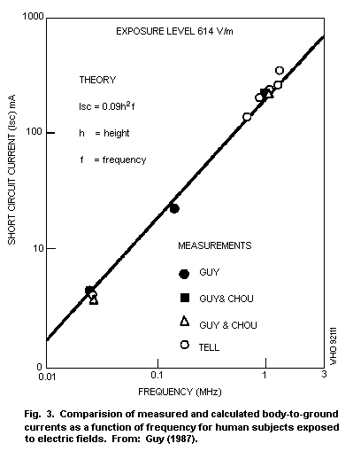 Calculations of SAR for exposure levels of 614 V/m, based on
measured electrical conductivity and current distribution, are
illustrated in Fig. 4 for exposure conditions where the feet are
grounded. The maximum SAR values were obtained from the average values
in each elliptical element by assuming that the current would be
shunted through fat, bone, and muscle tissues, according to the ratios
of the electrical conductivity of each tissue to the average
electrical conductivity of the entire elliptical element.
Calculations of SAR for exposure levels of 614 V/m, based on
measured electrical conductivity and current distribution, are
illustrated in Fig. 4 for exposure conditions where the feet are
grounded. The maximum SAR values were obtained from the average values
in each elliptical element by assuming that the current would be
shunted through fat, bone, and muscle tissues, according to the ratios
of the electrical conductivity of each tissue to the average
electrical conductivity of the entire elliptical element.
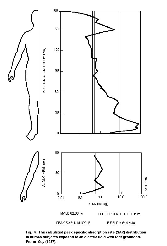 The peak SAR occurring in the muscle and blood vessels of the
ankle, when the feet are grounded, reaches a value of 100 W/kg. Gandhi
(1985) was the first to draw attention to this problem. Although these
SAR values are quite high, they occur in a relatively small volume and
the thermal consequences are difficult to predict.
In studies on the distribution of the electric field or the
absorbed power in different parts of the human body, it has been
demonstrated that, for fields of frequencies below 10 MHz, the
internal field strength increases in direct proportion to the
frequency for a given external electric field strength. Therefore, a
simple relationship exists between the internal and the external
electric field strengths, depending on the body part or organ
considered, on the electrical conductivity, and on the exposure
conditions.
A detailed evaluation of current density thresholds as a function
of frequency for various interactions, and an estimation of maximum
current densities in models of humans exposed to electric and magnetic
fields of frequencies of less than 100 kHz, have been reported
(Bernhardt, 1985). Envelope curves of current densities that are
required for cell stimulation, and those associated with endogenous
currents in brain tissue have been established for fields of
frequencies up to 100 kHz.
The current densities induced within the body by an external
electric field E and frequency f were calculated using the formula
J=KfE. The constant K depends on the part of the body considered
(Bernhardt, 1985). The longitudinal axis of the body parallel to the
external E field represents optimum coupling geometry and must be
considered as the "worst case".
The K values can be determined by two different methods. Data
from studies by different authors on absorption within the
quasi-static range can be used, or K can be determined by calculating
the current densities on the basis of the field strength measured on
the body-surface at 50/60 Hz. The K values, determined by entirely
different methods, coincide satisfactorily. The same value K=3.10-9
S/(Hz m) was obtained for the cardiac region and head, however for
other parts of the body the values of K may be larger (Kaune &
Phillips, 1980; Guy et al., 1982; Kaune & Forsythe, 1985). The surface
E field and current density data derived from human measurements
(Deno, 1977) and animal data (Kaune & Phillips, 1980) demonstrate that
the external unperturbed fields, which are almost always used to
specify exposure, must be scaled to equalize internal current
densities or surface E fields. This must be done in order to
extrapolate biological data from one species to another. This process
is complicated by the fact that the actual value of the scaling factor
depends on the internal quantity that is being scaled.
Currents in electrically-grounded people exposed to fields at
frequencies below 50 MHz have been measured (Guy & Chou, 1982; Gandhi
et al., 1985b, 1986). The resulting SARs in a small volume within the
ankle were estimated to be in the range of 200-540 W/kg for E fields
of 61.4 V/m in the range of frequencies 40-62.5 MHz. However, lower
values were found in a quantitative analysis by Dimbylow (1987, 1988).
The SAR in the wrist for contact with isolated metallic objects
in an RF field has been calculated as a function of contact current
for various frequencies used in broadcasting (Tell, 1990). The maximum
contact currents to maintain the SARs below 8 W/kg and 20 W/kg are
given in Table 5. The values in Table 5 are based on an assumed
effective wrist cross section of 11.1 cm2.
Table 5. Maximum contact currents to keep SARs in the wrist below
8 and 20 W/kga
Broadcast band Limiting current to control SAR (mA)
<8 W/kg <20 W/kg
AM (0.55-1.6 MHz) 75.1 119
Low VHF (54-88 MHz) 84.1 133
FM (88-l08 MHz) 87.3 138
High VHF (176-216 MHz) 93.6 148
Channel 14 (470-476 MHz) 99.7 158
Channel 20 (506-512 MHz) 100 159
Channel 66 (782-788 MHz) 124 197
a From: Tell (1990).
5.3 High-frequency range
The interaction of RF fields with matter can be described in
terms of its electrical properties, which are the macroscopic
reflection of interactions at the molecular or cellular level. The
basic interaction mechanisms, which are discussed in section 6,
involve relaxation phenomena due to the rotation of polar molecules,
such as water, amino acids, protein, lipids, interfacial space-charge
polarization due to non homogeneous structures (e.g., cell membranes),
and ionic conduction.
The internal fields can be quantified in various ways. The
magnetic permeability of tissue is practically equal to that of free
space, and all known and anticipated interactions occur through
mechanisms involving the electric field (including the current induced
by the magnetic field). Therefore, the electric field vector, or its
distribution throughout the exposed body, fully describes the exposure
field-tissue interactions. Some additional information may be needed
for full quantification, e.g., the frequency characteristics of the
exposure field, such as modulation characteristics and modulation
frequency.
A direct calculation of the expected temperature rise (DELTA T in
kelvin) in tissue exposed to RF fields for a time (t seconds) can be
made from the equation:
DELTA T = (SAR) t / C (Equation 5.6)
where C is the specific heat capacity expressed in J/kg K. This
equation, however, does not include terms to account for heat losses
via processes such as thermal conduction and convection. Although it
expresses the rate at which the electromagnetic energy is converted
into heat through well established interaction mechanisms, it provides
a valid quantitative measure of all the interaction mechanisms that
are dependent on the intensity of the internal electric field in a
non-linear manner. Some additional information may be relevant. For
instance, since some effects of RF fields modulated in amplitude at
ELF (extremely low frequencies) are dependent on the electric field
intensity (Adey, 1981), they could probably be expressed in terms of
the SAR, modulation characteristics, and the "zones" or windows of
amplitudes of the SAR that are biologically effective.
The SAR concept has proved to be a simple and useful tool in
quantifying the interactions of RF fields with living systems. It
enables comparison of experimentally observed biological effects for
various species under various exposure conditions and it provides the
only means, however imperfect, of extrapolating animal data to
potential hazards for humans exposed to RF. It also facilitates
planning and effective execution of therapeutic hyperthermic
treatment.
Dosimetry in bioelectromagnetic research has been developing in
two parallel but interacting complementary ways, the theoretical and
the experimental. RF dosimetry calculations can be performed by
solving Maxwell's equations for a given configuration approximating
the exposed object (an animal, a human being, a part of a human body)
and for given exposure conditions (e.g., a plane wave at a given
frequency, incident from a given direction). These data have been
collected and discussed in the Radiofrequency radiation dosimetry
handbook (Durney et al., 1986). However, even analyses of greatly
simplified models provide valuable information for quantifying
interactions of electromagnetic fields with biological systems. The
results obtained from simple models often provide valuable insight and
qualitative understanding that can facilitate the analysis of more
complex models.
Fig. 5 illustrates the average SAR as a function of frequency for
an average man exposed to a plane wave for three polarizations (Durney
et al., 1978; Durney, 1980). Various models used in the calculations
are also indicated.
From these data, the following conclusions can be drawn:
- the average SAR is a function of frequency;
- the average SAR depends on the wave polarization, and is greatest
for the E polarization (electric field is parallel to the long
axis of the body), except at higher frequencies, where it is
slightly greater for the H polarization (magnetic field (H) is
parallel to the long axis of the body);
- the average SARs for the E or K polarizations (when electric
field (E) or wave propagation direction (K), respectively, are
parallel to the long axis of the body) exhibit a maximum at
certain frequencies, called the resonant frequencies.
The peak SAR occurring in the muscle and blood vessels of the
ankle, when the feet are grounded, reaches a value of 100 W/kg. Gandhi
(1985) was the first to draw attention to this problem. Although these
SAR values are quite high, they occur in a relatively small volume and
the thermal consequences are difficult to predict.
In studies on the distribution of the electric field or the
absorbed power in different parts of the human body, it has been
demonstrated that, for fields of frequencies below 10 MHz, the
internal field strength increases in direct proportion to the
frequency for a given external electric field strength. Therefore, a
simple relationship exists between the internal and the external
electric field strengths, depending on the body part or organ
considered, on the electrical conductivity, and on the exposure
conditions.
A detailed evaluation of current density thresholds as a function
of frequency for various interactions, and an estimation of maximum
current densities in models of humans exposed to electric and magnetic
fields of frequencies of less than 100 kHz, have been reported
(Bernhardt, 1985). Envelope curves of current densities that are
required for cell stimulation, and those associated with endogenous
currents in brain tissue have been established for fields of
frequencies up to 100 kHz.
The current densities induced within the body by an external
electric field E and frequency f were calculated using the formula
J=KfE. The constant K depends on the part of the body considered
(Bernhardt, 1985). The longitudinal axis of the body parallel to the
external E field represents optimum coupling geometry and must be
considered as the "worst case".
The K values can be determined by two different methods. Data
from studies by different authors on absorption within the
quasi-static range can be used, or K can be determined by calculating
the current densities on the basis of the field strength measured on
the body-surface at 50/60 Hz. The K values, determined by entirely
different methods, coincide satisfactorily. The same value K=3.10-9
S/(Hz m) was obtained for the cardiac region and head, however for
other parts of the body the values of K may be larger (Kaune &
Phillips, 1980; Guy et al., 1982; Kaune & Forsythe, 1985). The surface
E field and current density data derived from human measurements
(Deno, 1977) and animal data (Kaune & Phillips, 1980) demonstrate that
the external unperturbed fields, which are almost always used to
specify exposure, must be scaled to equalize internal current
densities or surface E fields. This must be done in order to
extrapolate biological data from one species to another. This process
is complicated by the fact that the actual value of the scaling factor
depends on the internal quantity that is being scaled.
Currents in electrically-grounded people exposed to fields at
frequencies below 50 MHz have been measured (Guy & Chou, 1982; Gandhi
et al., 1985b, 1986). The resulting SARs in a small volume within the
ankle were estimated to be in the range of 200-540 W/kg for E fields
of 61.4 V/m in the range of frequencies 40-62.5 MHz. However, lower
values were found in a quantitative analysis by Dimbylow (1987, 1988).
The SAR in the wrist for contact with isolated metallic objects
in an RF field has been calculated as a function of contact current
for various frequencies used in broadcasting (Tell, 1990). The maximum
contact currents to maintain the SARs below 8 W/kg and 20 W/kg are
given in Table 5. The values in Table 5 are based on an assumed
effective wrist cross section of 11.1 cm2.
Table 5. Maximum contact currents to keep SARs in the wrist below
8 and 20 W/kga
Broadcast band Limiting current to control SAR (mA)
<8 W/kg <20 W/kg
AM (0.55-1.6 MHz) 75.1 119
Low VHF (54-88 MHz) 84.1 133
FM (88-l08 MHz) 87.3 138
High VHF (176-216 MHz) 93.6 148
Channel 14 (470-476 MHz) 99.7 158
Channel 20 (506-512 MHz) 100 159
Channel 66 (782-788 MHz) 124 197
a From: Tell (1990).
5.3 High-frequency range
The interaction of RF fields with matter can be described in
terms of its electrical properties, which are the macroscopic
reflection of interactions at the molecular or cellular level. The
basic interaction mechanisms, which are discussed in section 6,
involve relaxation phenomena due to the rotation of polar molecules,
such as water, amino acids, protein, lipids, interfacial space-charge
polarization due to non homogeneous structures (e.g., cell membranes),
and ionic conduction.
The internal fields can be quantified in various ways. The
magnetic permeability of tissue is practically equal to that of free
space, and all known and anticipated interactions occur through
mechanisms involving the electric field (including the current induced
by the magnetic field). Therefore, the electric field vector, or its
distribution throughout the exposed body, fully describes the exposure
field-tissue interactions. Some additional information may be needed
for full quantification, e.g., the frequency characteristics of the
exposure field, such as modulation characteristics and modulation
frequency.
A direct calculation of the expected temperature rise (DELTA T in
kelvin) in tissue exposed to RF fields for a time (t seconds) can be
made from the equation:
DELTA T = (SAR) t / C (Equation 5.6)
where C is the specific heat capacity expressed in J/kg K. This
equation, however, does not include terms to account for heat losses
via processes such as thermal conduction and convection. Although it
expresses the rate at which the electromagnetic energy is converted
into heat through well established interaction mechanisms, it provides
a valid quantitative measure of all the interaction mechanisms that
are dependent on the intensity of the internal electric field in a
non-linear manner. Some additional information may be relevant. For
instance, since some effects of RF fields modulated in amplitude at
ELF (extremely low frequencies) are dependent on the electric field
intensity (Adey, 1981), they could probably be expressed in terms of
the SAR, modulation characteristics, and the "zones" or windows of
amplitudes of the SAR that are biologically effective.
The SAR concept has proved to be a simple and useful tool in
quantifying the interactions of RF fields with living systems. It
enables comparison of experimentally observed biological effects for
various species under various exposure conditions and it provides the
only means, however imperfect, of extrapolating animal data to
potential hazards for humans exposed to RF. It also facilitates
planning and effective execution of therapeutic hyperthermic
treatment.
Dosimetry in bioelectromagnetic research has been developing in
two parallel but interacting complementary ways, the theoretical and
the experimental. RF dosimetry calculations can be performed by
solving Maxwell's equations for a given configuration approximating
the exposed object (an animal, a human being, a part of a human body)
and for given exposure conditions (e.g., a plane wave at a given
frequency, incident from a given direction). These data have been
collected and discussed in the Radiofrequency radiation dosimetry
handbook (Durney et al., 1986). However, even analyses of greatly
simplified models provide valuable information for quantifying
interactions of electromagnetic fields with biological systems. The
results obtained from simple models often provide valuable insight and
qualitative understanding that can facilitate the analysis of more
complex models.
Fig. 5 illustrates the average SAR as a function of frequency for
an average man exposed to a plane wave for three polarizations (Durney
et al., 1978; Durney, 1980). Various models used in the calculations
are also indicated.
From these data, the following conclusions can be drawn:
- the average SAR is a function of frequency;
- the average SAR depends on the wave polarization, and is greatest
for the E polarization (electric field is parallel to the long
axis of the body), except at higher frequencies, where it is
slightly greater for the H polarization (magnetic field (H) is
parallel to the long axis of the body);
- the average SARs for the E or K polarizations (when electric
field (E) or wave propagation direction (K), respectively, are
parallel to the long axis of the body) exhibit a maximum at
certain frequencies, called the resonant frequencies.
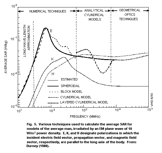
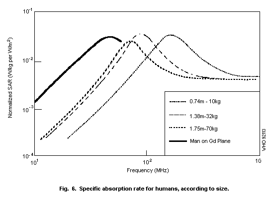 The frequency-dependent behaviour is illustrated in Fig. 6 for
several human sizes. The average whole-body SAR in W/kg is plotted as
a function of electromagnetic field frequency (MHz) for an incident
average power density of 1 W/m2.
Based on the absorption characteristics in the human body, the
radiofrequency range can be subdivided into four regions (IRPA,
1988a), as shown in Fig. 7:
(a) The sub-resonance range, less than 30 MHz, where surface
absorption dominates for the human trunk, but not for the neck
and legs, and where energy absorption increases rapidly with
frequency (in the neck and the legs significantly larger
absorptions may occur).
(b) The resonance range, extending from 30 MHz to about 300 MHz
for the whole body, and to even higher frequencies if partial
body resonances, more particularly in the head, are considered.
(c) The "hot-spot" range, extending from about 400 MHz up to about
3 GHz, where significant localized energy absorption can be
expected at incident power densities of about 100 W/m2; energy
absorption decreases when frequency increases and the sizes of
hot spots range from several cm at 915 MHz to about 1 cm at 3
GHz.
(d) The surface absorption range, greater than about 3 GHz, where
the temperature elevation is localized and restricted to the
surface of the body.
The average SAR varies with species, as illustrated in Fig. 8.
These data are of importance in extrapolation of the results from
experimental animal studies to human exposures. The average SAR varies
within one order of magnitude in the subresonance range, depending on
the separation of an average person from the electric ground plane
(with the highest SAR for a person on a ground plane).
Whole-body-average SARs have been measured for humans (Hill,
1984a,b,c; Hill & Walsh, 1985), and the spatial distribution of the
SARs in full-scale, realistic models of the human body (Kraszewski et
al., 1984; Stuchly M. et al., 1985, 1986; Stuchly S. et al., 1985).
The whole-body average SAR was measured for human volunteers exposed
to RF at a few frequencies between 3 and 41 MHz, which are below and
close to the resonant frequencies of adult humans. The exposure
conditions simulated free-space and grounded conditions in the
orientation that results in the greatest SARs (Hill, 1984a, b, c). At
all frequencies, the measured SARs exceeded the calculated values by
a factor of 2.7-3.9 in free space, and 4.3-4.4 for the grounded
condition.
The frequency-dependent behaviour is illustrated in Fig. 6 for
several human sizes. The average whole-body SAR in W/kg is plotted as
a function of electromagnetic field frequency (MHz) for an incident
average power density of 1 W/m2.
Based on the absorption characteristics in the human body, the
radiofrequency range can be subdivided into four regions (IRPA,
1988a), as shown in Fig. 7:
(a) The sub-resonance range, less than 30 MHz, where surface
absorption dominates for the human trunk, but not for the neck
and legs, and where energy absorption increases rapidly with
frequency (in the neck and the legs significantly larger
absorptions may occur).
(b) The resonance range, extending from 30 MHz to about 300 MHz
for the whole body, and to even higher frequencies if partial
body resonances, more particularly in the head, are considered.
(c) The "hot-spot" range, extending from about 400 MHz up to about
3 GHz, where significant localized energy absorption can be
expected at incident power densities of about 100 W/m2; energy
absorption decreases when frequency increases and the sizes of
hot spots range from several cm at 915 MHz to about 1 cm at 3
GHz.
(d) The surface absorption range, greater than about 3 GHz, where
the temperature elevation is localized and restricted to the
surface of the body.
The average SAR varies with species, as illustrated in Fig. 8.
These data are of importance in extrapolation of the results from
experimental animal studies to human exposures. The average SAR varies
within one order of magnitude in the subresonance range, depending on
the separation of an average person from the electric ground plane
(with the highest SAR for a person on a ground plane).
Whole-body-average SARs have been measured for humans (Hill,
1984a,b,c; Hill & Walsh, 1985), and the spatial distribution of the
SARs in full-scale, realistic models of the human body (Kraszewski et
al., 1984; Stuchly M. et al., 1985, 1986; Stuchly S. et al., 1985).
The whole-body average SAR was measured for human volunteers exposed
to RF at a few frequencies between 3 and 41 MHz, which are below and
close to the resonant frequencies of adult humans. The exposure
conditions simulated free-space and grounded conditions in the
orientation that results in the greatest SARs (Hill, 1984a, b, c). At
all frequencies, the measured SARs exceeded the calculated values by
a factor of 2.7-3.9 in free space, and 4.3-4.4 for the grounded
condition.
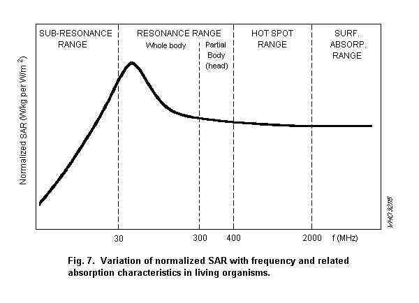
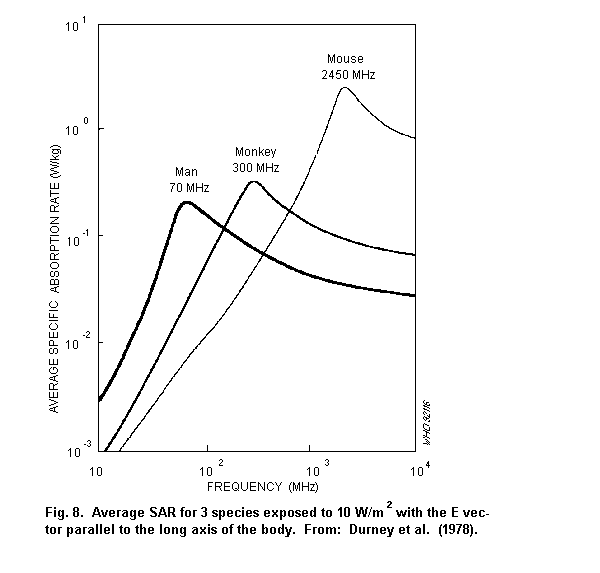 Similar differences between the calculated and measured SAR for
simple models were found on scaled-down models at 5-10 MHz (Guy,
1987). Spatial distributions of the SAR in models of the human body
have been investigated experimentally (Guy et al., 1984; Kraszewski et
al., 1984; Stuchly M. et al., 1986; Stuchly S. et al., 1987). Large
differences, typically by a factor of 10-30, between the measured SAR
values and those previously calculated using a block model, have been
observed (Stuchly M. et al., 1986) at frequencies above resonance.
However, despite the differences in spatial distributions, the ratios
of peak to whole-body average SARs predicted theoretically and
measured, were relatively small, except for the SAR at the body
surface. With reference to developing human exposure limits, these
results underscore the limitations of the theoretical methods of
prediction available at present.
The measurements on a full-scale model (Olsen, 1982; Stuchly S.
et al., 1986 ), on a scaled-down model of man (Guy et al., 1984), and
on a full-scale model of a monkey (Olsen & Griner, 1982) all indicated
that, for free space and the most absorbing orientation
(E-polarization), measured values are close to those predicted from
calculations at, and above, the resonant frequency (up to about 450
MHz).
Changes to the average SAR for important practical exposure
conditions (e.g., separation between the subject's feet and the ground
plane, the body position, articulations of the limbs, and two-body
interactions) have been investigated using human volunteers (Hill,
1984b, 1984c). Footwear reduces the average SAR with the degree of
reduction depending on the type of footwear and the frequency of the
exposure field.
Similar effects have been observed in body currents measured in
people exposed to HF and VHF antennas (Allen et al., 1988).
High local SARs also occur at frequencies around and below the
resonant frequency at locations such as the ankles (Gandhi et al.,
1985b, 1986) and the wrist (Guy & Chou, 1982). At frequencies above a
few GHz (millimetre waves), high local SARs are produced at the body
surface (Gandhi & Riazi, 1986). Exposures corresponding to 10 W/m2
may result in perception of heating.
Data have also been collected on the SAR distribution for
near-field exposures (Stuchly M. et al., 1985, 1986, 1987; Stuchly S.
et al., 1985, 1986). One of the most important findings is that the
SAR distributions are highly non-uniform, with typical ratios between
spatial peak and whole-body average SARs of the order of 150:1 to
200:1 (Stuchly S. et al., 1985). At all frequencies investigated, the
maximum SAR is at the body surface, with lower magnitude "hot spots"
located inside the body. Practically all the energy, however, is
deposited within about 20% of the body volume closest to the antenna.
Knowledge of these SARs can be used in specifying, for instance, the
maximum output power of portable transmitters that would be allowed
under a selected limit of the SAR.
5.4 Derivation of exposure limits from basic quantities
For the assessment of the possible health effects of
electromagnetic fields, it is useful to differentiate between basic
limits and derived limits.
Basic limits may be directly correlated with biological effects.
Using experimental data or related studies, a threshold exposure level
can be determined, above which adverse health effects are increasingly
likely, but below which no adverse effect occurs. The basic exposure
limit is based on this threshold level.
Since basic limits in terms of SAR or induced current density
cannot be measured easily in practical exposure situations, exposure
limits in conveniently measured quantities are derived from the basic
limit. These derived limits then indicate the acceptable limits in
terms of measured and/or calculated field parameters.
Three categories of basic limits have been identified and
quantitatively established.
1. The specific absorption rate (SAR) averaged over the whole body
or over parts of the body:
Whole body SAR is a widely accepted measure for relating adverse
effects to RF exposure, especially for frequencies above about 10
MHz. Local SAR values are necessary to evaluate and limit
excessive energy deposition in small parts of the body and to
avoid hot spots resulting from special exposure conditions.
Examples of such conditions are: a grounded individual exposed to
RF in the low MHz-range; individuals exposed in the near-field of
an antenna or individuals exposed at the higher end of the
frequency range, where the penetration depth of the RF is low.
2. The induced electric field strength or current density:
RF fields can induce sufficiently high current densities to
stimulate excitable tissue (nerve or muscle) or to produce other
potentially harmful effects, especially at frequencies below 100
kHz. The thresholds for biological effects are expressed in terms
of current density and are strongly frequency dependent.
3. Contact current between a person and a charged object:
A conductive object in an electric field can be energized by the
field. For field frequencies below 100 kHz, contact between the
object and a person may result in stimulation of electrically
excitable tissue with pain and more severe effects (burns), if
the current density is sufficiently high. For frequencies between
about 100 kHz and 100 MHz, the hazard of burns from contact
current will predominate.
Derived limits are necessary to provide a practical method to
evaluate a given RF exposure. Derived limits obtained from one of
these basic limits above include, e.g., electric and magnetic field
strength, power density, contact voltage of the conductive objects,
and short-circuit currents. The derived limits have to be calculated
in such a way that, even under worst-case conditions of field
exposure, the basic limits will not be exceeded. In many special
exposure conditions, e.g., in the near-field, very close (less than
0.5 wavelength) to an antenna, the assessment of possible health
effects may require separate measurements or calculations to
investigate whether the basic limit is exceeded.
6. INTERACTION MECHANISMS
6.1 General
Electromagnetic fields in the frequency range 300 Hz-300 GHz
interact with biological systems (humans and other animals) through
direct and indirect mechanisms. A direct interaction produces effects
in the exposed organisms directly from exposure to the electromagnetic
field. An indirect interaction is mediated through the presence of
other bodies in the electromagnetic field, and occurs as a result of
an interaction (usually physical contact) between the biological body
and another object, such as an automobile, fence, or even another
biological body.
Direct interactions that are well understood can be quantified in
terms of dosimetry, and can be considered as resulting from induced
currents and internal electric fields. The macroscopic spatial
distribution of these currents and fields within an exposed biological
body is of importance and is determined by theoretical and
experimental dosimetry. The spatial distributions of the currents and
fields within, and around, the cell are also important. As outlined
earlier, the patterns of induced currents and fields within biological
systems usually are highly non-uniform and depend on the geometry and
electrical properties of the exposed system, as well as on the field
frequency, and, for lower frequencies, the type of field, whether
electric or magnetic (where spatial separation of the electric and the
magnetic field is realistic). The extent to which the electric or
magnetic field plays a role is uncertain. However, apart from
differences due to different current distributions, the frequency of
the field clearly establishes the type of mechanism for the mechanisms
that are well understood.
For frequencies below about 100 kHz, an established interaction
mechanism is the stimulation of excitable tissues by induced currents.
For higher frequencies, thermal interactions predominate. At the lower
frequencies, much less of the electromagnetic field is absorbed by
biological systems. Thermal interactions occur at energy levels much
higher than interactions due to induced currents. Therefore, thermal
interactions are usually of little interest for fields at levels at
which people are exposed. Additionally, at frequencies below
approximately 1 kHz and at higher frequencies amplitude modulated at
extremely low frequencies (1-300 Hz), there is experimental evidence
that interactions occur through mechanisms other than thermal or cell
excitation. These mechanisms are not understood.
In the context of direct and indirect interaction mechanisms, the
electrical properties of tissues have to be considered. Macroscopic
electrical properties of tissues play a major role in defining induced
currents and fields and their patterns inside the body. Microscopic
electrical properties provide an insight into events at the molecular
and cellular level that result from exposure of the biological body to
an electromagnetic field.
A brief review of tissue electrical properties is presented in
this section, together with a discussion of direct and indirect
interaction mechanisms.
6.2 Electrical properties of cells and tissues
6.2.1 Permittivity
The interactions of an electric field with matter are described
in terms of the complex permittivity, epsilon*:
epsilon* = epsilon' - j epsilon" (Equation 6.1)
where epsilon' is the dielectric constant, epsilon" is the loss
factor, and j = square root -1.
Equation 6.1 is a representation in the complex plane of a
physical property, in this case the permittivity. Such representation
indicates two distinct properties. The dielectric constant, epsilon'
is a measure of the ability to store electric field energy. The loss
factor, epsilon", describes a fraction of energy dissipated in the
material per cycle.
The permittivity represents a combined macroscopic effect of
various molecular phenomena causing electrical polarization. It
includes contributions from relaxation phenomena due to molecules,
cells, and ion layers surrounding molecules. For convenience, it also
includes the contribution from ionic conductivity (movement of ions).
The contribution of each of the phenomena varies with frequency.
Frequently, the relative permittivity is used, i.e., the
permittivity normalized to that of free space (vacuum):
epsilon*r =
epsilon'r - j epsilon"r =
epsilon*/epsilono =
epsilon'/epsilono - j epsilon"/epsilono (Equation 6.2)
where epsilono is the permittivity of free space, 8.85 × 10⁻¹² F/m.
The loss factor, epsilon"r, is related to the electrical
conductivity of the material, sigma, in the following way:
epsilon"r = sigma/omega epsilono (Equation 6.3)
where omega = 2pi f, f is the frequency. The unit of electrical
conductivity is siemens per metre (S/m). The electrical conductivity
consists of two terms, the static electrical conductivity due to ionic
conduction, and the electrical conductivity due to various
polarizabilities.
Electrical properties of tissues change over a few orders of
magnitude with frequency in the range as shown in Fig. 9 (note the
logarithmic scale).
Similar differences between the calculated and measured SAR for
simple models were found on scaled-down models at 5-10 MHz (Guy,
1987). Spatial distributions of the SAR in models of the human body
have been investigated experimentally (Guy et al., 1984; Kraszewski et
al., 1984; Stuchly M. et al., 1986; Stuchly S. et al., 1987). Large
differences, typically by a factor of 10-30, between the measured SAR
values and those previously calculated using a block model, have been
observed (Stuchly M. et al., 1986) at frequencies above resonance.
However, despite the differences in spatial distributions, the ratios
of peak to whole-body average SARs predicted theoretically and
measured, were relatively small, except for the SAR at the body
surface. With reference to developing human exposure limits, these
results underscore the limitations of the theoretical methods of
prediction available at present.
The measurements on a full-scale model (Olsen, 1982; Stuchly S.
et al., 1986 ), on a scaled-down model of man (Guy et al., 1984), and
on a full-scale model of a monkey (Olsen & Griner, 1982) all indicated
that, for free space and the most absorbing orientation
(E-polarization), measured values are close to those predicted from
calculations at, and above, the resonant frequency (up to about 450
MHz).
Changes to the average SAR for important practical exposure
conditions (e.g., separation between the subject's feet and the ground
plane, the body position, articulations of the limbs, and two-body
interactions) have been investigated using human volunteers (Hill,
1984b, 1984c). Footwear reduces the average SAR with the degree of
reduction depending on the type of footwear and the frequency of the
exposure field.
Similar effects have been observed in body currents measured in
people exposed to HF and VHF antennas (Allen et al., 1988).
High local SARs also occur at frequencies around and below the
resonant frequency at locations such as the ankles (Gandhi et al.,
1985b, 1986) and the wrist (Guy & Chou, 1982). At frequencies above a
few GHz (millimetre waves), high local SARs are produced at the body
surface (Gandhi & Riazi, 1986). Exposures corresponding to 10 W/m2
may result in perception of heating.
Data have also been collected on the SAR distribution for
near-field exposures (Stuchly M. et al., 1985, 1986, 1987; Stuchly S.
et al., 1985, 1986). One of the most important findings is that the
SAR distributions are highly non-uniform, with typical ratios between
spatial peak and whole-body average SARs of the order of 150:1 to
200:1 (Stuchly S. et al., 1985). At all frequencies investigated, the
maximum SAR is at the body surface, with lower magnitude "hot spots"
located inside the body. Practically all the energy, however, is
deposited within about 20% of the body volume closest to the antenna.
Knowledge of these SARs can be used in specifying, for instance, the
maximum output power of portable transmitters that would be allowed
under a selected limit of the SAR.
5.4 Derivation of exposure limits from basic quantities
For the assessment of the possible health effects of
electromagnetic fields, it is useful to differentiate between basic
limits and derived limits.
Basic limits may be directly correlated with biological effects.
Using experimental data or related studies, a threshold exposure level
can be determined, above which adverse health effects are increasingly
likely, but below which no adverse effect occurs. The basic exposure
limit is based on this threshold level.
Since basic limits in terms of SAR or induced current density
cannot be measured easily in practical exposure situations, exposure
limits in conveniently measured quantities are derived from the basic
limit. These derived limits then indicate the acceptable limits in
terms of measured and/or calculated field parameters.
Three categories of basic limits have been identified and
quantitatively established.
1. The specific absorption rate (SAR) averaged over the whole body
or over parts of the body:
Whole body SAR is a widely accepted measure for relating adverse
effects to RF exposure, especially for frequencies above about 10
MHz. Local SAR values are necessary to evaluate and limit
excessive energy deposition in small parts of the body and to
avoid hot spots resulting from special exposure conditions.
Examples of such conditions are: a grounded individual exposed to
RF in the low MHz-range; individuals exposed in the near-field of
an antenna or individuals exposed at the higher end of the
frequency range, where the penetration depth of the RF is low.
2. The induced electric field strength or current density:
RF fields can induce sufficiently high current densities to
stimulate excitable tissue (nerve or muscle) or to produce other
potentially harmful effects, especially at frequencies below 100
kHz. The thresholds for biological effects are expressed in terms
of current density and are strongly frequency dependent.
3. Contact current between a person and a charged object:
A conductive object in an electric field can be energized by the
field. For field frequencies below 100 kHz, contact between the
object and a person may result in stimulation of electrically
excitable tissue with pain and more severe effects (burns), if
the current density is sufficiently high. For frequencies between
about 100 kHz and 100 MHz, the hazard of burns from contact
current will predominate.
Derived limits are necessary to provide a practical method to
evaluate a given RF exposure. Derived limits obtained from one of
these basic limits above include, e.g., electric and magnetic field
strength, power density, contact voltage of the conductive objects,
and short-circuit currents. The derived limits have to be calculated
in such a way that, even under worst-case conditions of field
exposure, the basic limits will not be exceeded. In many special
exposure conditions, e.g., in the near-field, very close (less than
0.5 wavelength) to an antenna, the assessment of possible health
effects may require separate measurements or calculations to
investigate whether the basic limit is exceeded.
6. INTERACTION MECHANISMS
6.1 General
Electromagnetic fields in the frequency range 300 Hz-300 GHz
interact with biological systems (humans and other animals) through
direct and indirect mechanisms. A direct interaction produces effects
in the exposed organisms directly from exposure to the electromagnetic
field. An indirect interaction is mediated through the presence of
other bodies in the electromagnetic field, and occurs as a result of
an interaction (usually physical contact) between the biological body
and another object, such as an automobile, fence, or even another
biological body.
Direct interactions that are well understood can be quantified in
terms of dosimetry, and can be considered as resulting from induced
currents and internal electric fields. The macroscopic spatial
distribution of these currents and fields within an exposed biological
body is of importance and is determined by theoretical and
experimental dosimetry. The spatial distributions of the currents and
fields within, and around, the cell are also important. As outlined
earlier, the patterns of induced currents and fields within biological
systems usually are highly non-uniform and depend on the geometry and
electrical properties of the exposed system, as well as on the field
frequency, and, for lower frequencies, the type of field, whether
electric or magnetic (where spatial separation of the electric and the
magnetic field is realistic). The extent to which the electric or
magnetic field plays a role is uncertain. However, apart from
differences due to different current distributions, the frequency of
the field clearly establishes the type of mechanism for the mechanisms
that are well understood.
For frequencies below about 100 kHz, an established interaction
mechanism is the stimulation of excitable tissues by induced currents.
For higher frequencies, thermal interactions predominate. At the lower
frequencies, much less of the electromagnetic field is absorbed by
biological systems. Thermal interactions occur at energy levels much
higher than interactions due to induced currents. Therefore, thermal
interactions are usually of little interest for fields at levels at
which people are exposed. Additionally, at frequencies below
approximately 1 kHz and at higher frequencies amplitude modulated at
extremely low frequencies (1-300 Hz), there is experimental evidence
that interactions occur through mechanisms other than thermal or cell
excitation. These mechanisms are not understood.
In the context of direct and indirect interaction mechanisms, the
electrical properties of tissues have to be considered. Macroscopic
electrical properties of tissues play a major role in defining induced
currents and fields and their patterns inside the body. Microscopic
electrical properties provide an insight into events at the molecular
and cellular level that result from exposure of the biological body to
an electromagnetic field.
A brief review of tissue electrical properties is presented in
this section, together with a discussion of direct and indirect
interaction mechanisms.
6.2 Electrical properties of cells and tissues
6.2.1 Permittivity
The interactions of an electric field with matter are described
in terms of the complex permittivity, epsilon*:
epsilon* = epsilon' - j epsilon" (Equation 6.1)
where epsilon' is the dielectric constant, epsilon" is the loss
factor, and j = square root -1.
Equation 6.1 is a representation in the complex plane of a
physical property, in this case the permittivity. Such representation
indicates two distinct properties. The dielectric constant, epsilon'
is a measure of the ability to store electric field energy. The loss
factor, epsilon", describes a fraction of energy dissipated in the
material per cycle.
The permittivity represents a combined macroscopic effect of
various molecular phenomena causing electrical polarization. It
includes contributions from relaxation phenomena due to molecules,
cells, and ion layers surrounding molecules. For convenience, it also
includes the contribution from ionic conductivity (movement of ions).
The contribution of each of the phenomena varies with frequency.
Frequently, the relative permittivity is used, i.e., the
permittivity normalized to that of free space (vacuum):
epsilon*r =
epsilon'r - j epsilon"r =
epsilon*/epsilono =
epsilon'/epsilono - j epsilon"/epsilono (Equation 6.2)
where epsilono is the permittivity of free space, 8.85 × 10⁻¹² F/m.
The loss factor, epsilon"r, is related to the electrical
conductivity of the material, sigma, in the following way:
epsilon"r = sigma/omega epsilono (Equation 6.3)
where omega = 2pi f, f is the frequency. The unit of electrical
conductivity is siemens per metre (S/m). The electrical conductivity
consists of two terms, the static electrical conductivity due to ionic
conduction, and the electrical conductivity due to various
polarizabilities.
Electrical properties of tissues change over a few orders of
magnitude with frequency in the range as shown in Fig. 9 (note the
logarithmic scale).
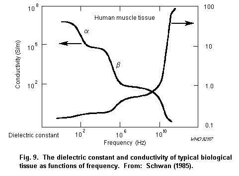 Biological tissues exhibit three strong relaxation phenomena (the
alpha-, ß-, and gamma-dispersion) and one weak (the delta-dispersion)
(Foster & Schwan, 1986, 1989). The molecular phenomena responsible for
the alpha-dispersion are the least understood.
Relaxation of counter-ions about the charged cellular structure,
intracellular structures, e.g., the tubular apparatus in muscle cells,
relaxational behaviour of membranes themselves, may all contribute to
this dispersion to various degrees. The ß-dispersion is mostly due to
membranes, which separate regions having different dielectric
constants and electrical conductivities, resulting in an interfacial
polarization causing the Maxwell-Wagner type relaxation. Smaller
contributions result from the relaxation of proteins. The
gamma-dispersion is due to free water relaxation and the
delta-dispersion results from relaxation of bound water, amino acid,
and charged side groups of proteins.
The alpha-dispersion occurs at frequencies that are usually below
10 kHz, the ß-dispersion at about 20 kHz-100 MHz, the delta-dispersion
at 100-1000 MHz, and the gamma-dispersion at 25 GHz (at 37 °C).
All the dispersions in most tissues occur over a broad range of
frequency, because of the highly non-uniform structure of tissues, and
usually with more than one specific interaction mechanism contributing
to the dispersion (Foster & Schwan, 1986, 1989; Stuchly & Stuchly,
1990).
The permittivity of cells and tissues has been extensively
studied and comprehensive reviews can be found (Foster & Schwan, 1986,
1989; Stuchly & Stuchly, 1990). A detailed description on the
molecular/cellular level of all the relaxation phenomena is provided
in a review by Foster & Schwan (1989).
Resonant dielectric absorption was reported in DNA solutions at
1-10 GHz (Edwards et al., 1984, 1985). Various theoretical hypotheses
were proposed to explain the resonances (Scott, 1985; Van Zandt,
1986). However, more careful measurements were performed by three
other research teams (Foster et al., 1987; Gabriel et al., 1987;
Maleev et al., 1987 ) and a part of the original team that found the
resonance (Rhee et al., 1988). None of the groups found resonant
behaviour of DNA in aqueous solutions. A lack of resonant behaviour is
in agreement with the earlier experimental data on the dielectric
properties of DNA (Takashima et al., 1984).
6.2.2 Non-linear effects
The bulk dielectric properties of tissues reflect the passive
properties of cells, e.g., the capacitance of cell membranes (Foster
& Schwan, 1989). The physiological response of the membrane to the
changes in the membrane potential, due to the applied field, results
in nonlinearity. These phenomena include changes in the membrane
conductance associated with gating and action potentials. An induced
potential across the membrane of the order of 10 mV or more is
required to produce firing of a resting nerve cell, which for a
membrane thickness of, for example, 50 nm corresponds to an electric
field strength of 200 kV/m. However, substantially lower electric
field strengths can induce changes in the firing pattern of pacemaker
cells (Sheppard et al., 1980; Wachtel, 1985). At high field strengths
(voltages across the membrane), pores are formed in the membrane, and,
eventually, at a few hundred mV across the membrane, breakdown occurs
(Foster & Schwan, 1989).
Muscle cells exhibit an anisotropic excitation, which is
consistent with the following phenomenon. The maximum voltage across
the membrane for spherical cells is related to the electric field
strength by the following relationship (Foster & Schwan, 1989):
Vm = 1.5 rE (Equation 6.4)
where r is the cell radius, and E is the electric field strength in
extracellular fluid (Fig. 10). For ellipsoidal cells, similar
equations have been derived by Bernhardt & Pauly (1973). Their results
show that electric fields axial to a cell induce a voltage across the
membrane that is proportional to the length of the cell and to the
extracellular electric field strength. Thus, asymmetrical muscle cells
exhibit dimension-dependent induced voltages, when exposed to electric
fields.
Gradients in the induced surface charge can also affect molecules
and cells in solution. Polar molecules (e.g., water, proteins) align
themselves with the field at high electric field strengths of the
order of 106 V/m. Also, non-spherical cells align themselves with
the field and form "pearl chains". The larger the cell, the lower the
field strength required for orientation and formation of pearl chains.
For instance, for a cell of radius 1 µm, an electric field of 10 kV/m
is required (Foster & Schwan, 1989).
Counter-ion polarization is likely to produce a nonlinear
dielectric response at moderate field strengths of the order of a few
hundred V/m in tissue for large cells, but the response is slow to
develop, and the relaxation frequency is a fraction of a hertz. There
have been relatively few studies on the nonlinear responses of the
counter-ion relaxation (Foster & Schwan, 1989).
6.2.3 Induced fields at the cellular level
Knowledge of the electric fields acting on specific parts of the
cell due to a certain electric field in tissue is important in
predicting cell stimulation. It is also important to evaluate the
possibility of interaction with the genetic apparatus, when fields of
sufficient strength are acting at the cell nucleus. A general analysis
of these fields was performed by Schwan (1984) and Foster & Schwan
(1989). The results of the analysis are illustrated in Fig. 11 showing
the plasma-membrane potential, the cytoplasm field strength, and the
nuclear membrane potential, as a function of frequency.
Biological tissues exhibit three strong relaxation phenomena (the
alpha-, ß-, and gamma-dispersion) and one weak (the delta-dispersion)
(Foster & Schwan, 1986, 1989). The molecular phenomena responsible for
the alpha-dispersion are the least understood.
Relaxation of counter-ions about the charged cellular structure,
intracellular structures, e.g., the tubular apparatus in muscle cells,
relaxational behaviour of membranes themselves, may all contribute to
this dispersion to various degrees. The ß-dispersion is mostly due to
membranes, which separate regions having different dielectric
constants and electrical conductivities, resulting in an interfacial
polarization causing the Maxwell-Wagner type relaxation. Smaller
contributions result from the relaxation of proteins. The
gamma-dispersion is due to free water relaxation and the
delta-dispersion results from relaxation of bound water, amino acid,
and charged side groups of proteins.
The alpha-dispersion occurs at frequencies that are usually below
10 kHz, the ß-dispersion at about 20 kHz-100 MHz, the delta-dispersion
at 100-1000 MHz, and the gamma-dispersion at 25 GHz (at 37 °C).
All the dispersions in most tissues occur over a broad range of
frequency, because of the highly non-uniform structure of tissues, and
usually with more than one specific interaction mechanism contributing
to the dispersion (Foster & Schwan, 1986, 1989; Stuchly & Stuchly,
1990).
The permittivity of cells and tissues has been extensively
studied and comprehensive reviews can be found (Foster & Schwan, 1986,
1989; Stuchly & Stuchly, 1990). A detailed description on the
molecular/cellular level of all the relaxation phenomena is provided
in a review by Foster & Schwan (1989).
Resonant dielectric absorption was reported in DNA solutions at
1-10 GHz (Edwards et al., 1984, 1985). Various theoretical hypotheses
were proposed to explain the resonances (Scott, 1985; Van Zandt,
1986). However, more careful measurements were performed by three
other research teams (Foster et al., 1987; Gabriel et al., 1987;
Maleev et al., 1987 ) and a part of the original team that found the
resonance (Rhee et al., 1988). None of the groups found resonant
behaviour of DNA in aqueous solutions. A lack of resonant behaviour is
in agreement with the earlier experimental data on the dielectric
properties of DNA (Takashima et al., 1984).
6.2.2 Non-linear effects
The bulk dielectric properties of tissues reflect the passive
properties of cells, e.g., the capacitance of cell membranes (Foster
& Schwan, 1989). The physiological response of the membrane to the
changes in the membrane potential, due to the applied field, results
in nonlinearity. These phenomena include changes in the membrane
conductance associated with gating and action potentials. An induced
potential across the membrane of the order of 10 mV or more is
required to produce firing of a resting nerve cell, which for a
membrane thickness of, for example, 50 nm corresponds to an electric
field strength of 200 kV/m. However, substantially lower electric
field strengths can induce changes in the firing pattern of pacemaker
cells (Sheppard et al., 1980; Wachtel, 1985). At high field strengths
(voltages across the membrane), pores are formed in the membrane, and,
eventually, at a few hundred mV across the membrane, breakdown occurs
(Foster & Schwan, 1989).
Muscle cells exhibit an anisotropic excitation, which is
consistent with the following phenomenon. The maximum voltage across
the membrane for spherical cells is related to the electric field
strength by the following relationship (Foster & Schwan, 1989):
Vm = 1.5 rE (Equation 6.4)
where r is the cell radius, and E is the electric field strength in
extracellular fluid (Fig. 10). For ellipsoidal cells, similar
equations have been derived by Bernhardt & Pauly (1973). Their results
show that electric fields axial to a cell induce a voltage across the
membrane that is proportional to the length of the cell and to the
extracellular electric field strength. Thus, asymmetrical muscle cells
exhibit dimension-dependent induced voltages, when exposed to electric
fields.
Gradients in the induced surface charge can also affect molecules
and cells in solution. Polar molecules (e.g., water, proteins) align
themselves with the field at high electric field strengths of the
order of 106 V/m. Also, non-spherical cells align themselves with
the field and form "pearl chains". The larger the cell, the lower the
field strength required for orientation and formation of pearl chains.
For instance, for a cell of radius 1 µm, an electric field of 10 kV/m
is required (Foster & Schwan, 1989).
Counter-ion polarization is likely to produce a nonlinear
dielectric response at moderate field strengths of the order of a few
hundred V/m in tissue for large cells, but the response is slow to
develop, and the relaxation frequency is a fraction of a hertz. There
have been relatively few studies on the nonlinear responses of the
counter-ion relaxation (Foster & Schwan, 1989).
6.2.3 Induced fields at the cellular level
Knowledge of the electric fields acting on specific parts of the
cell due to a certain electric field in tissue is important in
predicting cell stimulation. It is also important to evaluate the
possibility of interaction with the genetic apparatus, when fields of
sufficient strength are acting at the cell nucleus. A general analysis
of these fields was performed by Schwan (1984) and Foster & Schwan
(1989). The results of the analysis are illustrated in Fig. 11 showing
the plasma-membrane potential, the cytoplasm field strength, and the
nuclear membrane potential, as a function of frequency.
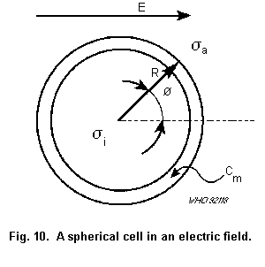 Data shown in Fig. 11 can be summarized as follows: below the
ß-dispersion for the cells, the plasma membrane shields the interior
of the cells; above the ß-frequencies for the plasma membrane and the
nucleus, the induced voltage drop across both membranes falls off as
the inverse of the frequency. The greatest potential is induced on the
nuclear membrane at frequencies between the ß-dispersion frequencies
for the plasma and the nuclear membranes, and this potential is
approximately equal to the product of the external electric field and
the nuclear radius (Foster & Schwan, 1989). Table 6 gives a summary of
induced fields in various parts of the cell and Fig. 11 gives the
induced membrane potentials and electric fields in various
compartments (Schwan, 1985).
Table 6. Summary of the coupling properties of external fields to cellular
membranes and compartments. fr is the beta-dispersion frequency of the plasma
cell membrane, where fn is the beta dispersion frequency of the nucleus and
other organelles. Approximate values of relaxation frequencies are given in
brackets. a
f<fr fr<f<fn f>fn
(approx 1 MHz) (approx 10 MHz)
Cell:
Membranes Polarized Not polarized Not polarized
Interior Doubly shielded Shielded Exposed
Organelles:
Membranes Not polarized Partially polarized Not polarized
Interior Doubly shielded Shielded Exposed
(Nucleic
acids)
Connecting
organelles:
Membranes Polarized Not polarized Not polarized
Interior Not exposed Exposed Exposed
a From: Schwan (1985).
Data shown in Fig. 11 can be summarized as follows: below the
ß-dispersion for the cells, the plasma membrane shields the interior
of the cells; above the ß-frequencies for the plasma membrane and the
nucleus, the induced voltage drop across both membranes falls off as
the inverse of the frequency. The greatest potential is induced on the
nuclear membrane at frequencies between the ß-dispersion frequencies
for the plasma and the nuclear membranes, and this potential is
approximately equal to the product of the external electric field and
the nuclear radius (Foster & Schwan, 1989). Table 6 gives a summary of
induced fields in various parts of the cell and Fig. 11 gives the
induced membrane potentials and electric fields in various
compartments (Schwan, 1985).
Table 6. Summary of the coupling properties of external fields to cellular
membranes and compartments. fr is the beta-dispersion frequency of the plasma
cell membrane, where fn is the beta dispersion frequency of the nucleus and
other organelles. Approximate values of relaxation frequencies are given in
brackets. a
f<fr fr<f<fn f>fn
(approx 1 MHz) (approx 10 MHz)
Cell:
Membranes Polarized Not polarized Not polarized
Interior Doubly shielded Shielded Exposed
Organelles:
Membranes Not polarized Partially polarized Not polarized
Interior Doubly shielded Shielded Exposed
(Nucleic
acids)
Connecting
organelles:
Membranes Polarized Not polarized Not polarized
Interior Not exposed Exposed Exposed
a From: Schwan (1985).
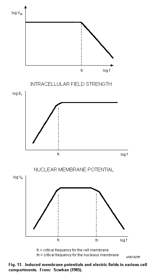 6.2.4 Body impedance
To determine the currents that flow when a person in an
electromagnetic field comes into contact or close proximity with an
isolated conducting object, it is important to consider the impedance
of the human body. The human body impedance can be considered as a
composite of the impedances of various parts through which the current
is flowing. For instance, for a finger contact with an automobile and
a current flowing to ground, the total impedance is the sum of the
following: the contact impedance, the finger impedance, the arm
impedance, the body (trunk plus legs) impedance and the capacitance to
ground. All these impedances are frequency dependent. Furthermore the
contact impedance depends on the surface area and condition (dry or
wet) of the contact surface, and at least at low frequencies probably
on contact voltage as documented by measurements at 60 Hz (Tenforde &
Kaune, 1987).
The complete body impedance can be represented by an equivalent
circuit consisting of a number of resistive and capacitive components,
some of them frequency dependent. Measurements of body impedance have
been performed at 60 Hz (Tenforde & Kaune, 1987) and from 10 kHz to 3
MHz (Gandhi et al., 1985a).
6.3 Direct interactions - strong fields
Well established interaction mechanisms for the direct effects of
electric and magnetic fields can be divided into two types, each
dependent on the field frequency. For frequencies below approximately
100 kHz, the interactions (stimulation) with excitable tissue are of
primary interest. Above about 100 kHz, the current density thresholds
for stimulation and other effects due to interactions with excitable
tissue are higher than those required to produce energy deposition
rates of about 1 W/kg. At such rates of energy deposition in tissue,
thermal interactions become important. In both frequency ranges, other
forms of interactions are also observed for induced currents and
fields below those associated with stimulation or heating.
6.3.1 Interactions with excitable tissues
In tissues, the induced electric fields are amplified across the
cell membranes. At sufficiently high field strengths, these affect the
electrical excitability of nerve and muscle cells. This inter-action
occurs up to hundreds of kilohertz (Lacourse et al., 1985), but
increasingly stronger fields are required above the ß-dispersion.
Changes in the membrane potential cause changes in the permeability to
ions, conformational changes in the embedded proteins, a number of ion
gates open, and eventually membrane depolarization results in an
action potential. Threshold current densities for subtle modulations
of excitable cells, and their biological significance, are less well
understood. There is a substantial amount of data on tissue
stimulation, extra-systole elicitation, and ventricular fibrillation.
These data, as summarized by Bernhardt (1985, 1986, 1988), are shown
in Fig. 12. The ventricular fibrillation thresholds are above those
needed for stimulation. Thresholds for the stimulation of excitable
tissue depend not only on the current density and frequency, but also
on the waveform. In the case of pulsed fields, they depend on pulse
duration and other parameters (Reilly, 1988).
6.2.4 Body impedance
To determine the currents that flow when a person in an
electromagnetic field comes into contact or close proximity with an
isolated conducting object, it is important to consider the impedance
of the human body. The human body impedance can be considered as a
composite of the impedances of various parts through which the current
is flowing. For instance, for a finger contact with an automobile and
a current flowing to ground, the total impedance is the sum of the
following: the contact impedance, the finger impedance, the arm
impedance, the body (trunk plus legs) impedance and the capacitance to
ground. All these impedances are frequency dependent. Furthermore the
contact impedance depends on the surface area and condition (dry or
wet) of the contact surface, and at least at low frequencies probably
on contact voltage as documented by measurements at 60 Hz (Tenforde &
Kaune, 1987).
The complete body impedance can be represented by an equivalent
circuit consisting of a number of resistive and capacitive components,
some of them frequency dependent. Measurements of body impedance have
been performed at 60 Hz (Tenforde & Kaune, 1987) and from 10 kHz to 3
MHz (Gandhi et al., 1985a).
6.3 Direct interactions - strong fields
Well established interaction mechanisms for the direct effects of
electric and magnetic fields can be divided into two types, each
dependent on the field frequency. For frequencies below approximately
100 kHz, the interactions (stimulation) with excitable tissue are of
primary interest. Above about 100 kHz, the current density thresholds
for stimulation and other effects due to interactions with excitable
tissue are higher than those required to produce energy deposition
rates of about 1 W/kg. At such rates of energy deposition in tissue,
thermal interactions become important. In both frequency ranges, other
forms of interactions are also observed for induced currents and
fields below those associated with stimulation or heating.
6.3.1 Interactions with excitable tissues
In tissues, the induced electric fields are amplified across the
cell membranes. At sufficiently high field strengths, these affect the
electrical excitability of nerve and muscle cells. This inter-action
occurs up to hundreds of kilohertz (Lacourse et al., 1985), but
increasingly stronger fields are required above the ß-dispersion.
Changes in the membrane potential cause changes in the permeability to
ions, conformational changes in the embedded proteins, a number of ion
gates open, and eventually membrane depolarization results in an
action potential. Threshold current densities for subtle modulations
of excitable cells, and their biological significance, are less well
understood. There is a substantial amount of data on tissue
stimulation, extra-systole elicitation, and ventricular fibrillation.
These data, as summarized by Bernhardt (1985, 1986, 1988), are shown
in Fig. 12. The ventricular fibrillation thresholds are above those
needed for stimulation. Thresholds for the stimulation of excitable
tissue depend not only on the current density and frequency, but also
on the waveform. In the case of pulsed fields, they depend on pulse
duration and other parameters (Reilly, 1988).
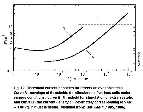 6.3.2 Thermal interactions
As described in section 5, exposure to an electromagnetic field
can result in a spatially nonuniform SAR in the body. The initial rate
of temperature increase, when heat losses are neglected, is directly
proportional to the SAR:
dT/dt = SAR/C (Equation 6.5)
where T is the temperature, t is time, and C is the specific heat
capacity of tissue.
At the molecular level, the phenomena involved in a conversion of
RF energy into thermal energy are the relaxation processes described
earlier. Deposition of RF energy in the body may not necessarily lead
to a proportional increase in its temperature, because of
thermoregulatory responses. Various mathematical models for human
thermoregulation have been applied to evaluate thermal interactions of
RF energy (Emery et al., 1976; Spiegel et al., 1980; Way et al., 1981;
Spiegel, 1982).
The rapid rate at which heating can occur, and a uniquely
non-uniform spatial pattern of energy deposition are important and
unique to thermal interactions of electromagnetic energy. The rate of
initial heating appears to be very important for pulsed fields. These
two features make biological responses due to electromagnetic thermal
loading unlike those due to other thermal agents. Thermal interactions
are not necessarily accompanied by significant local or whole-body
temperature increases.
In some thermal interactions, biological responses depend on the
temperature-time profile, where such a profile is achieved by RF
heating. In some other biological responses, the rate of temperature
change is the critical parameter while the total temperature rise may
be very small. Here again, RF energy (pulsed) can be very effective.
One of the most prominent, thermally-induced effects, where the
temperature increases are very small, is the microwave hearing effect
(Guy et al., 1975a; Lin, 1978). Exposure to one pulse of
electromagnetic energy results in the perception of a click, and
exposure to repeated pulses in a buzzing or hissing sound. The energy
threshold for human beings is very low (16 mJ/kg) and the resulting
temperature increase is estimated to be only about 5 × 10⁻⁶ °C (Guy et
al., 1975a). The simplified mechanism of interaction is as follows:
absorption of electromagnetic energy causes a rapid temperature
increase, which, in turn, produces thermal expansion pressure
initiating an acoustic wave that is detected by cochlea (Guy et al.,
1975a; Lin, 1978).
6.4 Direct interactions - weak fields
6.4.1 General
There is a growing body of data from studies indicating that
extremely low frequency fields (ELF) (Tenforde & Kaune, 1987; WHO,
1987) and RF amplitude modulated at ELF (Adey, 1981, 1988) interact
with various biological systems at energy levels significantly lower
than those needed for the stimulation of excitable tissues or for
thermal interactions. The mechanisms of these interactions are not
understood. Several mechanisms have been hypothesized, but these need
further development and testing, and possibly still other
considerations need to be taken into account to unravel the rather
complex mechanisms behind the observed interactions.
Pericellular currents induced by electromagnetic fields produce
electrochemical alterations in components of the cell membrane
surface. These changes are hypothesized to cause signals across the
cell membrane and produce intracellular alterations (Adey, 1981, 1988;
Tenforde & Kaune, 1987).
Weak field interactions are sometimes criticized and dismissed on
the grounds that the field intensities induced in the biological
systems that produce them are lower than those associated with thermal
noise. A recent analysis of noise and electric fields induced on a
simple model of cell membranes by Weaver & Astumian (1990) indicates
that induced fields of the order of 0.1-0.01 V/m are theoretically
detectable above the broad band noise level. Much smaller fields, of
the order of 10-4 V/m, are estimated to be detectable if only a narrow
frequency band response of the membrane or signal averaging are
assumed. The assumption of the narrow frequency band response is
consistent with some experimental data on biological responses. The
signal averaging is also supported by experimental work on
enzyme-catalysed reactions.
A description of some hypothetical interaction mechanisms for ELF
fields, which possibly also applies to the lower frequencies of
concern here (probably below 1000 Hz) and to RF fields modulated at
ELF can be found in Tenforde & Kaune (1987) and WHO (1987).
The hypothesized scheme of transductive coupling between induced
electric currents in the extracellular medium and the intracellular
events occurring in living cells is illustrated schematically in Fig.
13.
An alternative model involving magnetic-field induced changes in
specific molecular species associated with the plasma membrane has
been proposed by Blackman et al. (1988). In this model, as in others,
an amplification step must be involved. Conditions for the cellular
response may involve the induction of a weak electric field in the
extracellular fluid, a molecular change in the membranes to "trigger"
cooperative events within the cell membrane. The basic premise is that
the cell membrane exists in a metastable, non-equilibrium state that
can be significantly perturbed by weak stimuli. The stored energy is
released by this process as metabolic chemical energy through the
activation of ion pumps or enzymatic reactions within the membrane
(Fröhlich, 1968, 1977; Adey, 1981, 1983). This general model may also
be applicable to the results observed at 41 GHz (Grundler & Keilmann
1983, 1989). In this case, yeast growth rates have been affected at
SARs as low as 0.2 W/kg.
6.3.2 Thermal interactions
As described in section 5, exposure to an electromagnetic field
can result in a spatially nonuniform SAR in the body. The initial rate
of temperature increase, when heat losses are neglected, is directly
proportional to the SAR:
dT/dt = SAR/C (Equation 6.5)
where T is the temperature, t is time, and C is the specific heat
capacity of tissue.
At the molecular level, the phenomena involved in a conversion of
RF energy into thermal energy are the relaxation processes described
earlier. Deposition of RF energy in the body may not necessarily lead
to a proportional increase in its temperature, because of
thermoregulatory responses. Various mathematical models for human
thermoregulation have been applied to evaluate thermal interactions of
RF energy (Emery et al., 1976; Spiegel et al., 1980; Way et al., 1981;
Spiegel, 1982).
The rapid rate at which heating can occur, and a uniquely
non-uniform spatial pattern of energy deposition are important and
unique to thermal interactions of electromagnetic energy. The rate of
initial heating appears to be very important for pulsed fields. These
two features make biological responses due to electromagnetic thermal
loading unlike those due to other thermal agents. Thermal interactions
are not necessarily accompanied by significant local or whole-body
temperature increases.
In some thermal interactions, biological responses depend on the
temperature-time profile, where such a profile is achieved by RF
heating. In some other biological responses, the rate of temperature
change is the critical parameter while the total temperature rise may
be very small. Here again, RF energy (pulsed) can be very effective.
One of the most prominent, thermally-induced effects, where the
temperature increases are very small, is the microwave hearing effect
(Guy et al., 1975a; Lin, 1978). Exposure to one pulse of
electromagnetic energy results in the perception of a click, and
exposure to repeated pulses in a buzzing or hissing sound. The energy
threshold for human beings is very low (16 mJ/kg) and the resulting
temperature increase is estimated to be only about 5 × 10⁻⁶ °C (Guy et
al., 1975a). The simplified mechanism of interaction is as follows:
absorption of electromagnetic energy causes a rapid temperature
increase, which, in turn, produces thermal expansion pressure
initiating an acoustic wave that is detected by cochlea (Guy et al.,
1975a; Lin, 1978).
6.4 Direct interactions - weak fields
6.4.1 General
There is a growing body of data from studies indicating that
extremely low frequency fields (ELF) (Tenforde & Kaune, 1987; WHO,
1987) and RF amplitude modulated at ELF (Adey, 1981, 1988) interact
with various biological systems at energy levels significantly lower
than those needed for the stimulation of excitable tissues or for
thermal interactions. The mechanisms of these interactions are not
understood. Several mechanisms have been hypothesized, but these need
further development and testing, and possibly still other
considerations need to be taken into account to unravel the rather
complex mechanisms behind the observed interactions.
Pericellular currents induced by electromagnetic fields produce
electrochemical alterations in components of the cell membrane
surface. These changes are hypothesized to cause signals across the
cell membrane and produce intracellular alterations (Adey, 1981, 1988;
Tenforde & Kaune, 1987).
Weak field interactions are sometimes criticized and dismissed on
the grounds that the field intensities induced in the biological
systems that produce them are lower than those associated with thermal
noise. A recent analysis of noise and electric fields induced on a
simple model of cell membranes by Weaver & Astumian (1990) indicates
that induced fields of the order of 0.1-0.01 V/m are theoretically
detectable above the broad band noise level. Much smaller fields, of
the order of 10-4 V/m, are estimated to be detectable if only a narrow
frequency band response of the membrane or signal averaging are
assumed. The assumption of the narrow frequency band response is
consistent with some experimental data on biological responses. The
signal averaging is also supported by experimental work on
enzyme-catalysed reactions.
A description of some hypothetical interaction mechanisms for ELF
fields, which possibly also applies to the lower frequencies of
concern here (probably below 1000 Hz) and to RF fields modulated at
ELF can be found in Tenforde & Kaune (1987) and WHO (1987).
The hypothesized scheme of transductive coupling between induced
electric currents in the extracellular medium and the intracellular
events occurring in living cells is illustrated schematically in Fig.
13.
An alternative model involving magnetic-field induced changes in
specific molecular species associated with the plasma membrane has
been proposed by Blackman et al. (1988). In this model, as in others,
an amplification step must be involved. Conditions for the cellular
response may involve the induction of a weak electric field in the
extracellular fluid, a molecular change in the membranes to "trigger"
cooperative events within the cell membrane. The basic premise is that
the cell membrane exists in a metastable, non-equilibrium state that
can be significantly perturbed by weak stimuli. The stored energy is
released by this process as metabolic chemical energy through the
activation of ion pumps or enzymatic reactions within the membrane
(Fröhlich, 1968, 1977; Adey, 1981, 1983). This general model may also
be applicable to the results observed at 41 GHz (Grundler & Keilmann
1983, 1989). In this case, yeast growth rates have been affected at
SARs as low as 0.2 W/kg.
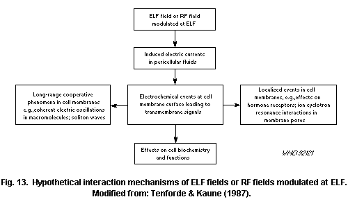 6.4.2 Microelectrophoretic motion
Recent experimental evidence has given some support to the
concept that the interactions of ELF fields with living cells occur at
specific loci on the cell membrane. A model of membrane interactions
in which a microelectrophoretic motion induced in the cell membrane by
weak ELF electric fields influences the average distance between
charged ligands and the cell-surface receptors to which they are bound
was proposed by Chiabrera et al. (1984). In this theoretical model,
the effect of the imposed electric field is to decrease the mean
lifetime of the ligand-receptor complexes on the membrane surface. The
authors proposed that this effect could influence various biological
phenomena, such as the activation of lymphocytes by antigens and
various lectins, and the gating mechanisms that control the membrane
transport of various types of cations, such as calcium.
6.4.3 Ion-resonance conditions
Some experimental evidence suggests that effects occur at
specific frequencies for ELF fields and static magnetic fields with
strengths comparable to that of the geomagnetic field. Theoretically
frequencies up to 1 kHz or higher, depending on the ion involved, can
be effective under these conditions. It is proposed that the frequency
of interaction is related to the ion characteristics and the static
magnetic flux density according to the following relationship:
f = kBq/m (Equation 6.6)
where: f is the resonant frequency, k is a constant (integer), q is
the ion charge, m is the ion mass, and B is the constant magnetic flux
density. Some of the earlier models, such as the cyclotron resonance
(Liboff, 1985; McLeod & Liboff, 1986), suffered from serious
limitations (Halle 1988). Other models appear worthy of closer
scrutiny (Lednev, 1990; Male & Edwards, 1990).
Overall, the experimental data for q/m effects on ion binding to
the membrane or enzyme surfaces and on cation transport through cell
membrane pores are intriguing, but there is a clear need for
refinements in the theoretical description of this phenomenon and to
substantiate the experimental results. Whether, and how, any of the
resonance models (Chiabrera et al., 1984; Liboff, 1985; McLeod &
Liboff, 1986; Lednev, 1990; Male & Edmonds, 1990) can be applied to RF
fields amplitude modulated at ELF has not yet been considered or
tested.
6.4.4 Calcium ion exchange
An observed change in the EEG pattern of cats exposed to 147 MHz
fields amplitude modulated at ELF, prompted further investigation with
an isolated chick-brain tissue preparation, to determine whether the
presence of the peripheral nervous system was required to elicit a
change in the central nervous system. Statistically significant
increases in labelled calcium ion efflux were observed in isolated
tissues exposed to 10-20 W/m2, 147 MHz fields amplitude-modulated
at frequencies from 6-20 Hz, but levels remained the same as control
levels at modulation frequencies of less than 6 Hz or greater than 20
Hz. No effect on calcium ion efflux was observed from exposure to
unmodulated RF fields (Bawin et al., 1975). The SAR was less than
0.004 W/kg. This field-induced effect is of interest because it occurs
at SARs too low to implicate heating, and because calcium ions play a
prominent role in the transductive coupling of many cell
membrane-mediated responses. Thus, this in vitro result provides a
means of interrogating the function and processes occurring at the
cell membrane and of identifying possible subtle mechanisms of
interaction of RF fields.
Using 50, 147, and 450 MHz carrier waves, this work has been
replicated and extended with one or more modulation frequency or power
density windows being reported (Blackman et al., 1979, 1980a,b, 1985,
1989; Sheppard et al., 1979). A power density window centred on 8.3
W/m2 (0.00l4 W/kg) has been reported. Six power density windows were
observed for 16 Hz modulated 50 MHz, with five of the windows
separated by a geometric relationship that may reveal a characteristic
of the underlying mechanism (Blackman 1980a,b, 1985, 1989).
Lee et al. (1987) reported enhanced release of calcium ions from
chick-brain tissue exposed in two power density regions of 147 MHz
fields, modulated at 16 Hz, only when specific temperature conditions
were instituted in the preparation of the tissue. The temperature
conditions during sample preparation were also shown to affect the
relative direction of the efflux and to control the sensitivity of the
brain tissue samples to ELF signals (Blackman et al., 1991). The
release of calcium ions from a rat synaptosomal preparation was also
reported to be affected by 450 MHz, amplitude modulated at 16 Hz, at
10 W/m2 (Lin-Liu & Adey, 1982).
Exposures at 315 Hz and at 405 Hz, at intensities of 15 V/m and
60 nT, were reported to enhance calcium efflux, whereas intensities
between, above, and immediately below these values did not (Blackman
et al., 1988). The 315 Hz exposure was dependent on the perpendicular
flux density and orientation of the DC magnetic field of the earth
(Blackman et al., 1990). Additional work at lower frequencies suggests
that the DC magnetic field may have a direct influence on which
frequencies are effective (Blackman et al., 1985).
Some investigators have reported null results with brain tissue
preparations. Shelton & Merritt (1981) did not observe any changes in
the release of calcium ions from an in vitro rat brain tissue
preparation exposed to 1 GHz, pulse modulated at 16 or 32 Hz, at 5,
l0, 20, or 150 W/m2. Similarly, no effects were observed with rat
tissue labelled in vivo and exposed in vitro or in vivo to 1 GHz
or 2.06 GHz, pulse modulated at several ELF and power density
combinations (Merritt et al., 1982). Null effects were also reported
by Albert et al. (1987) using chick brain tissue exposed to a few
power densities of 147 MHz, amplitude modulated at 16 Hz, under anoxic
and under modified media conditions designed to supply more oxygen to
the tissue.
In none of these null-effect experiments did the authors
reproduce the exposure conditions used by Bawin or Blackman,
particularly the medium composition, power density, sinusoidal
modulation, or number of samples per experiment.
Increases in calcium ion efflux have been reported in two other
biological preparations. Isolated frog hearts showed enhanced calcium
ion efflux at SARs of 0.00015 and 0.0003 W/kg when exposed to 240 MHz,
amplitude modulated at 16 Hz (Schwartz et al., 1990). Human
neuroblastoma cells exposed in culture to amplitude modulated 147 and
915 MHz at SARs of 0.005 and 0.05 W/kg displayed maximal calcium ion
efflux at modulation frequencies around 16 and 60 Hz (Dutta et al.,
1984, 1989). The latter experiment was conducted under natural,
cell-culture growth conditions and suggests that anoxia is not an
absolute requirement for sensitivity of nervous system derived cells
to RF fields modulated at ELF frequencies.
Overall, the exposure-induced release of calcium ions from
tissues should be viewed as contributing to the characterization of
exposure conditions required to elicit a response, and, thus, to the
development of an underlying mechanism of action. The efflux assay
system may ultimately be useful in defining the various aspects of the
physical and biological exposure conditions that sensitize and affect
membrane responses to electromagnetic field exposure. It should be
emphasized that insufficient information is available to define the
weak field interactions. Furthermore, the reported effects cannot be
characterized as a potential adverse effect on health, since little or
no confirmed information has been gathered that suggests this effect
occurs in animals or humans.
6.5 Indirect interactions
Electromagnetic fields, at frequencies below about 100 MHz,
interact with biological bodies through electrical charges induced on
ungrounded or poorly grounded metallic objects, such as cars, trucks,
cranes, wires, and fences.
Two types of interaction may occur:
(a) a spark discharge before a person touches the object;
(b) the passage of current to ground through a person coming into
contact with such objects; the magnitude of these currents depends on
the total charge on the object. This charge, in turn, depends on the
frequency and electric field strength, the object geometry and
capacitance, and the person's impedance to ground.
Above a certain threshold, the current to ground is perceived by
the person as a tingling or prickling sensation in the finger or hand
touching the charged object, for frequencies below about 100 kHz, and
as heat at higher frequencies. A severe shock can be experienced at
levels much higher than this threshold. The threshold currents depend
on frequency, surface of contact area, and the individual. The
thresholds for effects (perception, shock, etc.) are generally higher
for men than for women and children, though there are also individual
differences.
All effects due to induced charges on objects are defined below
in order of increasing severity:
Perception - The person is just able to detect the stimulus. There
is a difference in the current perception threshold for touch and grip
contact.
Annoyance - The person would consider the sensation to be a mild
irritant, if it were to occur repeatedly.
Startle - If a person receives one exposure, it is sufficient to
motivate the person to avoid situations that would lead to a similar
experience.
The remaining reactions apply only to contact of alternating
currents at frequencies below 100 kHz.
Let-go - A person cannot let go of a gripped conductor as long as
the stimulus persists, because of uncontrollable muscle contraction.
If a person is exposed to prolonged currents, somewhat above the
let-go level, through the chest, breathing becomes difficult and,
eventually, the person may become exhausted and die.
Respiratory tetanus - A person is unable to breathe as long as the
stimulus is applied, owing to the contraction of the muscle
responsible for breathing.
Fibrillation - Uncoordinated asynchronous heart contractions produce
no blood pumping action.
Threshold currents for their occurrence are given in Table 7.
Fig. 14 and 15 show threshold currents for perception and let-go, for
different percentages of the population at lower frequencies.
Thresholds for perception and pain (well below the let-go) were
evaluated for nearly 200 men and 200 women and also estimated for
10-year-old children (Chatterjee et al., 1986). The thresholds are
lower for finger contact than for grasping contact. Fig. 16 and 17
show perception and pain for finger contact (Chatterjee et al., 1986).
The stimuli in both cases are tingling/pricking at frequencies below
about 100 kHz and heat/warmth at higher frequencies.
Currents flowing from an object to ground through a person who
touches the object can be reduced if shoes are worn (Chatterjee et
al., 1986). Electric charge induced on various objects and, therefore,
contact currents for people, can be calculated for a known electric
field strength. Results of such calculations are shown in Fig. 18 and
19 for finger contact for males, females, and children, respectively.
RF burns can occur when current enters through a small
cross-section of the body, such as a finger, when the finger contacts
an electrically charged object. Another interaction that may occur at
lower frequencies is a transient discharge, which occurs between a
person and a charged object either by direct contact or through an air
gap (Tenforde & Kaune, 1987).
Table 7. Threshold currents (mA) for various effects at frequencies ranging from 50 Hz to 3 MHz
(experimental data for 50% of men, women, and children)
Effect Subject Threshold current (mA) at various frequencies
50/60 300 1000 10 30 100 300 1 3
Hz Hz Hz kHz kHz kHz kHz MHz MHz
Touch perception men 0.36 (0.47) (0.79) 4 15 40 40 40 40
(finger contact) women 0.24 (0.31) (0.53) 3.2 12 35 35 35 35
children 0.18 0.24 0.40 2.5 8 25 25 25 25
Grip perception men 1.1 1.3 2.2 15 50 300 300 300 300
women 0.7 0.9 1.5 10 35 200 200 200 200
children 0.55 0.65 1.1 9 30 150 150 150 150
Shock, not painful men 1.8 (2.3) (3.2) 17(10) (25) (25)
(grasping contact) women 1.2 1.5 2.1 11 16.7 16.7
children 0.9 1.1 1.6 8.5 12.5 12.5
Pain men (1.8) (2.4) (3.3) 10 30 55 50 50 50
(finger contact) women 1.2 1.6 2.2 6.5 23 47 45 40 40
children 0.9 1.2 1.6 6 18 33 30 28 28
Shock, painful; muscle men 9 (11.7) (16.2) 55 (126) (126)
control (let-go threshold women 6 7.8 10.8 37 84 84
for 0.5% of population) children 4.5 5.9 8.1 27 63 63
Burn (finger contact) men 200 200
Painful shock, men 16 18 24 75(88) (224) (224)
let-go threshold women 10.5 12 16 50 150 150
children 8 9 12 37 112 112
Table 7 (contd).
Effect Subject Threshold current (mA) at various frequencies
50/60 300 1000 10 30 100 300 1 3
Hz Hz Hz kHz kHz kHz kHz MHz MHz
Severe shock, men 23 (30) (41) 94(126) (320) (320)
breathing difficulty women 15 20 27 63 214 214
children 12 15 20.5 47 160 160
a From Dalziel 1954a,b; Deno, 1974; Guy & Chou, 1982; Guy, 1985; Chatterjee et el., 1986). Data in
brackets were calculated by using the frequency factors for perception thresholds and for pain end
let-go thresholds, given in IEC Publication 479. Date in italics were calculated by assuming
thresholds for women two-thirds of that of men and thresholds for children one-half of that for men
(IEEE, 1978; Guy, 1985).
6.4.2 Microelectrophoretic motion
Recent experimental evidence has given some support to the
concept that the interactions of ELF fields with living cells occur at
specific loci on the cell membrane. A model of membrane interactions
in which a microelectrophoretic motion induced in the cell membrane by
weak ELF electric fields influences the average distance between
charged ligands and the cell-surface receptors to which they are bound
was proposed by Chiabrera et al. (1984). In this theoretical model,
the effect of the imposed electric field is to decrease the mean
lifetime of the ligand-receptor complexes on the membrane surface. The
authors proposed that this effect could influence various biological
phenomena, such as the activation of lymphocytes by antigens and
various lectins, and the gating mechanisms that control the membrane
transport of various types of cations, such as calcium.
6.4.3 Ion-resonance conditions
Some experimental evidence suggests that effects occur at
specific frequencies for ELF fields and static magnetic fields with
strengths comparable to that of the geomagnetic field. Theoretically
frequencies up to 1 kHz or higher, depending on the ion involved, can
be effective under these conditions. It is proposed that the frequency
of interaction is related to the ion characteristics and the static
magnetic flux density according to the following relationship:
f = kBq/m (Equation 6.6)
where: f is the resonant frequency, k is a constant (integer), q is
the ion charge, m is the ion mass, and B is the constant magnetic flux
density. Some of the earlier models, such as the cyclotron resonance
(Liboff, 1985; McLeod & Liboff, 1986), suffered from serious
limitations (Halle 1988). Other models appear worthy of closer
scrutiny (Lednev, 1990; Male & Edwards, 1990).
Overall, the experimental data for q/m effects on ion binding to
the membrane or enzyme surfaces and on cation transport through cell
membrane pores are intriguing, but there is a clear need for
refinements in the theoretical description of this phenomenon and to
substantiate the experimental results. Whether, and how, any of the
resonance models (Chiabrera et al., 1984; Liboff, 1985; McLeod &
Liboff, 1986; Lednev, 1990; Male & Edmonds, 1990) can be applied to RF
fields amplitude modulated at ELF has not yet been considered or
tested.
6.4.4 Calcium ion exchange
An observed change in the EEG pattern of cats exposed to 147 MHz
fields amplitude modulated at ELF, prompted further investigation with
an isolated chick-brain tissue preparation, to determine whether the
presence of the peripheral nervous system was required to elicit a
change in the central nervous system. Statistically significant
increases in labelled calcium ion efflux were observed in isolated
tissues exposed to 10-20 W/m2, 147 MHz fields amplitude-modulated
at frequencies from 6-20 Hz, but levels remained the same as control
levels at modulation frequencies of less than 6 Hz or greater than 20
Hz. No effect on calcium ion efflux was observed from exposure to
unmodulated RF fields (Bawin et al., 1975). The SAR was less than
0.004 W/kg. This field-induced effect is of interest because it occurs
at SARs too low to implicate heating, and because calcium ions play a
prominent role in the transductive coupling of many cell
membrane-mediated responses. Thus, this in vitro result provides a
means of interrogating the function and processes occurring at the
cell membrane and of identifying possible subtle mechanisms of
interaction of RF fields.
Using 50, 147, and 450 MHz carrier waves, this work has been
replicated and extended with one or more modulation frequency or power
density windows being reported (Blackman et al., 1979, 1980a,b, 1985,
1989; Sheppard et al., 1979). A power density window centred on 8.3
W/m2 (0.00l4 W/kg) has been reported. Six power density windows were
observed for 16 Hz modulated 50 MHz, with five of the windows
separated by a geometric relationship that may reveal a characteristic
of the underlying mechanism (Blackman 1980a,b, 1985, 1989).
Lee et al. (1987) reported enhanced release of calcium ions from
chick-brain tissue exposed in two power density regions of 147 MHz
fields, modulated at 16 Hz, only when specific temperature conditions
were instituted in the preparation of the tissue. The temperature
conditions during sample preparation were also shown to affect the
relative direction of the efflux and to control the sensitivity of the
brain tissue samples to ELF signals (Blackman et al., 1991). The
release of calcium ions from a rat synaptosomal preparation was also
reported to be affected by 450 MHz, amplitude modulated at 16 Hz, at
10 W/m2 (Lin-Liu & Adey, 1982).
Exposures at 315 Hz and at 405 Hz, at intensities of 15 V/m and
60 nT, were reported to enhance calcium efflux, whereas intensities
between, above, and immediately below these values did not (Blackman
et al., 1988). The 315 Hz exposure was dependent on the perpendicular
flux density and orientation of the DC magnetic field of the earth
(Blackman et al., 1990). Additional work at lower frequencies suggests
that the DC magnetic field may have a direct influence on which
frequencies are effective (Blackman et al., 1985).
Some investigators have reported null results with brain tissue
preparations. Shelton & Merritt (1981) did not observe any changes in
the release of calcium ions from an in vitro rat brain tissue
preparation exposed to 1 GHz, pulse modulated at 16 or 32 Hz, at 5,
l0, 20, or 150 W/m2. Similarly, no effects were observed with rat
tissue labelled in vivo and exposed in vitro or in vivo to 1 GHz
or 2.06 GHz, pulse modulated at several ELF and power density
combinations (Merritt et al., 1982). Null effects were also reported
by Albert et al. (1987) using chick brain tissue exposed to a few
power densities of 147 MHz, amplitude modulated at 16 Hz, under anoxic
and under modified media conditions designed to supply more oxygen to
the tissue.
In none of these null-effect experiments did the authors
reproduce the exposure conditions used by Bawin or Blackman,
particularly the medium composition, power density, sinusoidal
modulation, or number of samples per experiment.
Increases in calcium ion efflux have been reported in two other
biological preparations. Isolated frog hearts showed enhanced calcium
ion efflux at SARs of 0.00015 and 0.0003 W/kg when exposed to 240 MHz,
amplitude modulated at 16 Hz (Schwartz et al., 1990). Human
neuroblastoma cells exposed in culture to amplitude modulated 147 and
915 MHz at SARs of 0.005 and 0.05 W/kg displayed maximal calcium ion
efflux at modulation frequencies around 16 and 60 Hz (Dutta et al.,
1984, 1989). The latter experiment was conducted under natural,
cell-culture growth conditions and suggests that anoxia is not an
absolute requirement for sensitivity of nervous system derived cells
to RF fields modulated at ELF frequencies.
Overall, the exposure-induced release of calcium ions from
tissues should be viewed as contributing to the characterization of
exposure conditions required to elicit a response, and, thus, to the
development of an underlying mechanism of action. The efflux assay
system may ultimately be useful in defining the various aspects of the
physical and biological exposure conditions that sensitize and affect
membrane responses to electromagnetic field exposure. It should be
emphasized that insufficient information is available to define the
weak field interactions. Furthermore, the reported effects cannot be
characterized as a potential adverse effect on health, since little or
no confirmed information has been gathered that suggests this effect
occurs in animals or humans.
6.5 Indirect interactions
Electromagnetic fields, at frequencies below about 100 MHz,
interact with biological bodies through electrical charges induced on
ungrounded or poorly grounded metallic objects, such as cars, trucks,
cranes, wires, and fences.
Two types of interaction may occur:
(a) a spark discharge before a person touches the object;
(b) the passage of current to ground through a person coming into
contact with such objects; the magnitude of these currents depends on
the total charge on the object. This charge, in turn, depends on the
frequency and electric field strength, the object geometry and
capacitance, and the person's impedance to ground.
Above a certain threshold, the current to ground is perceived by
the person as a tingling or prickling sensation in the finger or hand
touching the charged object, for frequencies below about 100 kHz, and
as heat at higher frequencies. A severe shock can be experienced at
levels much higher than this threshold. The threshold currents depend
on frequency, surface of contact area, and the individual. The
thresholds for effects (perception, shock, etc.) are generally higher
for men than for women and children, though there are also individual
differences.
All effects due to induced charges on objects are defined below
in order of increasing severity:
Perception - The person is just able to detect the stimulus. There
is a difference in the current perception threshold for touch and grip
contact.
Annoyance - The person would consider the sensation to be a mild
irritant, if it were to occur repeatedly.
Startle - If a person receives one exposure, it is sufficient to
motivate the person to avoid situations that would lead to a similar
experience.
The remaining reactions apply only to contact of alternating
currents at frequencies below 100 kHz.
Let-go - A person cannot let go of a gripped conductor as long as
the stimulus persists, because of uncontrollable muscle contraction.
If a person is exposed to prolonged currents, somewhat above the
let-go level, through the chest, breathing becomes difficult and,
eventually, the person may become exhausted and die.
Respiratory tetanus - A person is unable to breathe as long as the
stimulus is applied, owing to the contraction of the muscle
responsible for breathing.
Fibrillation - Uncoordinated asynchronous heart contractions produce
no blood pumping action.
Threshold currents for their occurrence are given in Table 7.
Fig. 14 and 15 show threshold currents for perception and let-go, for
different percentages of the population at lower frequencies.
Thresholds for perception and pain (well below the let-go) were
evaluated for nearly 200 men and 200 women and also estimated for
10-year-old children (Chatterjee et al., 1986). The thresholds are
lower for finger contact than for grasping contact. Fig. 16 and 17
show perception and pain for finger contact (Chatterjee et al., 1986).
The stimuli in both cases are tingling/pricking at frequencies below
about 100 kHz and heat/warmth at higher frequencies.
Currents flowing from an object to ground through a person who
touches the object can be reduced if shoes are worn (Chatterjee et
al., 1986). Electric charge induced on various objects and, therefore,
contact currents for people, can be calculated for a known electric
field strength. Results of such calculations are shown in Fig. 18 and
19 for finger contact for males, females, and children, respectively.
RF burns can occur when current enters through a small
cross-section of the body, such as a finger, when the finger contacts
an electrically charged object. Another interaction that may occur at
lower frequencies is a transient discharge, which occurs between a
person and a charged object either by direct contact or through an air
gap (Tenforde & Kaune, 1987).
Table 7. Threshold currents (mA) for various effects at frequencies ranging from 50 Hz to 3 MHz
(experimental data for 50% of men, women, and children)
Effect Subject Threshold current (mA) at various frequencies
50/60 300 1000 10 30 100 300 1 3
Hz Hz Hz kHz kHz kHz kHz MHz MHz
Touch perception men 0.36 (0.47) (0.79) 4 15 40 40 40 40
(finger contact) women 0.24 (0.31) (0.53) 3.2 12 35 35 35 35
children 0.18 0.24 0.40 2.5 8 25 25 25 25
Grip perception men 1.1 1.3 2.2 15 50 300 300 300 300
women 0.7 0.9 1.5 10 35 200 200 200 200
children 0.55 0.65 1.1 9 30 150 150 150 150
Shock, not painful men 1.8 (2.3) (3.2) 17(10) (25) (25)
(grasping contact) women 1.2 1.5 2.1 11 16.7 16.7
children 0.9 1.1 1.6 8.5 12.5 12.5
Pain men (1.8) (2.4) (3.3) 10 30 55 50 50 50
(finger contact) women 1.2 1.6 2.2 6.5 23 47 45 40 40
children 0.9 1.2 1.6 6 18 33 30 28 28
Shock, painful; muscle men 9 (11.7) (16.2) 55 (126) (126)
control (let-go threshold women 6 7.8 10.8 37 84 84
for 0.5% of population) children 4.5 5.9 8.1 27 63 63
Burn (finger contact) men 200 200
Painful shock, men 16 18 24 75(88) (224) (224)
let-go threshold women 10.5 12 16 50 150 150
children 8 9 12 37 112 112
Table 7 (contd).
Effect Subject Threshold current (mA) at various frequencies
50/60 300 1000 10 30 100 300 1 3
Hz Hz Hz kHz kHz kHz kHz MHz MHz
Severe shock, men 23 (30) (41) 94(126) (320) (320)
breathing difficulty women 15 20 27 63 214 214
children 12 15 20.5 47 160 160
a From Dalziel 1954a,b; Deno, 1974; Guy & Chou, 1982; Guy, 1985; Chatterjee et el., 1986). Data in
brackets were calculated by using the frequency factors for perception thresholds and for pain end
let-go thresholds, given in IEC Publication 479. Date in italics were calculated by assuming
thresholds for women two-thirds of that of men and thresholds for children one-half of that for men
(IEEE, 1978; Guy, 1985).
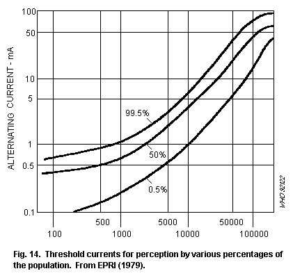
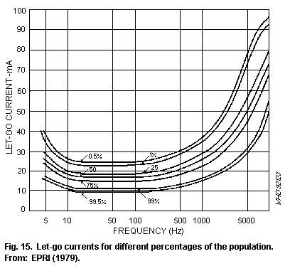
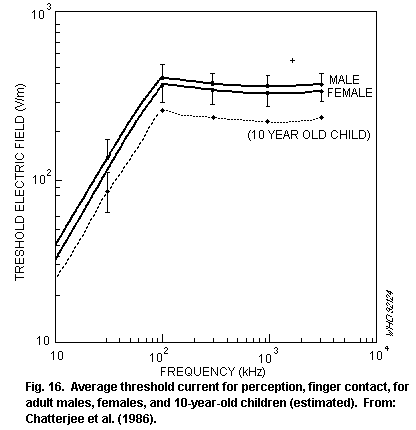
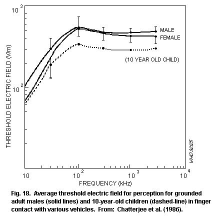
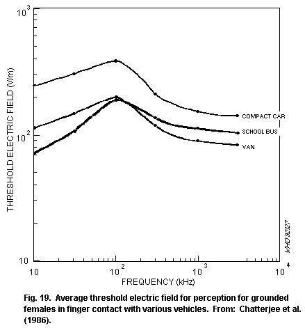 7. CELLULAR AND ANIMAL STUDIES
7.1 Introduction
Numerous reviews and monographs dealing with the biological
effects of electromagnetic fields have been published including: WHO
(1981); Grandolfo et al. (1983); USEPA (1984); Akoev (1986); NCRP
(1986); Polk & Postow (1988); Francescretti et al. (1989); WHO (1989);
Adey (1989, 1990); Saunders et al. (1991). The purpose of this section
is to provide an overview of the biological effects that are relevant
to considerations of the health and safety of exposed people.
The available scientific data are unevenly distributed within the
very broad range of frequencies that this publication covers.
Considerable numbers of in vitro and experimental animal studies
have been performed in the mega- and gigahertz range. Relatively few
scientific reports of effects in the kilohertz range can be found and
data are particularly sparse for the range between 300 Hz and about 10
kHz.
7.2 Macromolecules and cell systems
Studies of isolated (in vitro) components of a biological
system offer possible insights into the mechanisms of RF action. In
vitro systems are simple, allowing biological variables to be
controlled and subtle effects to be identified without being masked by
the homeostatic responses of the whole organism.
In addition, the precise control of the temperature of in vitro
preparations during exposure should make it possible for thermal and
athermal interactions to be clearly distinguished, though thermal
gradients cannot be entirely eliminated from such systems. Effects to
be tested in vivo (whole animal) can be identified from these
studies.
From their review of RF effects on macromolecular and cellular
systems, NCRP (1986) concluded that RF fields, at least continuous
waves at frequencies above 5 MHz, have little, if any, effect on
biopolymers, cell organelles, and microorganisms, other than effects
associated with elevated temperatures. Likewise, they concluded that
the effects of RF fields on the genetic material of cells have not
been convincingly demonstrated, unless related to elevations of
temperature.
More recently, Cleary (1989) noted that there was strong evidence
from a number of in vitro experiments for the involvement of
non-thermal RF interactions, as well as heating. He concluded that
effects that may be attributed to RF-specific interactions include
altered potassium and sodium ion transport across red blood cell
membranes, changes in membrane calcium ion fluxes, decreased
non-cAMP-dependent protein kinase activity, inhibition of T-lymphocyte
cytotoxicity, biphasic effects on lymphocyte proliferation, changes in
brain cell energy metabolism, altered firing rates and resting
potentials of neurons, and effects on cell transformation rate. Many
of these responses are discussed below.
7.2.1 Effects on cell membranes
The cell membrane has been suggested as a likely site for the
interaction of RF fields (Adey, 1981; Cleary, 1987). Several studies
(summarized in Table 8) have focused on effects on membrane
permeability and integrity.
Baranski et al (1971) reported increased cation permeability and
decreased osmotic resistance in rabbit erythrocytes exposed to 3 GHz
for up to 3 h at power densities as low as 10 W/m2; higher power
densities produced effects of greater magnitude. Using thermal
controls heated in a waterbath to the same temperature as exposed
cells, Hamrick & Zinkl (1975) were unable to replicate these effects.
Liu et al. (1979) attributed observed increases in cation permeability
of erythrocytes to heating.
More recently however, it has been reported in several studies
that exposure to RF fields caused specific increases in the cation
permeability of the cell membrane. The results of these studies have
been consistent with a sensitivity of the cell membrane at particular
temperature-dependent energetic states; in some studies, effects have
been reported only at apparent membrane phase transition temperatures
(between 8 °C and 36 °C). Membranes loaded with cholesterol to
eliminate the phase transition were unaffected by microwaves (Liburdy
& Vanek, 1987). RF-induced changes in the activity of the
membrane-bound enzyme Na/K ATPase have been suggested as a possible
mechanism (Allis & Sinha-Robinson, 1987), but similar permeability
changes have been reported in membranes with no associated protein
(Liburdy & Magin, 1985).
Table 8. Membrane studies (in vitro)
Exposure condition Effect Reference
3 GHz (CW) at Increased K+ efflux Baranski et al.
10-100 W/m2, for up and decreased osmotic (1974)
to 3 h resistance in rabbit
erythrocytes compared
with room temperature
controls (increased
effect at higher
power densities)
2.45 or 3 GHz (CW) at No effects on K+ efflux Hamrick & Zinkl
40-750 W/m2 or osmotic resistance (1975)
(3-57 W/kg), for up in rabbit erythrocytes to 3 h
compared with thermal
controls
Table 8 (continued)
Exposure condition Effect Reference
2.45, 3 and 3.95 GHz Increased K+ ion and Liu et al. (1979)
(CW) at up to haemoglobin release
200 W/kg (26 - 44 °C) and osmotic lysis by
rabbit, canine, and
human erythrocytes;
similar effects with
conventional heating
2.45 GHz, at up to Increased passive Olcerst et al.
390 W/kg, for 1 h efflux of Na22 (1980)
and Rb86 from rabbit
erythrocytes compared
with thermal controls,
only at the transition
temperatures for efflux
(8-13 °C, 22.5 °C and 36 °C)
8.42 GHz, CW or pulse Increased K+ efflux Cleary et al.
modulated, for 2 h, at from rabbit erythrocytes (1982)
up to 90 W/kg relative to thermal (23-28 °C)
controls at around 24 °C
2.45 GHz (CW) at Increased Na+ efflux Fisher et al.
2-3 W/kg for up to from human erythrocytes (1982)
2 h (7-35 °C) compared with thermal
controls at 22-25 °C
2.45 GHz (CW) at Increased passive Na+ Liburdy & Penn (1984)
60 W/kg, for 30 min transport and protein
(15-24 °C) shedding from rabbit
erythrocytes compared with
thermal controls, only at
membrane phase transition
temperatures of 17.7-19.5 °C
2.45 GHz (CW) up to Increased Na+ Liburdy & Vanek
100 W/kg, for up to permeability of rabbit (1987)
60 min (13-43 °C) erythrocytes compared
with thermal controls, only
at 17.7-19.5 °C; response
abolished in cholesterol-
loaded membranes with no
apparent phase transition
Table 8 (continued)
Exposure condition Effect Reference
1.0 GHz (CW) at up to No effect on membrane Allis & Sinha (1981)
15 W/kg, for up to fluidity of human 5 h (15-40 °C)
erythrocytes, as measured by
lateral diffusion of
lipophilic dye
2.45 GHz (CW) 6 W/kg, Inhibition of Na/K Allis & Sinha-Robinson
for 20 min (23-27 °C) ATPase activity in human (1987)
erythrocyte ghosts, only at
25 °C
7.2.2 Effects on haematopoietic tissue
A summary of in vitro studies conducted to determine
haematopoietic and immunological end points is shown in Table 9. In
general, these studies show that RF exposure, under
temperature-controlled conditions, at SARs up to 28 W/kg have no
effects on cell survival or mitogen-stimulated lymphoblastoid
transformations.
In some studies, effects are reported at levels too low to
involve significant heating, or at certain RF modulation frequencies.
In one unreplicated study, depressed phagocytosis was reported in
RF-exposed mouse macrophages. A slight rise in temperature in the
culture medium would have tended to increase activity. T-lymphocyte
cytotoxicity was depressed during low-level exposure to 450 MHz RF
modulated at frequencies of 16 and 60 Hz, but not at other
frequencies. In other studies, a lack of effects of sinusoidal or
pulse-modulated RF fields on B-lymphocyte capping in mouse spleen
cells, viability, and DNA synthesis in human mononuclear lymphocytes
has been reported.
Table 9. Haematopoietic and immunological studies (in vitro)
Exposure conditions Effect on exposed group Reference
Colony-forming ability
2.45 GHz (CW) up to Dose-related, reduced Lin et al.
2 kW/kg, for 15 min colony-forming ability of (1979)
mouse bone marrow
cells - temperature kept
constant; direct effect of RF
on haematopoietic precursor
Table 9 (continued)
Exposure conditions Effect on exposed group Reference
2.45 GHz (CW), cells; spleen colonies in Rotkovska et al.
2.4 kW/m2, for 5 min; radiation-depleted (1987)
rise in temperature recipients rose when tempera-
of mouse bone marrow ture rose to between 33 and
suspension was from 40 °C, but fell above 41 °C
20 to 45 °C
Mitogen responses
2.45 GHz (CW), 19 W/kg, No changes in cell viability Smialowicz
for 1-4 h, temperature or blastogenic responses (1976)
controlled at 37 °C of mouse spleen lymphocytes
to several mitogens
2.45 GHz (CW), up to No effects on spontaneous Hamrick &
28 W/kg for up to 44 h, or mitogen-stimulated Fox (1977)
constant temperature. of transformation of rat
37 °C lymphocytes
2.45 GHz (CW) up to No effects on human Roberts et al.
4 W/kg, temperature rise leukocytes viability or on (1983)
of 0.9 °C unstimulated or
mitogen-stimulated
lymphoblastoid transformation
Modulated RF
450 MHz, 15 W/m2, Suppression of mouse T- Lyle et al.
sinusoidal-modulated lymphocyte cytotoxicity, (1983)
at 3, 16, 40, 60, 80, peak at 60 Hz (20%)
100 Hz
147 MHz, pulse- No change in mouse spleen Sultan et al.
modulated at 9, 16, B-lymphocyte capping (1983b)
60 Hz, 1.1-480 W/m2 as temperature maintained
constant
2.45 GHz, pulse- No effects on human Roberts et al.
modulated at 16 or lymphocyte viability, (1984)
60 Hz, up to 4 W/kg unstimulated or
mitogen-stimulated DNA
synthesis, or total protein
synthesis
Table 9 (continued)
Exposure conditions Effect on exposed group Reference
Pulsed 9 GHz (1000 pps) Decrease in no. of Bottreau et al.
200 W/m2 amplitude- plaque-forming cells (1987)
modulated at 16 MHz and cytotoxicity of
(100% mod.) and 16 Hz NK cells in mice
(5% mod.) plus 0.8 Hz
magnetic field 60 mT, 12 h
per day, for 5 days
Other
2.45 GHz (CW), 500 Depression of phagocytosis Mayers &
W/m2, for 30 min, in peritoneal mouse Habershaw
temperature rise of 2.5 °C macrophages (1973)
but below optimum
temperature for phagocytic
activity
2.45 GHz (CW), for 30 min, Ability of normal mouse B- Sultan et al.
up to 1 kW/m2 (SAR lymphocytes to form a "cap" (1983a)
up to 45 W/kg) on the plasma membrane of
antigen-antibody complex
reduces with increasing
temperature; if temperature
kept constant, no difference
between exposed and control
cells
7.2.3 Isolated cerebral tissue, peripheral nerve tissue, and heart
preparations
Studies carried out on calcium ion exchange in chick cerebral
tissue preparations and other tissues exposed to RF fields, amplitude
modulated at ELF frequencies, are described in section 6 on
interaction mechanisms.
Table 10. Peripheral nervous tissue studies
Exposure conditions Effect on exposed group Reference
2.45 GHz (CW or No effect on compound Chou & Guy
pulsed), 1 kW/kg, for action potential in (1973);
several minutes, functioning isolated frog Courtney et al.
temperature controlled and cat nerves or (1975)
peripheral autonomic
ganglia of rabbits
Table 10 (continued)
Exposure conditions Effect on exposed group Reference
1.5 or 2.45 GHz (CW), Reversible changes in Wachtel et al.
few minutes above firing pattern of (1975)
threshold of approx. pacemaker neurons of
5 W/kg Aplysia
2.45 GHz (CW or Prolongation of refractory McRee & Wachtel
pulsed), temperature period of isolated frog (1980, 1982)
controlled above a sciatic nerves
threshold of 5-10 W/kg,
for 30 min
Studies conducted on peripheral nerve tissue are summarized in
Table 10. Most effects of RF exposure on the properties of isolated
nerve preparations can be ascribed to heating. For example, the
changes seen in the firing pattern of pacemaker neurons in Aplysia,
exposed at 5 W/kg or above (Wachtel et al., 1975), were considered to
be consistent with heating (Seamen, 1977). However, in two studies,
changes were reported in the properties of frog nerves exposed above
5-10 W/kg, under constant temperature conditions. These changes were
not induced by exposure to infrared radiation, suggesting an athermal
response. The authors noted, however, that, even under
temperature-controlled conditions, thermal gradients were difficult to
eliminate.
Several studies (reviewed by Liddle & Blackman (1984) and NCRP
(1986) have been performed on isolated heart preparations. Decreases
in heart rate (bradycardia) have been reported in isolated turtle,
frog, and rat heart preparations exposed to RF at intensities as low
as 15 W/m2 (NCRP, 1986). However, Clapman & Cain (1975) indicated
that at least some of the effects observed with these preparations may
have been caused by currents induced in electrodes in contact with the
myocardia. Some support for this comes from the work of Yee et al.
(1984), though a later study (Yee et al., 1988) also implicated the
low temperatures and oxygen levels used in these experiments.
7.2.4 Mutagenic effects
Numerous tests have been carried out to examine the potential
mutagenic action of RF field exposure. In general, no changes in
mutation rate have been observed, except in cases where substantial
temperature increases may also have occurred (USEPA, 1984; NCRP 1986).
Studies of the chromosomal effects resulting from RF exposure of
somatic cells are summarized in Table 11. Most well-conducted studies
report a lack of effect on chromosome aberration frequencies or sister
chromatid exchange rates, even when RF exposure produces mild
hyperthermic conditions. Increased aberration frequencies were
reported in one isolated, long-term study of rat kangaroo cells
exposed for 50 passages (over 320 days) to 2.45 GHz at 15 W/kg.
However, these results may have been confounded by temperature and
senescence (aging) in the cell populations.
Table 11. Mutagenic effects in somatic cells
Exposure conditions Effect on exposed group Reference
20 kHz sawtooth magnetic Non-significant (P=0.06) Nordessen et
field, 16 µT pk-pk, increase in chromosome al. (1989)
for 72 h aberration frequency
in human amniotic cells,
DNA synthesis reduced
2.45 GHz (CW), up to 200 Human blood lymphocytes Lloyd et al.
W/kg, for 20 min showed no increase in (1984, 1986)
temperature rose from unstable chromosome or
4 °C or 23 °C to 36 °C sister chromatid exchanges
during exposure, second
experiment temperature
rose from 37 °C to 40 °C.
2.45 GHz (CW) 15 W/kg, Increased chromosome Yee (1982)
for up to 320 days aberrations and polyploidy
(50 passages) and decreased growth rate
in rat kangaroo RH5 and
RH16 cells
7.2.5 Cancer-related studies
Experiments on cell systems exposed to RF that have end points
related to cancer are shown in Table 12. Cellular transformation
studies are important assays of potential carcinogenicity, in which
the potential is examined of a suspect carcinogen to abolish contact
inhibition, an important regulator of cell division. They are,
however, very susceptible to factors such as variation in growth
media. Balcer-Kubiczek & Harrison (1985, 1989) reported enhanced
transformation rates in mouse fibroblasts after RF exposure for 24 h
at 4.4 W/kg (alone or combined with X-radiation), followed by
treatment with the chemical promotor TPA. These experiments are not
conclusive; there were inconsistencies between the studies in plating
efficiency and in the response to RF combined with X-radiation. The
authors also noted that the transformation rates were susceptible to
temperature changes. However, these studies are important and should
be replicated.
Table 12. Cancer-related studies (in vitro)
Exposure conditions Effect on exposed group Reference
2.45 GHz (CW), 4.4 W/kg RF reduced plating efficiency of Balcer -
for 24 h, temperature mouse embryo fibroblasts to half, Kubiczek &
constant at 37 °C but no effect on transformation Harrison
rate was induced by treatment with (1985)
benzopyrene or X-rays alone. Exposure
of cells to RF and X-rays, then
tumour promotor (phorbal ester TPA),
caused a several-fold increase in
transformation frequency compared
with cells exposed to X-rays and
treated with TPA
2.45 GHz (CW), 4.4 W/kg Increased mouse embryo fibroblast Balcer-
for 24 h, temperature transformation rate per surviving Kubiczek &
constant at 37 °C cell, in cells exposed to RF with, Harrison
or without X-rays, and then treated (1989)
with TPA. In contrast to 1985
study, no effect on cell plating
efficiency or difference in
transformation response to
combined X-ray, RF and TPA compared
with X-ray & TPA alone
450 MHz, pulse- No effect on human lymphocyte Byus et al.
modulated at 3-100 Hz, cAMP-dependent protein kinase (1984)
10 W/m2, for up to activity. cAMP-independent
60 min kinase activity fell to less than
50% of control levels after
15-30 min exposure, then returned
to control levels at 45-60 min.
Reduced enzyme activity occurred
at 16, 40, 60 Hz modulation, not
at 3, 6, 80 or 100 Hz, or
unmodulated carrier
450 MHz amplitude- Increased ornithine decarboxylase Byus et al.
modulated, 10 W/m2, for (ODC) at 10, 16, 20 Hz modulation (1988)
1 h by Reuber H35 hepatoma cells, CHO
cells, and human melanoma cells;
RF (modulated at 16 Hz) exposure of
CHO and hepatoma cells potentiated a
TPA induced increase in ODC, but not
in DNA synthesis in TPA-treated cells
Protein kinases and ornithine decarboxylase are enzymes important
in normal and neoplastic cell growth and division. Byus et al. (1984)
reported an effect of exposure to amplitude-modulated RF on
cAMP-independent kinase, but no effect on cAMP-dependent kinase,
normally implicated in cellular responses leading to proliferation.
Amplitude-modulated RF exposure was also found to enhance ornithine
decarboxylase activity in several different cell lines (Byus et al.,
1988), though only by a small amount compared with chemical promotors.
No effect was seen on DNA synthesis (assayed 14 h after exposure),
which is a subsequent step in the promotional sequence. It is not
possible to draw any conclusions with respect to cancer from these
studies.
7.2.6 Summary and conclusions: in vitro studies
The results of in vitro studies, conducted so far, suggest that
the cell membrane is a site of interaction of RF fields and that
alterations in membrane permeability can result, as well as changes in
membrane cation fluxes, changes in the activity of certain enzymes,
and suppression of some immune responses. RF fields are not mutagenic;
an effect on cellular proliferation, particularly in relation to
tumour promotion, by interactions other than tisue heating, has not
been established. Evidence is presented that some effects may result
from athermal interactions, particularly in response to
amplitude-modulated fields. However, in many other cases, there is
great difficulty in eliminating thermal gradients within exposure
samples exposed at high levels.
7.3 Animal studies
While in vitro studies are important in determining the
mechanisms of interaction and identifying appropriate biological
end-points and exposure conditions to be tested in whole animals, they
cannot serve as a basis for health risk assessment in humans. Whole
animal studies are necessary in order to evaluate the integrated
response of various systems of the body that serve to maintain
homeostasis, the condition necessary for the proper functioning of the
body. Three bodily systems can be identified as of particular
importance in this respect: the nervous, endocrine, and immune
systems. The coordinated interdependent interaction of these systems
in response to chemical and physical stimuli provides a great capacity
for adaptation and compensation in response to changes in
environmental or internal bodily conditions.
Local hyperthermia, caused by exposure to strong RF fields, and
damage to morphological structures of the above systems, can lead, in
turn, to physiological deregulation. Exposure to weaker RF fields with
minimal thermal loading can result in adaptive and compensatory shifts
of these homeostatic mechanisms.
Another important end-point in the consideration of human health
and safety concerns the possible effects on reproduction, and on
pre-and post-natal development. In this context, the induction of
mutagenic changes in germ cells by RF exposure might result in
hereditary effects in offspring. In somatic cells, such changes could
be associated with the induction of cancers.
The effects of exposure to RF fields on these various biological
end-points is described in the following sections. It is important to
note that, as far as thermal responses are concerned, experimental
interpretation can be confounded by differences in ambient
temperature, relative humidity, and air flow. In addition, the thermal
load induced by a given SAR is different in different animals,
generally increasing with body weight in small animal species. These
two points have been evaluated by Gordon et al. (1986) and Gordon
(1987), who argue for a conservative extrapolation of thermal effects
from laboratory animals to humans.
7.3.1 Nervous system
Studies of the effects of RF exposure on the nervous system are
shown in Table 13. Results of early studies suggested that the
blood-brain barrier (which regulates cerebro-spinal fluid composition)
was possibly susceptible to RF field exposure. For example, Frey et
al. (1975) reported the penetration of the blood-brain barrier of
anaesthetised rats by fluorescein after low-level, pulsed or CW
exposure. Oscar & Hawkins (1977) reported increased permeability to
radiolabelled saccharides after exposure of anaesthetized rats to
low-level RF. However, later work (reviewed by Blackwell & Saunders,
1986; NCRP 1986) indicated that these responses may have been
confounded by various factors, including alteration in cerebral blood
flow, the effect of the anaesthetic, and changes in renal clearance.
The uptake of horseradish peroxidase by brain tissue is less
susceptible to these factors. Increased uptake reported in conscious
Chinese hamsters after exposure at 2 W/kg (Albert, 1977); decreased
uptake has been reported at higher SARs (Williams et al., 1984b,d).
More recently, changes in blood-brain barrier permeability have been
reported after exposure to MRI field conditions; however, the evidence
for an effect is contradictory, at present (Prato et al., 1990; Ross
et al., 1990).
Table 13. Nervous system effects
Exposure conditions Effect on exposed group Reference
450 MHz (amp. mod. Altered exchange rate of Ca++ Adey et al.
16 Hz), for 60 min, to during and after exposure of (1982)
30 W/m2 (33 V/m, SAR: cat cortex
0.29 W/kg)
2.06 GHz (CW or pulsed No change in Ca++ mobility Merritt et al.
18, 6, 32 Hz), 5-100 in rat cerebral tissue (1982)
W/m2 (SAR 0.12-2.4 W/kg)
Table 13 (continued)
Exposure conditions Effect on exposed group Reference
2.45 GHz pulsed (2 µs Decreased choline uptake Lai et al.
pulses at 500 Hz) or CW in the rat brain tissue; (1988)
for 45 min (SAR 0.6 W/kg) effect depended on exposure
parameters
2.45 GHz (pulsed - 2 µs Decreased choline uptake Lai et al.
pulses at 500 Hz) for in the rat brain tissue at (1989)
45 min. (SAR 0.3-1.2 W/kg) 0.45 W/kg and above
915 MHz (CW), 10-400 W/m2, Decreased latency of late Johnson & Guy
for 15 min exposure of head components only evoked (1972)
(SAR threshold 2.5-5 W/kg) potentials in thalamus of
cats
147 MHz (amplitude-modu- Altered EEG responses in cats Bawin et al.
lated 1-25 Hz) 10 W/m2 exposed to field modulated (1973, 1974)
(approx SAR 0.015 W/kg) at EEG frequencies
2.95 GHz, single or EEG of rabbits unaffected by Baranski &
repeated exposure up to acute exposure; desynchron- Edelwejn (1975)
50 W/m2 (SAR 1 W/kg) ization of EEG from long-term
2 h/day for 3-4 months exposure; pulsed (1 µs pulses
at 1200 Hz) more effective for
changes than CW
3 GHz (1 µs pulses at Transient enhancement of EEG Servantie &
500-699 Hz) 50 W/m2 (SAR at frequency of pulse repetition Etienne (1975)
1 W/kg) in rats, for rate, persisted after exposure
10 days ceased
1-10 MHz (amplitude-modu- Sustained changes in EEG after Takashima et
lated 14-16 Hz) E field 2-3 weeks of exposure of al. (1979)
500 V/m, 2 h/day, for rabbits
6 weeks
1-30 MHz (amplitude-modu- No effect
lated 60 Hz) single exposure,
for 3 h
500 MHz - 3GHz 25-50 W/m2, No effects on EEG in rats and Klein
for 15 days, at 0.5-1 W/kg monkeys et al. (1985)
4 GHz, CW or amplitude- Slight changes in EEG pattern, Mangel et al.
modulated at 16 Hz particularly at 16.8 W/kg (1990)
(70% mod.), for 30 min amplitude-modulated RF
(SAR in cortex 8.4, 16.8, and 42W/Kg CW
or 42 W/kg)
Pulsed RF fields appear to have various effects on the nervous
system. Exposure to very high peak power pulses is reported to
suppress startle reflex and evoke body movements in conscious mice
(Wachtel et al., 1988; 1989). For evoked body movement, each pulse (10
µs in duration) produced a mid-brain specific absorption of around 200
J/kg, corresponding to an SAR of 20 MW/kg and was estimated to lead to
a rise in mid-brain temperature of 0.05 °C. Pulsed fields were only
about twice as effective as CW suggesting that the effect is unlikely
to be due to thermoelastic mechanisms.
Pulsed RF exposure of rats for 45 min at SARs as low as 0.45 W/kg
has been shown to affect the sodium-dependent, high affinity choline
uptake (an indicator of cholinergic activity) in different parts of
the brain (Lai et al., 1989). In a previous study, Lai et al. (1988)
found that the effect varied with different exposure parameters.
Further work (Lai et al., 1990) revealed that the concentration of
benzodiazepine receptor (involved in anxiety and stress responses) in
the brain of rats exposed for 45 min to pulsed 2.45 GHz or whole body
SARs of 0.6 W/kg was increased in some parts of the brain, immediately
after exposure. However, the effect diminished with repeated exposure
over a 10-day period. The authors suggested that the data support the
hypothesis that low-level RF exposure is a mild nonspecific stressor.
There are a number of responses (heat, noise) that can be regarded as
nonspecific stressors. This set of studies needs further elaboration
to identify the extent and mechanisms of the stress involved, before
its implication for health risk can be assessed. High levels of RF,
sufficient to raise spinal or thalamic temperatures by several degrees
Celsius, decreased the latency of late components of thalamic evoked
potentials.
Exposure to low levels of amplitude-modulated RF has been
reported to alter brain activity (measured using
electroencephalography) and to affect calcium ion mobility in the
cortex. Exposure to 147 MHz fields, amplitude-modulated between 1 and
25 Hz, has been reported to affect the ability of cats to produce
selected EEG rhythms. Changes have also been reported in the EEG
frequency spectrum in rabbits exposed to long-term 1-10 MHz,
amplitude-modulated at 14-16 Hz.
Small changes in EEG patterns, particularly earlier studies on
desynchronisation, were reported in rats and rabbits, after exposure
to an SAR at around 1 W/kg (Baranski & Edelwejn, 1975; Servantie &
Etienne, 1975). However, later studies reported a lack of effect.
The exposure of cats at about 0.3 W/kg to 450 MHz,
amplitude-modulated at 16 Hz, has been reported to alter calcium ion
mobility in the cortex (measured as the efflux of labelled calcium
ions from the cortex surface) (Adey et al., 1982). In contrast,
exposure at between 0.12 and 2.4 W/kg to 2.06 GHz, pulse-modulated at
8, 16, or 32 Hz, was reported to have no effect on calcium ion
exchange in the rat cortex (Merritt et al., 1982).
Exposure to RF has been shown by several authors to influence the
effects of various neuroactive drugs (see Table 14). Acute and
long-term exposure have been reported to potentiate the effects of
stimulant and convulsant compounds (Baranski & Edelwejn, 1974;
Servantie et al., 1974). Thermally significant exposures have been
reported to decrease the period of barbituate-induced anaesthesia in
mice and rabbits; Blackwell (1980) suggested thermally enhanced
redistribution from brain tissue as a probable mechanism.
7.3.2 Ocular effects
The lens of the eye is potentially sensitive to RF exposure,
because it lacks a blood supply and so has a reduced ability to
dissipate heat compared withto other tissues. In addition, the fibres
that make up the bulk of the lens have only a limited capacity for
repair and tend to accumulate the effects of minor insults.
Table 14. Nervous system effects with drugs
Exposure conditions Effect on exposed group Reference
1.7 or 2.45 GHz (CW) Rabbits injected with sodium Cleary &
up to 500 W/m2 (up to pentobarbital and exposed to RF Wangemann
10 W/kg) showed reduced sleeping times; (1976)
correlated with increased
rectal temperature
2.45 GHz (CW), 250 W/m2 SAR dependent reduction in Blackwell
and above (SARs hexobarbital-induced (1980)
>17 W/kg) (rectal sleeping time in mice during
temperature rise 3 °C) RF exposure
3 GHz (CW) 70 W/m2 Variable effect on Baranski &
(1.2 W/kg) for 3 h/day, chlorpromazine and pentylen- Edelwejn
for 200 h exposure etetrazol changes in EEG (1974)
activity in rabbits
3 GHz (pulsed 1 µs at Variable latency of response Servantie et
525 Hz), for unspecified to pentylenetetrazol al. (1974)
duration, each day for induction of convulsion
8-35 days, at 5 W/kg activity
9.3 GHz (CW), 7-28 W/m2 No differences in EEG from Goldstein &
0.6 W/kg, for 5 min normal sodium pentobarbital Sisko (1974)
anaesthetic action
Most experimental work on the RF induction of cataracts (see
Tables 15 and 16) has been carried out using near-field exposures at
2.45 GHz, to selectively irradiate the eye or the side of the head, in
order to avoid whole-body thermal stress. The intense exposures used
in these studies have often led to other effects, such as lacrimation
and oedema of surrounding tissue.
Exposure has usually been well above perception threshold and the
animals have usually been anaesthetised. In most studies, the rabbit
has been used as the experimental animal model, because the dimensions
of its eye approach those of the human eye.
Different conditions of exposure can affect the type of opacity
formed or be ineffective in inducing any permanent change. The
efficacy with which the applied RF field can induce cataracts depends
on the depth of penetration and hence the frequency. Below 1.5 GHz,
the dimensions of the orbit-eye combination are too small to result in
local field concentration. Above about 10 GHz, penetration decreases
and power absorption becomes increasingly restricted to the
superficial tissue (NCRP, 1986).
Table 15. Ocular effects from acute exposure
Exposure conditions Effect on exposed group Reference
Rabbits
2.45 GHz (CW); 4.2 kW/m2, Posterior cortical Carpenter &
for 5 min, or 1.5 kW/m2, opacities within a Van Ummerson
for 60 min week; first visible (1968)
changes (milky bands)
1-2 days after exposure
2.45 GHz (CW); up to Ultrastructural changes Williams et
2.5 kW/m2 repetitive in lenses seen with al. (1975)
exposure microscope; slit lamp
picture appeared normal
2.45 GHz (CW); single acute Threshold exposure to Guy et al.
exposure of 1.5 kW/m2, for produce lens cataract (1975b)
up to 100 min (SAR peak in
vitreous of 138 W/kg, 43 °C
peak)
2.45 GHz (CW); SAR 100 Cataract in rabbit Kramar et al.
100 W/kg, after >140 min (1978)
3 GHz (CW); (far-field) No lenticular changes, Appleton
5 kW/m2, for 30 min periorbital burns et al. (1975)
107 GHz or 35 GHz, Keratitis in cornea; Rosenthal
for 60 min, at 400 W/m2 damage more immediate et al. (1976)
but recovery quicker
at 107 GHz
Table 15 (continued)
Exposure conditions Effect on exposed group Reference
Monkeys
2.45 GHz; 5 kW/m2, for No cataract in rhesus Kramar et al.
60 min monkey after 13 months (1978)
In general, field intensities associated with the acute induction
of cataracts in the rabbit are of such magnitude that they are lethal
if applied to the whole animal. Studies on the acute exposure of
rabbits' eyes suggest the existence of an RF exposure threshold for
the production of a cataract. This is best shown in the data of Kramar
et al. (1978) given graphically in Fig. 20. The threshold power
density to produce a cataract is approximately 1500 W/m2 for at
least 1 h.
7. CELLULAR AND ANIMAL STUDIES
7.1 Introduction
Numerous reviews and monographs dealing with the biological
effects of electromagnetic fields have been published including: WHO
(1981); Grandolfo et al. (1983); USEPA (1984); Akoev (1986); NCRP
(1986); Polk & Postow (1988); Francescretti et al. (1989); WHO (1989);
Adey (1989, 1990); Saunders et al. (1991). The purpose of this section
is to provide an overview of the biological effects that are relevant
to considerations of the health and safety of exposed people.
The available scientific data are unevenly distributed within the
very broad range of frequencies that this publication covers.
Considerable numbers of in vitro and experimental animal studies
have been performed in the mega- and gigahertz range. Relatively few
scientific reports of effects in the kilohertz range can be found and
data are particularly sparse for the range between 300 Hz and about 10
kHz.
7.2 Macromolecules and cell systems
Studies of isolated (in vitro) components of a biological
system offer possible insights into the mechanisms of RF action. In
vitro systems are simple, allowing biological variables to be
controlled and subtle effects to be identified without being masked by
the homeostatic responses of the whole organism.
In addition, the precise control of the temperature of in vitro
preparations during exposure should make it possible for thermal and
athermal interactions to be clearly distinguished, though thermal
gradients cannot be entirely eliminated from such systems. Effects to
be tested in vivo (whole animal) can be identified from these
studies.
From their review of RF effects on macromolecular and cellular
systems, NCRP (1986) concluded that RF fields, at least continuous
waves at frequencies above 5 MHz, have little, if any, effect on
biopolymers, cell organelles, and microorganisms, other than effects
associated with elevated temperatures. Likewise, they concluded that
the effects of RF fields on the genetic material of cells have not
been convincingly demonstrated, unless related to elevations of
temperature.
More recently, Cleary (1989) noted that there was strong evidence
from a number of in vitro experiments for the involvement of
non-thermal RF interactions, as well as heating. He concluded that
effects that may be attributed to RF-specific interactions include
altered potassium and sodium ion transport across red blood cell
membranes, changes in membrane calcium ion fluxes, decreased
non-cAMP-dependent protein kinase activity, inhibition of T-lymphocyte
cytotoxicity, biphasic effects on lymphocyte proliferation, changes in
brain cell energy metabolism, altered firing rates and resting
potentials of neurons, and effects on cell transformation rate. Many
of these responses are discussed below.
7.2.1 Effects on cell membranes
The cell membrane has been suggested as a likely site for the
interaction of RF fields (Adey, 1981; Cleary, 1987). Several studies
(summarized in Table 8) have focused on effects on membrane
permeability and integrity.
Baranski et al (1971) reported increased cation permeability and
decreased osmotic resistance in rabbit erythrocytes exposed to 3 GHz
for up to 3 h at power densities as low as 10 W/m2; higher power
densities produced effects of greater magnitude. Using thermal
controls heated in a waterbath to the same temperature as exposed
cells, Hamrick & Zinkl (1975) were unable to replicate these effects.
Liu et al. (1979) attributed observed increases in cation permeability
of erythrocytes to heating.
More recently however, it has been reported in several studies
that exposure to RF fields caused specific increases in the cation
permeability of the cell membrane. The results of these studies have
been consistent with a sensitivity of the cell membrane at particular
temperature-dependent energetic states; in some studies, effects have
been reported only at apparent membrane phase transition temperatures
(between 8 °C and 36 °C). Membranes loaded with cholesterol to
eliminate the phase transition were unaffected by microwaves (Liburdy
& Vanek, 1987). RF-induced changes in the activity of the
membrane-bound enzyme Na/K ATPase have been suggested as a possible
mechanism (Allis & Sinha-Robinson, 1987), but similar permeability
changes have been reported in membranes with no associated protein
(Liburdy & Magin, 1985).
Table 8. Membrane studies (in vitro)
Exposure condition Effect Reference
3 GHz (CW) at Increased K+ efflux Baranski et al.
10-100 W/m2, for up and decreased osmotic (1974)
to 3 h resistance in rabbit
erythrocytes compared
with room temperature
controls (increased
effect at higher
power densities)
2.45 or 3 GHz (CW) at No effects on K+ efflux Hamrick & Zinkl
40-750 W/m2 or osmotic resistance (1975)
(3-57 W/kg), for up in rabbit erythrocytes to 3 h
compared with thermal
controls
Table 8 (continued)
Exposure condition Effect Reference
2.45, 3 and 3.95 GHz Increased K+ ion and Liu et al. (1979)
(CW) at up to haemoglobin release
200 W/kg (26 - 44 °C) and osmotic lysis by
rabbit, canine, and
human erythrocytes;
similar effects with
conventional heating
2.45 GHz, at up to Increased passive Olcerst et al.
390 W/kg, for 1 h efflux of Na22 (1980)
and Rb86 from rabbit
erythrocytes compared
with thermal controls,
only at the transition
temperatures for efflux
(8-13 °C, 22.5 °C and 36 °C)
8.42 GHz, CW or pulse Increased K+ efflux Cleary et al.
modulated, for 2 h, at from rabbit erythrocytes (1982)
up to 90 W/kg relative to thermal (23-28 °C)
controls at around 24 °C
2.45 GHz (CW) at Increased Na+ efflux Fisher et al.
2-3 W/kg for up to from human erythrocytes (1982)
2 h (7-35 °C) compared with thermal
controls at 22-25 °C
2.45 GHz (CW) at Increased passive Na+ Liburdy & Penn (1984)
60 W/kg, for 30 min transport and protein
(15-24 °C) shedding from rabbit
erythrocytes compared with
thermal controls, only at
membrane phase transition
temperatures of 17.7-19.5 °C
2.45 GHz (CW) up to Increased Na+ Liburdy & Vanek
100 W/kg, for up to permeability of rabbit (1987)
60 min (13-43 °C) erythrocytes compared
with thermal controls, only
at 17.7-19.5 °C; response
abolished in cholesterol-
loaded membranes with no
apparent phase transition
Table 8 (continued)
Exposure condition Effect Reference
1.0 GHz (CW) at up to No effect on membrane Allis & Sinha (1981)
15 W/kg, for up to fluidity of human 5 h (15-40 °C)
erythrocytes, as measured by
lateral diffusion of
lipophilic dye
2.45 GHz (CW) 6 W/kg, Inhibition of Na/K Allis & Sinha-Robinson
for 20 min (23-27 °C) ATPase activity in human (1987)
erythrocyte ghosts, only at
25 °C
7.2.2 Effects on haematopoietic tissue
A summary of in vitro studies conducted to determine
haematopoietic and immunological end points is shown in Table 9. In
general, these studies show that RF exposure, under
temperature-controlled conditions, at SARs up to 28 W/kg have no
effects on cell survival or mitogen-stimulated lymphoblastoid
transformations.
In some studies, effects are reported at levels too low to
involve significant heating, or at certain RF modulation frequencies.
In one unreplicated study, depressed phagocytosis was reported in
RF-exposed mouse macrophages. A slight rise in temperature in the
culture medium would have tended to increase activity. T-lymphocyte
cytotoxicity was depressed during low-level exposure to 450 MHz RF
modulated at frequencies of 16 and 60 Hz, but not at other
frequencies. In other studies, a lack of effects of sinusoidal or
pulse-modulated RF fields on B-lymphocyte capping in mouse spleen
cells, viability, and DNA synthesis in human mononuclear lymphocytes
has been reported.
Table 9. Haematopoietic and immunological studies (in vitro)
Exposure conditions Effect on exposed group Reference
Colony-forming ability
2.45 GHz (CW) up to Dose-related, reduced Lin et al.
2 kW/kg, for 15 min colony-forming ability of (1979)
mouse bone marrow
cells - temperature kept
constant; direct effect of RF
on haematopoietic precursor
Table 9 (continued)
Exposure conditions Effect on exposed group Reference
2.45 GHz (CW), cells; spleen colonies in Rotkovska et al.
2.4 kW/m2, for 5 min; radiation-depleted (1987)
rise in temperature recipients rose when tempera-
of mouse bone marrow ture rose to between 33 and
suspension was from 40 °C, but fell above 41 °C
20 to 45 °C
Mitogen responses
2.45 GHz (CW), 19 W/kg, No changes in cell viability Smialowicz
for 1-4 h, temperature or blastogenic responses (1976)
controlled at 37 °C of mouse spleen lymphocytes
to several mitogens
2.45 GHz (CW), up to No effects on spontaneous Hamrick &
28 W/kg for up to 44 h, or mitogen-stimulated Fox (1977)
constant temperature. of transformation of rat
37 °C lymphocytes
2.45 GHz (CW) up to No effects on human Roberts et al.
4 W/kg, temperature rise leukocytes viability or on (1983)
of 0.9 °C unstimulated or
mitogen-stimulated
lymphoblastoid transformation
Modulated RF
450 MHz, 15 W/m2, Suppression of mouse T- Lyle et al.
sinusoidal-modulated lymphocyte cytotoxicity, (1983)
at 3, 16, 40, 60, 80, peak at 60 Hz (20%)
100 Hz
147 MHz, pulse- No change in mouse spleen Sultan et al.
modulated at 9, 16, B-lymphocyte capping (1983b)
60 Hz, 1.1-480 W/m2 as temperature maintained
constant
2.45 GHz, pulse- No effects on human Roberts et al.
modulated at 16 or lymphocyte viability, (1984)
60 Hz, up to 4 W/kg unstimulated or
mitogen-stimulated DNA
synthesis, or total protein
synthesis
Table 9 (continued)
Exposure conditions Effect on exposed group Reference
Pulsed 9 GHz (1000 pps) Decrease in no. of Bottreau et al.
200 W/m2 amplitude- plaque-forming cells (1987)
modulated at 16 MHz and cytotoxicity of
(100% mod.) and 16 Hz NK cells in mice
(5% mod.) plus 0.8 Hz
magnetic field 60 mT, 12 h
per day, for 5 days
Other
2.45 GHz (CW), 500 Depression of phagocytosis Mayers &
W/m2, for 30 min, in peritoneal mouse Habershaw
temperature rise of 2.5 °C macrophages (1973)
but below optimum
temperature for phagocytic
activity
2.45 GHz (CW), for 30 min, Ability of normal mouse B- Sultan et al.
up to 1 kW/m2 (SAR lymphocytes to form a "cap" (1983a)
up to 45 W/kg) on the plasma membrane of
antigen-antibody complex
reduces with increasing
temperature; if temperature
kept constant, no difference
between exposed and control
cells
7.2.3 Isolated cerebral tissue, peripheral nerve tissue, and heart
preparations
Studies carried out on calcium ion exchange in chick cerebral
tissue preparations and other tissues exposed to RF fields, amplitude
modulated at ELF frequencies, are described in section 6 on
interaction mechanisms.
Table 10. Peripheral nervous tissue studies
Exposure conditions Effect on exposed group Reference
2.45 GHz (CW or No effect on compound Chou & Guy
pulsed), 1 kW/kg, for action potential in (1973);
several minutes, functioning isolated frog Courtney et al.
temperature controlled and cat nerves or (1975)
peripheral autonomic
ganglia of rabbits
Table 10 (continued)
Exposure conditions Effect on exposed group Reference
1.5 or 2.45 GHz (CW), Reversible changes in Wachtel et al.
few minutes above firing pattern of (1975)
threshold of approx. pacemaker neurons of
5 W/kg Aplysia
2.45 GHz (CW or Prolongation of refractory McRee & Wachtel
pulsed), temperature period of isolated frog (1980, 1982)
controlled above a sciatic nerves
threshold of 5-10 W/kg,
for 30 min
Studies conducted on peripheral nerve tissue are summarized in
Table 10. Most effects of RF exposure on the properties of isolated
nerve preparations can be ascribed to heating. For example, the
changes seen in the firing pattern of pacemaker neurons in Aplysia,
exposed at 5 W/kg or above (Wachtel et al., 1975), were considered to
be consistent with heating (Seamen, 1977). However, in two studies,
changes were reported in the properties of frog nerves exposed above
5-10 W/kg, under constant temperature conditions. These changes were
not induced by exposure to infrared radiation, suggesting an athermal
response. The authors noted, however, that, even under
temperature-controlled conditions, thermal gradients were difficult to
eliminate.
Several studies (reviewed by Liddle & Blackman (1984) and NCRP
(1986) have been performed on isolated heart preparations. Decreases
in heart rate (bradycardia) have been reported in isolated turtle,
frog, and rat heart preparations exposed to RF at intensities as low
as 15 W/m2 (NCRP, 1986). However, Clapman & Cain (1975) indicated
that at least some of the effects observed with these preparations may
have been caused by currents induced in electrodes in contact with the
myocardia. Some support for this comes from the work of Yee et al.
(1984), though a later study (Yee et al., 1988) also implicated the
low temperatures and oxygen levels used in these experiments.
7.2.4 Mutagenic effects
Numerous tests have been carried out to examine the potential
mutagenic action of RF field exposure. In general, no changes in
mutation rate have been observed, except in cases where substantial
temperature increases may also have occurred (USEPA, 1984; NCRP 1986).
Studies of the chromosomal effects resulting from RF exposure of
somatic cells are summarized in Table 11. Most well-conducted studies
report a lack of effect on chromosome aberration frequencies or sister
chromatid exchange rates, even when RF exposure produces mild
hyperthermic conditions. Increased aberration frequencies were
reported in one isolated, long-term study of rat kangaroo cells
exposed for 50 passages (over 320 days) to 2.45 GHz at 15 W/kg.
However, these results may have been confounded by temperature and
senescence (aging) in the cell populations.
Table 11. Mutagenic effects in somatic cells
Exposure conditions Effect on exposed group Reference
20 kHz sawtooth magnetic Non-significant (P=0.06) Nordessen et
field, 16 µT pk-pk, increase in chromosome al. (1989)
for 72 h aberration frequency
in human amniotic cells,
DNA synthesis reduced
2.45 GHz (CW), up to 200 Human blood lymphocytes Lloyd et al.
W/kg, for 20 min showed no increase in (1984, 1986)
temperature rose from unstable chromosome or
4 °C or 23 °C to 36 °C sister chromatid exchanges
during exposure, second
experiment temperature
rose from 37 °C to 40 °C.
2.45 GHz (CW) 15 W/kg, Increased chromosome Yee (1982)
for up to 320 days aberrations and polyploidy
(50 passages) and decreased growth rate
in rat kangaroo RH5 and
RH16 cells
7.2.5 Cancer-related studies
Experiments on cell systems exposed to RF that have end points
related to cancer are shown in Table 12. Cellular transformation
studies are important assays of potential carcinogenicity, in which
the potential is examined of a suspect carcinogen to abolish contact
inhibition, an important regulator of cell division. They are,
however, very susceptible to factors such as variation in growth
media. Balcer-Kubiczek & Harrison (1985, 1989) reported enhanced
transformation rates in mouse fibroblasts after RF exposure for 24 h
at 4.4 W/kg (alone or combined with X-radiation), followed by
treatment with the chemical promotor TPA. These experiments are not
conclusive; there were inconsistencies between the studies in plating
efficiency and in the response to RF combined with X-radiation. The
authors also noted that the transformation rates were susceptible to
temperature changes. However, these studies are important and should
be replicated.
Table 12. Cancer-related studies (in vitro)
Exposure conditions Effect on exposed group Reference
2.45 GHz (CW), 4.4 W/kg RF reduced plating efficiency of Balcer -
for 24 h, temperature mouse embryo fibroblasts to half, Kubiczek &
constant at 37 °C but no effect on transformation Harrison
rate was induced by treatment with (1985)
benzopyrene or X-rays alone. Exposure
of cells to RF and X-rays, then
tumour promotor (phorbal ester TPA),
caused a several-fold increase in
transformation frequency compared
with cells exposed to X-rays and
treated with TPA
2.45 GHz (CW), 4.4 W/kg Increased mouse embryo fibroblast Balcer-
for 24 h, temperature transformation rate per surviving Kubiczek &
constant at 37 °C cell, in cells exposed to RF with, Harrison
or without X-rays, and then treated (1989)
with TPA. In contrast to 1985
study, no effect on cell plating
efficiency or difference in
transformation response to
combined X-ray, RF and TPA compared
with X-ray & TPA alone
450 MHz, pulse- No effect on human lymphocyte Byus et al.
modulated at 3-100 Hz, cAMP-dependent protein kinase (1984)
10 W/m2, for up to activity. cAMP-independent
60 min kinase activity fell to less than
50% of control levels after
15-30 min exposure, then returned
to control levels at 45-60 min.
Reduced enzyme activity occurred
at 16, 40, 60 Hz modulation, not
at 3, 6, 80 or 100 Hz, or
unmodulated carrier
450 MHz amplitude- Increased ornithine decarboxylase Byus et al.
modulated, 10 W/m2, for (ODC) at 10, 16, 20 Hz modulation (1988)
1 h by Reuber H35 hepatoma cells, CHO
cells, and human melanoma cells;
RF (modulated at 16 Hz) exposure of
CHO and hepatoma cells potentiated a
TPA induced increase in ODC, but not
in DNA synthesis in TPA-treated cells
Protein kinases and ornithine decarboxylase are enzymes important
in normal and neoplastic cell growth and division. Byus et al. (1984)
reported an effect of exposure to amplitude-modulated RF on
cAMP-independent kinase, but no effect on cAMP-dependent kinase,
normally implicated in cellular responses leading to proliferation.
Amplitude-modulated RF exposure was also found to enhance ornithine
decarboxylase activity in several different cell lines (Byus et al.,
1988), though only by a small amount compared with chemical promotors.
No effect was seen on DNA synthesis (assayed 14 h after exposure),
which is a subsequent step in the promotional sequence. It is not
possible to draw any conclusions with respect to cancer from these
studies.
7.2.6 Summary and conclusions: in vitro studies
The results of in vitro studies, conducted so far, suggest that
the cell membrane is a site of interaction of RF fields and that
alterations in membrane permeability can result, as well as changes in
membrane cation fluxes, changes in the activity of certain enzymes,
and suppression of some immune responses. RF fields are not mutagenic;
an effect on cellular proliferation, particularly in relation to
tumour promotion, by interactions other than tisue heating, has not
been established. Evidence is presented that some effects may result
from athermal interactions, particularly in response to
amplitude-modulated fields. However, in many other cases, there is
great difficulty in eliminating thermal gradients within exposure
samples exposed at high levels.
7.3 Animal studies
While in vitro studies are important in determining the
mechanisms of interaction and identifying appropriate biological
end-points and exposure conditions to be tested in whole animals, they
cannot serve as a basis for health risk assessment in humans. Whole
animal studies are necessary in order to evaluate the integrated
response of various systems of the body that serve to maintain
homeostasis, the condition necessary for the proper functioning of the
body. Three bodily systems can be identified as of particular
importance in this respect: the nervous, endocrine, and immune
systems. The coordinated interdependent interaction of these systems
in response to chemical and physical stimuli provides a great capacity
for adaptation and compensation in response to changes in
environmental or internal bodily conditions.
Local hyperthermia, caused by exposure to strong RF fields, and
damage to morphological structures of the above systems, can lead, in
turn, to physiological deregulation. Exposure to weaker RF fields with
minimal thermal loading can result in adaptive and compensatory shifts
of these homeostatic mechanisms.
Another important end-point in the consideration of human health
and safety concerns the possible effects on reproduction, and on
pre-and post-natal development. In this context, the induction of
mutagenic changes in germ cells by RF exposure might result in
hereditary effects in offspring. In somatic cells, such changes could
be associated with the induction of cancers.
The effects of exposure to RF fields on these various biological
end-points is described in the following sections. It is important to
note that, as far as thermal responses are concerned, experimental
interpretation can be confounded by differences in ambient
temperature, relative humidity, and air flow. In addition, the thermal
load induced by a given SAR is different in different animals,
generally increasing with body weight in small animal species. These
two points have been evaluated by Gordon et al. (1986) and Gordon
(1987), who argue for a conservative extrapolation of thermal effects
from laboratory animals to humans.
7.3.1 Nervous system
Studies of the effects of RF exposure on the nervous system are
shown in Table 13. Results of early studies suggested that the
blood-brain barrier (which regulates cerebro-spinal fluid composition)
was possibly susceptible to RF field exposure. For example, Frey et
al. (1975) reported the penetration of the blood-brain barrier of
anaesthetised rats by fluorescein after low-level, pulsed or CW
exposure. Oscar & Hawkins (1977) reported increased permeability to
radiolabelled saccharides after exposure of anaesthetized rats to
low-level RF. However, later work (reviewed by Blackwell & Saunders,
1986; NCRP 1986) indicated that these responses may have been
confounded by various factors, including alteration in cerebral blood
flow, the effect of the anaesthetic, and changes in renal clearance.
The uptake of horseradish peroxidase by brain tissue is less
susceptible to these factors. Increased uptake reported in conscious
Chinese hamsters after exposure at 2 W/kg (Albert, 1977); decreased
uptake has been reported at higher SARs (Williams et al., 1984b,d).
More recently, changes in blood-brain barrier permeability have been
reported after exposure to MRI field conditions; however, the evidence
for an effect is contradictory, at present (Prato et al., 1990; Ross
et al., 1990).
Table 13. Nervous system effects
Exposure conditions Effect on exposed group Reference
450 MHz (amp. mod. Altered exchange rate of Ca++ Adey et al.
16 Hz), for 60 min, to during and after exposure of (1982)
30 W/m2 (33 V/m, SAR: cat cortex
0.29 W/kg)
2.06 GHz (CW or pulsed No change in Ca++ mobility Merritt et al.
18, 6, 32 Hz), 5-100 in rat cerebral tissue (1982)
W/m2 (SAR 0.12-2.4 W/kg)
Table 13 (continued)
Exposure conditions Effect on exposed group Reference
2.45 GHz pulsed (2 µs Decreased choline uptake Lai et al.
pulses at 500 Hz) or CW in the rat brain tissue; (1988)
for 45 min (SAR 0.6 W/kg) effect depended on exposure
parameters
2.45 GHz (pulsed - 2 µs Decreased choline uptake Lai et al.
pulses at 500 Hz) for in the rat brain tissue at (1989)
45 min. (SAR 0.3-1.2 W/kg) 0.45 W/kg and above
915 MHz (CW), 10-400 W/m2, Decreased latency of late Johnson & Guy
for 15 min exposure of head components only evoked (1972)
(SAR threshold 2.5-5 W/kg) potentials in thalamus of
cats
147 MHz (amplitude-modu- Altered EEG responses in cats Bawin et al.
lated 1-25 Hz) 10 W/m2 exposed to field modulated (1973, 1974)
(approx SAR 0.015 W/kg) at EEG frequencies
2.95 GHz, single or EEG of rabbits unaffected by Baranski &
repeated exposure up to acute exposure; desynchron- Edelwejn (1975)
50 W/m2 (SAR 1 W/kg) ization of EEG from long-term
2 h/day for 3-4 months exposure; pulsed (1 µs pulses
at 1200 Hz) more effective for
changes than CW
3 GHz (1 µs pulses at Transient enhancement of EEG Servantie &
500-699 Hz) 50 W/m2 (SAR at frequency of pulse repetition Etienne (1975)
1 W/kg) in rats, for rate, persisted after exposure
10 days ceased
1-10 MHz (amplitude-modu- Sustained changes in EEG after Takashima et
lated 14-16 Hz) E field 2-3 weeks of exposure of al. (1979)
500 V/m, 2 h/day, for rabbits
6 weeks
1-30 MHz (amplitude-modu- No effect
lated 60 Hz) single exposure,
for 3 h
500 MHz - 3GHz 25-50 W/m2, No effects on EEG in rats and Klein
for 15 days, at 0.5-1 W/kg monkeys et al. (1985)
4 GHz, CW or amplitude- Slight changes in EEG pattern, Mangel et al.
modulated at 16 Hz particularly at 16.8 W/kg (1990)
(70% mod.), for 30 min amplitude-modulated RF
(SAR in cortex 8.4, 16.8, and 42W/Kg CW
or 42 W/kg)
Pulsed RF fields appear to have various effects on the nervous
system. Exposure to very high peak power pulses is reported to
suppress startle reflex and evoke body movements in conscious mice
(Wachtel et al., 1988; 1989). For evoked body movement, each pulse (10
µs in duration) produced a mid-brain specific absorption of around 200
J/kg, corresponding to an SAR of 20 MW/kg and was estimated to lead to
a rise in mid-brain temperature of 0.05 °C. Pulsed fields were only
about twice as effective as CW suggesting that the effect is unlikely
to be due to thermoelastic mechanisms.
Pulsed RF exposure of rats for 45 min at SARs as low as 0.45 W/kg
has been shown to affect the sodium-dependent, high affinity choline
uptake (an indicator of cholinergic activity) in different parts of
the brain (Lai et al., 1989). In a previous study, Lai et al. (1988)
found that the effect varied with different exposure parameters.
Further work (Lai et al., 1990) revealed that the concentration of
benzodiazepine receptor (involved in anxiety and stress responses) in
the brain of rats exposed for 45 min to pulsed 2.45 GHz or whole body
SARs of 0.6 W/kg was increased in some parts of the brain, immediately
after exposure. However, the effect diminished with repeated exposure
over a 10-day period. The authors suggested that the data support the
hypothesis that low-level RF exposure is a mild nonspecific stressor.
There are a number of responses (heat, noise) that can be regarded as
nonspecific stressors. This set of studies needs further elaboration
to identify the extent and mechanisms of the stress involved, before
its implication for health risk can be assessed. High levels of RF,
sufficient to raise spinal or thalamic temperatures by several degrees
Celsius, decreased the latency of late components of thalamic evoked
potentials.
Exposure to low levels of amplitude-modulated RF has been
reported to alter brain activity (measured using
electroencephalography) and to affect calcium ion mobility in the
cortex. Exposure to 147 MHz fields, amplitude-modulated between 1 and
25 Hz, has been reported to affect the ability of cats to produce
selected EEG rhythms. Changes have also been reported in the EEG
frequency spectrum in rabbits exposed to long-term 1-10 MHz,
amplitude-modulated at 14-16 Hz.
Small changes in EEG patterns, particularly earlier studies on
desynchronisation, were reported in rats and rabbits, after exposure
to an SAR at around 1 W/kg (Baranski & Edelwejn, 1975; Servantie &
Etienne, 1975). However, later studies reported a lack of effect.
The exposure of cats at about 0.3 W/kg to 450 MHz,
amplitude-modulated at 16 Hz, has been reported to alter calcium ion
mobility in the cortex (measured as the efflux of labelled calcium
ions from the cortex surface) (Adey et al., 1982). In contrast,
exposure at between 0.12 and 2.4 W/kg to 2.06 GHz, pulse-modulated at
8, 16, or 32 Hz, was reported to have no effect on calcium ion
exchange in the rat cortex (Merritt et al., 1982).
Exposure to RF has been shown by several authors to influence the
effects of various neuroactive drugs (see Table 14). Acute and
long-term exposure have been reported to potentiate the effects of
stimulant and convulsant compounds (Baranski & Edelwejn, 1974;
Servantie et al., 1974). Thermally significant exposures have been
reported to decrease the period of barbituate-induced anaesthesia in
mice and rabbits; Blackwell (1980) suggested thermally enhanced
redistribution from brain tissue as a probable mechanism.
7.3.2 Ocular effects
The lens of the eye is potentially sensitive to RF exposure,
because it lacks a blood supply and so has a reduced ability to
dissipate heat compared withto other tissues. In addition, the fibres
that make up the bulk of the lens have only a limited capacity for
repair and tend to accumulate the effects of minor insults.
Table 14. Nervous system effects with drugs
Exposure conditions Effect on exposed group Reference
1.7 or 2.45 GHz (CW) Rabbits injected with sodium Cleary &
up to 500 W/m2 (up to pentobarbital and exposed to RF Wangemann
10 W/kg) showed reduced sleeping times; (1976)
correlated with increased
rectal temperature
2.45 GHz (CW), 250 W/m2 SAR dependent reduction in Blackwell
and above (SARs hexobarbital-induced (1980)
>17 W/kg) (rectal sleeping time in mice during
temperature rise 3 °C) RF exposure
3 GHz (CW) 70 W/m2 Variable effect on Baranski &
(1.2 W/kg) for 3 h/day, chlorpromazine and pentylen- Edelwejn
for 200 h exposure etetrazol changes in EEG (1974)
activity in rabbits
3 GHz (pulsed 1 µs at Variable latency of response Servantie et
525 Hz), for unspecified to pentylenetetrazol al. (1974)
duration, each day for induction of convulsion
8-35 days, at 5 W/kg activity
9.3 GHz (CW), 7-28 W/m2 No differences in EEG from Goldstein &
0.6 W/kg, for 5 min normal sodium pentobarbital Sisko (1974)
anaesthetic action
Most experimental work on the RF induction of cataracts (see
Tables 15 and 16) has been carried out using near-field exposures at
2.45 GHz, to selectively irradiate the eye or the side of the head, in
order to avoid whole-body thermal stress. The intense exposures used
in these studies have often led to other effects, such as lacrimation
and oedema of surrounding tissue.
Exposure has usually been well above perception threshold and the
animals have usually been anaesthetised. In most studies, the rabbit
has been used as the experimental animal model, because the dimensions
of its eye approach those of the human eye.
Different conditions of exposure can affect the type of opacity
formed or be ineffective in inducing any permanent change. The
efficacy with which the applied RF field can induce cataracts depends
on the depth of penetration and hence the frequency. Below 1.5 GHz,
the dimensions of the orbit-eye combination are too small to result in
local field concentration. Above about 10 GHz, penetration decreases
and power absorption becomes increasingly restricted to the
superficial tissue (NCRP, 1986).
Table 15. Ocular effects from acute exposure
Exposure conditions Effect on exposed group Reference
Rabbits
2.45 GHz (CW); 4.2 kW/m2, Posterior cortical Carpenter &
for 5 min, or 1.5 kW/m2, opacities within a Van Ummerson
for 60 min week; first visible (1968)
changes (milky bands)
1-2 days after exposure
2.45 GHz (CW); up to Ultrastructural changes Williams et
2.5 kW/m2 repetitive in lenses seen with al. (1975)
exposure microscope; slit lamp
picture appeared normal
2.45 GHz (CW); single acute Threshold exposure to Guy et al.
exposure of 1.5 kW/m2, for produce lens cataract (1975b)
up to 100 min (SAR peak in
vitreous of 138 W/kg, 43 °C
peak)
2.45 GHz (CW); SAR 100 Cataract in rabbit Kramar et al.
100 W/kg, after >140 min (1978)
3 GHz (CW); (far-field) No lenticular changes, Appleton
5 kW/m2, for 30 min periorbital burns et al. (1975)
107 GHz or 35 GHz, Keratitis in cornea; Rosenthal
for 60 min, at 400 W/m2 damage more immediate et al. (1976)
but recovery quicker
at 107 GHz
Table 15 (continued)
Exposure conditions Effect on exposed group Reference
Monkeys
2.45 GHz; 5 kW/m2, for No cataract in rhesus Kramar et al.
60 min monkey after 13 months (1978)
In general, field intensities associated with the acute induction
of cataracts in the rabbit are of such magnitude that they are lethal
if applied to the whole animal. Studies on the acute exposure of
rabbits' eyes suggest the existence of an RF exposure threshold for
the production of a cataract. This is best shown in the data of Kramar
et al. (1978) given graphically in Fig. 20. The threshold power
density to produce a cataract is approximately 1500 W/m2 for at
least 1 h.
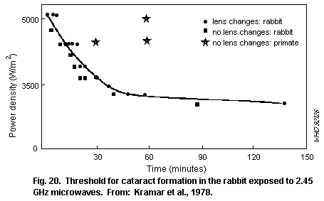 The possibility of a cumulative effect of repeated subthreshold
exposure leading to the development of a cataract has been examined,
as shown in Table 16. Subthreshold exposures of rabbit eyes caused
reversible changes, and damage accumulated only when exposure was
repeated before repair had occurred. However, these exposures were
only just below the single acute exposure threshold (EPA, 1984).
Long-term, whole-body exposures in the far-field at lower levels of
power density have not produced any lens opacities.
Opacities were induced in the eyes of anaesthetized primates
after exposures well above threshold levels for rabbits, or after
long-term exposure of conscious primates (to 9.3 GHz) at up to 1500
W/m2. It has been suggested that the difference in acute response
between rabbits and monkeys reflects structural differences in the
face and lens and, hence, energy deposition and heating in the eye.
Table 16. Ocular effects from repeated short-term threshold exposure
Exposure conditions Effect on exposed group Reference
Rabbits
2.45 GHz (CW), at 4.2 kW/m2 Various degrees of lens Carpenter et al.
opacity : (1960a,b)
4 min for 4 days - in all rabbits
4 exposures at 1-week intervals - in 70% of rabbits
4 exposures at 2-week intervals - in 40% of rabbits
3-min exposure, 5 times in week - few opacities formed
3-min exposure, 5 times in 5 weeks - no opacities formed
2.45 GHz (CW), at 100 W/m2 (SAR No lens opacities for up Ferri & Hagan
1.5 W/kg), for 8 h/day, 5 days/week to 3 months after (1976)
for 17 weeks exposure
2.45 GHz (CW), 1.8 kW/m2, for Cataracts in 8 out of Carpenter et al.
1 h repeated up to 20 times 10 rabbits (1974),
1.5 kW/m2, for 1 h, Cataracts in 4 out of Carpenter
repeated up to 32 times 10 rabbits (1979)
1.2 kW/m2, for 1 h, Cataracts in 1 out of
repeated up to 24 times 9 rabbits
2.45 GHz (CW), 100 W/m2 No changes in rabbits Guy et al.
(SAR to head 17 W/kg max), eyes (1980)
23 h/day for 180 days
Monkeys
9.3 GHz (CW), 1.5 kW/m2, No cataract or corneal McAffee
10 h/day for over 3 months lesions found in et al. (1979)
macaque monkey
2.45 GHz (pulsed-10 µs pulses Endothelial cell damage Kues et al.
at 100 Hz), 100 W/m2, to the corneas of (1985, 1988)
2.6 W/kg, 4 h/day for 3 days, monkeys, leakage of iris
2.45 GHz (CW), 200 W/m2, vasculature; damage
6.3 W/kg, 4 h/day for 3 days greater in timolol
maleate-treated eyes
Table 16 (continued)
Exposure conditions Effect on exposed group Reference
2.45 GHz (10 µs pulses Leakage of iris vascu- Monahan et al.
at 100 Hz), 0-100 W/m2, lature in timolol maleate- (1988)
4 h/day for 3 days treated eyes of anaesthet-
ized adult rhesus and cyno-
molgus monkeys; damage
observed at 10 W/m2, but
not at 2 W/m2
Histological evaluation of the irises of monkeys, exposed in
vivo, long-term to 100 W/m2 pulsed 2.45 GHz at an SAR to the eye
of 2.6 W/kg, indicated an increased vascular leakage (Kues et al.,
1988). The leakage was increased in exposed animals whose eyes were
pretreated with the ophthalmic drug timolol maleate (timolol maleate
is used by people with glaucoma to lower the intraocular pressure by
reducing the production of aqueous humour). In an extension of this
study, Monahan et al. (1988) observed vascular leakage in timolol
maleate-treated monkey eyes at power densities as low as 10 W/m2 (an
SAR of only 0.26 W/kg).
The authors suggested that serum protein leakage could have
contributed to the corneal endothelial lesions observed in an earlier
paper (Kues et al., 1985). More recently, the authors briefly reported
that exposure to 50 or 100 W/m2 pulsed 2.45 GHz over a 10-week
period resulted in degenerative changes in the retinal layer (Kues &
McLeod, 1990). Timolol maleatic pretreatment increased the severity of
the responses. Although requiring further study, these results, if
established, could have important implications for the development of
standards.
7.3.3 Auditory perception
Auditory perception of pulsed RF exposure by animals is well
established (see Table 17). For short pulses, thresholds are dependent
on the energy density per pulse (Guy et al., 1975a, Chou et al., 1985)
rather than the average power density, indicating a thermo-elastic
interaction.
Table 17. Perception
Exposure conditions Effect on exposed group Reference
918 MHz (10 µs pulses at Pulsed RF produced similar Johnson et al.
10 Hz), peak SAR auditory stimulus for rat be- (1977)
75 W/kg havioural response as a 7.5
kHz tone repeated at 10 Hz
Table 17 (continued)
Exposure conditions Effect on exposed group Reference
2.45 GHz, pulses with Threshold auditory percep- Guy et al.
width less than 30 µs tion of pulsed RF fields (1975a)
10-16 mJ/kg: for cats Chou et al.
0.9-1.8 mJ/kg: for rats (1985)
2.45 GHz (CW), 0.6-2.4 Threshold for perception of King et al.
W/kg, for 1 min the RF field in rats (1971)
Threshold specific energy densities for pulses shorter than 30 µs
were reported as 10-16 mJ/kg for cats and 0.9-1.8 mJ/kg for rats. CW
fields are ineffective in generating the rapid thermoelastic expansion
necessary for this effect, but can be perceived if temperature sensors
in the skin are stimulated; the perception threshold has been reported
to lie around 0.6-2.4 W/kg (King et al., 1971).
7.3.4 Behaviour
7.3.4.1 Thermoregulation
Exposure to thermally significant levels of RF will induce a heat
load in addition to metabolic heat production (and other sources of
heat) and will elicit the various physiological and behavioural
mechanisms animals use to regulate body temperature. The thresholds
for such responses, given in Table 18, are dependent on the
relationships between the total heat load, heat-loss mechanisms, which
depend on ambient conditions, and small changes in heat storage. In
cool environments, animals compensate for RF-induced body heating by
lowering their rate of metabolic heat production. The threshold
response of squirrel monkeys, exposed for 10-15 minutes to 2.45 GHz,
varied between about 0.5 and 5 W/kg, depending on ambient temperature.
Food intake is also reduced in proportion to SAR in animals exposed
long-term to RF; a threshold response for rats occurs at around 2-3
W/kg.
Other thermoregulatory responses to RF heating include
vasodilation, which increases skin thermal conductance, and sweating.
Thresholds of between about 0.3 and 3 W/kg have been described in rats
and monkeys. Similar responses have been reported in mice (Stern et
al., 1979). Thresholds for behavioural thermoregulation, in which
animals selected cooler environmental temperatures or selected shorter
durations of infrared heating in response to microwave radiation of
around 1 W/kg, have been described in rats and monkeys. Mice were
shown to select a cooler environment by moving along a temperature
gradient above a threshold SAR of 7 W/kg (Gordon, 1983). The threshold
SAR necessary to activate a given thermoregulatory response or raise
body temperature varied inversely with body mass (Gordon 1987). Thus,
SAR dose-response data must be interpreted carefully when considering
the extrapolation from experimental animals to humans.
Table 18. Thermoregulation
Exposure conditions Effect on exposed group Reference
Heat production/food intake
2.45 GHz (CW) Reduce endogenous heat Ho & Edwards
production to compensate (1977); Phillips
for RF body heating by rats et al. (1975)
2.45 GHz (CW), up to Threshold RF exposure to Adair & Adams
1.5 W/kg reduce squirrel monkey (1982)
metabolic heat production
0.6-0.9 W/kg
2.45 GHz (CW), 0.7 W/kg, No change in food or water D'Andrea et al.
7 h/day for 98 days intake or weight in rats (1986b)
915 MHz (CW) Food intake by rats Lovely et al.
- up to 2 W/kg - not reduced (1977, 1983)
- at 3.2 W/kg - decreased consumption
918 MHz (CW), 3.6 W/kg, Decreased food consumption, Moe et al.
but no change in water intake (1976)
or body weight in rats
Vasomotor/behavioural regulation
2.45 GHz (CW), 5-min Threshold for detectable Adair & Adams
sessions, 1 W/kg changes in thermal conduct- (1980a)
ance of skin in squirrel
monkeys; power density to
cause vasodilation related
to ambient temperature
225 MHz (CW), 1.4 W/kg Threshold for metabolic and Lotz & Saxton
vasomotor responses in (1987)
rhesus monkeys
2.45 GHz (CW), 1.2 W/kg Threshold for sweat response Adair (1983b)
(ambient temperature of from foot in squirrel monkeys;
36 °C) increased threshold with
decreased ambient temperature
2.45 GHz (CW), 10-220 Threshold of approx. Adair & Adams
W/m2, for 10 min 1.2 W/kg for initiation of (1980b)
thermoregulatory behaviour
in squirrel monkeys
Table 18 (continued)
Exposure conditions Effect on exposed group Reference
450 MHz (CW), for Threshold of approx. Adair & Adams
10-180 min 1.2 W/kg for initiation of (1988)
thermoregulatory behaviour
in squirrel monkeys
2.45 GHz (CW), 1 W/kg Threshold for initiation of Stern et al.
thermoregulatory behaviour (1979)
in rats
2.45 GHz (CW) at 7 W/kg Threshold for movement from Gordon (1983)
in waveguide with preferred normal ambient
temperature gradient temperature
225 MHz (CW), for Heat poorly dissipated Lotz & Saxton
6×10-min exposure or by rhesus monkeys at (1988)
120-min exposure, 255 MHz compared with
12-100 W/m2, 1.29 GHz
0.35-2.85 W/kg
1.2 GHz Rats: Frey & Feld
20 W/m2 (CW): No avoidance of RF field (1975)
2 W/m2 (pulsed): Avoided RF fields
The thermoregulatory responses elicited by RF exposure have been
reviewed by Adair, 1988. They were found to be similar to those
elicited by exposure to conventional radiant or conductive heat
sources. However, the overall thermoregulatory response of an animal
to RF exposure will depend on the distribution of RF energy absorption
and, thus, on the RF frequency. At frequencies below about 10 GHz, RF
radiation is more deeply penetrating than, for example, infrared
radiation, and is thus less effective in stimulating the superficial
temperature sensitive receptors involved in local (and whole-body)
thermoregulatory responses (Adair, 1983a).
The effects of the distribution of RF absorption on
thermoregulatory efficacy is particularly marked during exposure at
frequencies near whole-body resonance. For example, although
qualitatively similar, the thermoregulatory responses of squirrel and
rhesus monkeys were less effective in preventing a rise in skin and
body temperatures during exposure at resonance than during exposure at
supra-resonant frequencies (Adair & Adams, 1988; Lotz & Saxton, 1988).
7.3.4.2 Activity (spontaneous movement)
Acute and long-term exposure of rats has been reported to reduce
their spontaneous locomotor activity (e.g., Moe et al., 1976, Mitchell
et al., 1988). The rats reduced activity to lower their endogenous
heat production. In a lifetime study, activity levels were also
reduced after 6 weeks continuous exposure of young rats at up to 0.4
W/kg, but values returned to control levels during subsequent
exposure. No other effects on open-field behaviour were reported
during 25 months exposure. Table 19 includes a summary of reports on
the activity of RF-exposed rats.
7.3.4.3 Learned behaviours
Operant techniques that require behavioural responses, such as
certain rates of lever pressing in response to a visual or auditory
cue, provide a means of assessing the performance of specific learned
tasks in a highly quantified and standardized manner. Such studies are
summarized in Table 20. It is important to note, however, that
threshold values for changes in behaviour will depend on many factors,
such as the complexity of the task being performed. To quote single
threshold values for a range of tasks is an oversimplification.
In rodents acutely exposed to RF, thresholds for the disruption
of operant behaviour have been reported to lie between 2.5 and 8 W/kg,
with concomitant rises in rectal temperature of around 1 °C. Deficits
in performance have been reported following long-term exposure to 2.45
GHz at 2.3 W/kg. The acquisition of a learned task by rats appears
more sensitive to disruption than performance. Thresholds have been
estimated of between 0.14 and 0.7 W/kg for long-term exposure to
continuous wave RF at 2.45 GHz and between 0.7 and 1.7 W/kg for acute
exposure to pulsed 2.8 GHz RF. Auditory effects were avoided by
testing after the exposure; however, the data were sometimes variable.
The responses of primates have been less extensively
investigated. Operant task performance by rhesus and squirrel monkeys
has been reduced by acute exposure to above resonant frequencies
(1.3-5.8 GHz) at SARs of between 4 and 5 W/kg. Exposure of rhesus
monkeys at whole-body resonance (225 MHz) resulted in reduced task
performance at only 2.5 W/kg. However, both thresholds corresponded to
raised body temperatures of about 1 °C; the lower threshold for body
resonance presumably reflected the deeper heating and less efficient
thermoregulation noted in the previous section.
The effects of drugs on behaviour were augmented after
pulsed-wave RF exposures of 30 min at an average SAR of 0.2 W/kg
(Thomas et al., 1979).
Table 19. Effects on behaviour - activity
Exposure conditions Effect on exposed group Reference
2.45 GHz (pulsed), 6.3 No differences in the Hunt et al.
W/kg, for 30 min activity of rats (1975)
Table 19 (continued)
Exposure conditions Effect on exposed group Reference
2.45 GHz (CW), 2.7 W/kg, Rats less responsive Mitchell et
for 7 h to novel acoustic stimuli, al. (1988)
no effect on acquisition or
retention of passive
avoidance task, reduced
locomotion and rearing
918 MHz (CW), 3.6-4.2 Lower activity and different Moe et al.
W/kg, 10 h/night for temporal distribution of (1976)
21 nights activity of rats
2.45 GHz (CW), 1.2 W/kg, Reduced activity of rats D'Andrea et
8 h/day, 5 days/week, for after exposure, but locomotor al. (1979)
16 weeks measures unaffected over
long term
3 or 10.7 GHz (CW), up to Activity and stereotypic Roberti et
approx. 0.3 W/kg, for behaviour (rearing, sniffing al. (1975)
185 h continuously etc.) of rats unaffected by
RF exposure
2.45 GHz (CW), 7 h/day for Decreased activity in rats D'Andrea et
up to 14 weeks, 0.7 W/kg 30 days after exposure, al. (1986b)
intermittent exposures increased sensitivity to mild
(25 W/m2) AC shock
2.45 GHz (10 µs pulses Except for first session when Guy et al. square
at Hz, square wave- general activity reduced, no (1985);
modulated at 8 Hz), up to difference in behavioural Johnson et
0.4 W/kg, from 2 to 27 responses to lifetime al. (1983)
months continuous exposure exposure of rats
Table 20. Effects on behaviour - operant performance
Exposure conditions Effect on exposed group Reference
Rats
2.45 GHz (CW), 0.14 W/kg, Variable changes in rate D'Andrea et
7 h/day, for up to 14 weeks of acquisition of operant al. (1986a);
5 W/m2 tasks, not confirmed by DeWitt et
DeWitt et al. (1987) al. (1987)
Table 20 (continued)
Exposure conditions Effect on exposed group Reference
2.45 GHz (CW), 2.5-8 W/kg, Threshold for performance Sanza &
for 60 min disruption in exposed de Lorge
rats (1977);
de Lorge &
Ezell (1980)
360, 480, 500 MHz (CW), Threshold for reduced D'Andrea et
>4 W/kg 250 W/m2, for up performance in rats; rectal al. (1976)
to 25 min temp rise >1 °C
600 MHz (CW), >6 W/kg, Threshold to stop pressing D'Andrea et
for up to 55 min level for food al. (1977)
2.45 GHz (CW), 2.3 W/kg Impaired operant performance Mitchell et
(mean) for 110, 5-h in exposed rats al. (1977)
sessions over 22 weeks
2.37 MHz (CW), 10 or After 10 days exposure, Shandala et
50 W/m2, 7 h/day, for increased learning of al. (1977)
up to 90 days avoidance task;
up to 90 days decreased
retention and reaquisition
2.45 GHz (CW) at 100, Reduced performance Gage (1979a)
150, or 200 W/m2, for fixed ratio alternating
15 h (SAR 3, 4.5, or 6 W/kg) operant schedule by rats
or 300 W/m2 for 55 min
(ambient temperature, 22 °C)
2.45 GHz (CW) at 50, 100, Reduced performance random Gage (1979b)
150 W/m2 for 15 h (SAR interval operant schedule
1, 2 or 3 W/kg). by rats
(ambient temperature, 28 °C)
2.45 GHz (pulsed, 1 µs Impaired performance on Thomas et al.
pulses at 500 pps), 2-6 discrimination tasks (1976)
W/kg, for 30 min
2.8 GHz (pulsed, 2 µs Threshold for decreased Schrot et al.
pulses at 500 pps), acquisition of response (1980)
1.7 W/kg, for 30 min sequence schedule
Monkeys
2.45 GHz (CW), 5 W/kg, for Reduced performance and de Lorge (1976)
up to 2 h increased response time,
rectal temperature increased
by 2 °C in rhesus monkeys
Table 20 (continued)
Exposure conditions Effect on exposed group Reference
2.45 GHz (CW), > 2.75 W/kg, Reduced performance of de Lorge
for 60 min observing task in squirrel (1979)
monkeys; correlated with
rectal temperature increase
225 MHz (CW) 2.5 W/kg or Threshold for impairing de Lorge
1.3 or 5.8 GHz (pulsed) performance of observing- (1984)
4-5 W/kg response tasks; rectal
temperature rise >1 °C in
rhesus monkey
1.2 GHz (CW), 1.6 W/kg Performance of visual Scholl &
repeated 120-min exposures tracking task by rhesus Allen (1979)
of head monkey unaffected
7.3.5 Endocrine system
An extensive literature describes the endocrine responses of
various species to RF exposure (Table 21). The endocrine responses to
acute RF exposure are generally consistent with the acute responses to
non-specific stressors, such as heat, or with changes in metabolism
caused by hyperthermia (Roberts et al., 1986).
It has been reported in several papers that plasma corticosterone
levels in rats were significantly enhanced by exposure above a
threshold level that decreased with increasing duration of exposure.
Similar effects were found in cortisol levels in primates. The
response seems to be modulated in amplitude by the circadian rhythm of
cortisol (or corticosterone) levels.
Stressful stimuli are known to depress circulating plasma levels
of growth hormone and thyroxin hormones in rodents. Plasma levels of
these hormones have been similarly reduced by whole-body exposure of
rats to RF. In one study, a threshold response for changes in serum
growth hormone levels was reported to be as low as 0.2 W/kg. In
contrast, no significant effects on growth hormone or thyroxin levels
has been seen in primates.
No effects on the endocrine system were seen in a lifetime study
on rats exposed from 2 up to 27 months of age at SARs of up to 0.4
W/kg.
Table 21. Endocrine system effects
Exposure conditions Effect on exposed group Reference
Corticosterone/cortisol
Rats
2.45 GHz (CW); 500 W/m2, Threshold for significant Lotz &
up to 10 W/kg, for up increase in plasma Michaelson
to 60 min or corticosterone levels; (1978)
200 W/m2, 3.2 W/kg, for Increased (0.7-1.5 °C)
120 min colon temperature needed
for effect
2.45 GHz (CW), 600 W/m2, Plasma corticosterone Lotz &
9.6 W/kg, for 60 min, or levels not increased in Michaelson
500 W/m2, 8.3 W/kg, for hypophysectomized rats (1979)
60 min for drug-injected or rats injected with
rats dexamethasone (suppresses
ACTH release)
2.45 GHz (CW), 100 W/m2, No change in plasma Parker
approx 2.5 W/kg, for 16 h corticosterone level (1973)
or rectal temperature
2.45 GHz (CW), up to Alteration in normal Lu et al.
400 W/m2, up to 8.4 W/kg circadian elevation in (1981)
for 4 or 8 h corticosterone levels
918 MHz (CW), 100 W/m2, No change in rectal Moe et al.
up to 4.2 W/kg, temperature or in basal or (1976)
10 h/day, for 21 days ether stress-induced serum
corticosterone levels
918 MHz (CW); 25 W/m2, No change in rectal Lovely et
1 W/kg (ave), 10 h/day, temperature or serum al. (1977)
for 91 days corticosterone levels
Monkeys
1.29 GHz (pulsed), Increased serum cortisol Lotz &
3-4 W/kg, for 4 h levels and increased rectal Podgorski
temperature (0.7-1.6 °C) (1982)
but no change in serum growth
hormone levels or thyroxin in
rhesus monkeys
Table 21 (continued)
Exposure conditions Effect on exposed group Reference
1.29 GHz (pulsed) 380 W/m2 Increased serum cortisol Lotz (1983)
4.1 W/kg, for 8 h levels when rhesus monkeys
were exposed during day, but
no change when exposed at night;
rectal temperature rose by
similar amount
255 MHz (CW), 50 W/m2, No change in serum Lotz (1985)
3.4 W/kg, for 4 h cortisol level;
rectal temperature increase of
1.5-2 °C in rhesus monkeys
Growth/thyroid hormones
2.45 GHz (CW), 90-360 W/m2 Decrease in serum growth Michaelson et
SAR up to 7.5 W/kg, for hormone levels in young al. (1975)
10-150 min rats only when exposed
to 7.5 W/kg for at least
60 min; colon temperature rose
to more than 40 °C
2.45 GHz (CW), 500 W/m2, Threshold to induce changes Lu et al.
10.5 W/kg, for 1 h, or in serum growth hormone levels (1980b)
10 W/m2, 0.2 W/kg was dependent on baseline
for 2 h growth hormone level in rats at
time of exposure; no change in
thyroxin level; no effect with
exposure > 4 h
2.45 GHz (CW), Increased thyroxin and tri- Magin et al.
58-190 W/kg, for 120 min iodothyronine secretion (1977a,b)
when dog thyroid exposed;
increased levels proportional
to temperature increase
2.45 GHz (CW), 200 W/m2 or Depressed circulating Lu et al.
higher, 4.2-5 W/kg, for thyroxin and TSH levels in (1977, 1980b)
4 or 8 h rats; rectal temperature
rose to about 40 °C
Table 21 (continued)
Exposure conditions Effect on exposed group Reference
Other
2.8 GHz (CW), 100 W/m2, for Increased luteinising Mikolajczyk
6 h/day, 6 days/week for hormone, no change in (1976)
6 weeks follicle-stimulating or
gonadotrophic
hormone levels in rats;
2.45 GHz (pulsed), no differences in plasma Johnson et al.
4.8 W/m2, 0.15-0.4 W/kg, endocrine levels between (1983)
continuous exposure of exposed and control animals Guy et al (1985)
rats from 2 to 27 months of
age (lifetime exposure)
7.3.6 Haematopoietic and immune systems
In a large number of studies, haematological effects have been
found in animals exposed to RF, mainly when a significant rise in body
temperature has been observed. Few effects have been reported in the
absence of a detectable increase in temperature, as shown in Table 22.
Athermal responses have not been established.
Smialowicz (1984) reviewed earlier studies and did not find any
consistent effects of RF exposure on peripheral blood cells in
developing rats. No consistent changes were found in erythrocyte,
leukocyte, or differential leukocyte cell counts in rats exposed pre-
and post-natally to RF fields.
RF exposure has been reported to affect various components of the
immune system. Whilst both stimulatory and inhibitory responses have
been reported, these have been mostly transient in nature and usually
attributable to thermal stress.
Several authors have noted that exposure to thermogenic levels of
RF will result in increased levels of circulating neutrophils and
decreased levels of circulating lymphocytes (see Fig. 21 from Liburdy
(1979) and Table 23). A lack of effect of low-level exposure on
circulating blood cell count in rats has been reported in other
studies. On the basis of his results, Liburdy (1979, 1980) suggested
that whole-body RF exposure induces heat stress that activates the
hypothalamic-hypophyseal-adrenal axis to trigger the release of
adrenal steroids into the blood, leading to the transient changes in
blood cell counts and other haematopoietic and immunological changes
associated with RF exposure.
Table 22. Haematopoietic system effects
Exposure conditions Effect on exposed group Reference
Circulating blood cells
800 MHz, 430 W/m2 (average), No change in erythrocyte Spalding et al.
2 h/day, 5 day/week, for count, haemocrit, or (1971)
35 weeks, SAR estimated haemoglobin concentration
at less than 1.5 W/kg in mice; 4 exposed mice died
2.95 GHz, 30 W/m2 Decreased erythrocyte Siekierzynski
(average), for 158 h production in rabbits; (1972)
(CW or pulsed) pulsed exposure more
effective
2.4 GHz (CW) 100 W/m2 Increased erythrocyte Djordjevic &
for 2 h/day, for up to count; 1 °C rise in Kolak (1973)
30 days, SAR approx rectal temperature in rats
2 W/kg
26 MHz(CW) SAR 13 W/kg, Decreased peripheral Liburdy (1977)
for up to 3 h; rectal temper- lymphocytes, increased
ature rose by 2-4 °C neutrophils in mice
2.45 GHz (CW) No effect on peripheral Smialowicz
300 W/m2, for 30 min/day blood cell count in et al.
for 22 days, SAR mice (1979a)
22 W/kg
2.4 GHz (CW) 50 W/m2, No effect on peripheral Djordjevic
1 h/day for 90 days, blood cell count in et al. (1977)
SAR approx 1 W/kg rats
970 MHz (CW) SAR No effect on blood count Smialowicz
2.5 W/kg, 22 h/day for in rats et al. (1981a)
70 days
2.45 GHz (CW) SAR No effect on peripheral Galvin et al.
2.2 W/kg, for 8 h blood cell count in rats (1982)
20 MHz (CW) SAR No effect on blood cells Wong et al.
0.3 W/kg, 6 h/day for up in rats (1985)
to 6 weeks
2.45 GHz (CW), 5 W/kg No consistent changes Smialowicz
100 MHz (CW), 3 W/kg in erythrocyte or et al. (1979b,
425 MHz (CW), 7 W/kg leukocyte counts in 1981b, 1982)
rats exposed pre- or
postnatally, for up Smialowicz
to 41 days et al. (1982)
Table 22 (continued)
Exposure conditions Effect on exposed group Reference
Bone marrow cells
2.95 GHz (CW) 10 W/m2, Shift in circadian Czerski et al.
for 4 h/day for 14 days rhythm of division of (1974a);
in guinea-pig; 4 h at blast cells in bone Czerski
5 W/m2 in mice marrow and lymphocytes; (1975)
no statistical analysis,
hence, response suggestive
only
2.45 GHz (CW), 150 W/m2 Reduced ability of Huang &
SAR 11 W/kg, 30 min/day mouse bone marrow cells Mold (1980)
for 9 days to form myeloid or
erythroid colonies in vitro
2.88 GHz (pulsed) Significant but Ragan et al.
SAR 4.5 W/kg, 7.5 h/day inconsistent alterations (1983)
for 360 days in bone marrow, blood cell
and serum protein values
in mice
General long-term studies
2.45 GHz (CW), SAR 41 parameters measured, McRee et al.
1.5 W/kg, only 3 of which changed; (1980)
23 h/day for 180 days Lower eosinophil, serum
albumin, and calcium
levels in rabbits
2.45 GHz (pulsed) SAR No effect on haematology Guy et al.
0.15-0.4 W/kg, or serum chemistry parameters (1985)
for 25 months in rats
Exposure to thermogenic levels of RF fields has been shown to
cause several effects including a depression of natural killer cell
activity, implicated, for example, in tumour cell cytolysis, and
macrophage activation. One group of workers (Wiktor-Jedrzejczak et
al., 1977a,b, 1980) reported an increase in the number of lymphocytes
bearing a surface marker (complement receptor) in mice exposed to high
levels of microwave radiation. Smialowicz et al. (1979a) were unable
to replicate this effect using a different strain of mouse. This
difference in response between the two strains may be due to the
presence of a single gene located on chromosome 5 (Schlagel et al.,
1980, 1982). At present, this remains an unresolved issue.
Table 23. Immune system effects
Exposure conditions Effect on exposed group Reference
Mitogen response - blast transformation
2.45 GHz (CW) SAR Transient increase in Huang et al.
21 W/kg, 15 min/day, transformation rate of (1977)
for 5 days peripheral blood lymphocytes
(to lymphoblasts) in Chinese
hamsters, decreased mitotic
frequency in mitogen-stimulated
lymphocytes
2.45 GHz (CW) up to Altered mitogen response Huang &
150 W/m2 SAR 11 W/kg, of T- and B- lymphocytes Mold (1980)
30 min/day, for 17 days in Balb/c mice
2.45 GHz (CW) up to No effect on mitogen Smialowicz
300 W/m2 SAR 22 W/kg, response of T- and B- et al. (1979a,
30 min/day, for cells in Balb/c mice 1983)
22 days, or 11 W/kg, or CBA/J mice
1.5 h/day, for 9 days
10.5, 19.27, 26.6 MHz Enhanced mitogen response Prince et al.
(CW) up to 2 W/kg, for in lymphocytes from rhesus (1972)
30 min monkeys; rectal temperature
increased up to 2.5 °C.
Surface (complement receptor) marker
2.45 GHz (CW) up to Increased lymphocytes Wiktor-
15 W/kg, for 30 min with surface marker Jedrzejczak
(complement receptor) et al.
in CBA/J mice (1977a,b,1980)
2.45 GHz (CW) up to No increase in complement- Smialowicz
22 W/kg, for 30 min receptor positive lymphocytes et al. (1979a)
on 22 consecutive days in Balb/c mice
2.45 GHz (CW) 14 W/kg Increase in complement- Schlagel et al.
receptor positive lymphocytes (1980, 1982)
in >12-week-old CBA/J mice;
no effect in BALB/c mice
2.45 GHz (CW) 28 W/kg Increase in complement- Smialowicz
receptor positive lymphocytes et al. (1981c)
in 16-week-old CBA/J mice
Table 23 (continued)
Exposure conditions Effect on exposed group Reference
Lymphocyte circulation
26 MHz (CW); 5.6 W/kg, for Reduced mouse peripheral Liburdy (1979)
15 min (single or repeated) lymphocytes; increased neutro-
in warm air environment; phils, T- and B- cells
rectal temperatures rose in spleen, elevated
by 2-3 °C corticosteroid levels
2.6 GHz (CW), for 1 h Lymphocyte circulation, Liburdy (1980)
lung, spleen, and bone
marrow - changes only
when rectal temperature
of mice increased;
at 19 W/kg: Altered significantly;
at 3.8 W/kg: Not affected
Macrophage/NK T-cell activity
2.45 GHz (CW); SAR Activation of macrophages Rama Rao et al.
13 W/kg in hamsters (depressed (1983)
killer T-cell activity)
2.45 GHz (CW) for 1 h Natural killer T-cell Yang et al.
activity in hamster: (1983)
(Changes due to heat stress?)
at 13 W/kg, colon Transient decrease
temperature >3 °C:
at 8 W/kg: Unchanged
2.45 GHz (CW); 21 W/kg; Transient decrease in Smialowicz
Increased rectal killer T-cell activity; et al. (1983)
temperature increased macrophage
activity in mice
2.45 GHz (CW); 22 W/kg No change in killer Huang & Mold
5×30 min; no significant T-cell activity in mice; (1980)
rectal temperature increase increased macrophage
activity
Table 23 (continued)
Exposure conditions Effect on exposed group Reference
Antibody response
9 GHz (pulsed); 100 W/m2 Stimulation of antibody Liddle et al.
(average) SAR 4.7 W/kg, response and increased (1980)
2 h/day for 5 days survival time of mice
injected with pneumococcal
polysaccharide
2.375 GHz (CW); 0.1, Appearance of circulating Shandala &
0.5, 5.0 W/m2, for autoantibodies in rats Vinogradov
7 h/day for 45 days against brain and liver (1982, 1990)
tissue and antibodies against
fetal tissue in pregnant dams
only at 5.0 W/m2
As above, except SAR No effect on normal Liddle et al.
0.47 W/kg antibody response and survival (1986)
2.45 GHz (CW), for 1 h Primary antibody response of Rama Rao et al.
spleen lymphocytes to sheep (1985)
RBCs in hamsters:
8 - 13 W/kg increased
< 8 W/kg no change
Long-term: Juveniles/adults
2.45 GHz (CW), up to Increased lymphocyte Smialowicz
5 W/kg, for up to 41 days response to T- and B- et al. (1979b)
of age mitogens in rats
425 MHz (CW), up to Same as above Smialowicz
7 W/kg, for up to 41 days et al. (1982)
of age
100 MHz (CW), SAR No effect on blood Smialowicz
2-3 W/kg, for 4 h/day, cell count, mitogen et al. (1981b)
until 97 days of age or antibody response in rats
2.45 GHz (10 µs pulses, No significant differences Guy et al.
800 Hz) 4.5 W/m2, in immunological (1985)
SAR 0.15-0.4 W/kg, parameters in rats;
up to 27 months of age transient change in lymphocyte
count and responsiveness at
13 months
The results of studies on the developing immune system, shown in
Table 23, may indicate an effect of the higher SARs on lymphocyte
responsiveness. This effect is consistent with other reports and with
observations of increased lymphocyte activity elicited by conventional
heating (Roberts, 1979).
A lifetime exposure study (Guy et al., 1985) in which rats were
exposed to up to 0.4 W/kg between 2 and 27 months of age did not
reveal any effects on haematological or immunological parameters,
except for a transient change in the number and responsiveness of B-
and T-lymphocytes to specific mitogens after 13 months exposure.
The possibility of a cumulative effect of repeated subthreshold
exposure leading to the development of a cataract has been examined,
as shown in Table 16. Subthreshold exposures of rabbit eyes caused
reversible changes, and damage accumulated only when exposure was
repeated before repair had occurred. However, these exposures were
only just below the single acute exposure threshold (EPA, 1984).
Long-term, whole-body exposures in the far-field at lower levels of
power density have not produced any lens opacities.
Opacities were induced in the eyes of anaesthetized primates
after exposures well above threshold levels for rabbits, or after
long-term exposure of conscious primates (to 9.3 GHz) at up to 1500
W/m2. It has been suggested that the difference in acute response
between rabbits and monkeys reflects structural differences in the
face and lens and, hence, energy deposition and heating in the eye.
Table 16. Ocular effects from repeated short-term threshold exposure
Exposure conditions Effect on exposed group Reference
Rabbits
2.45 GHz (CW), at 4.2 kW/m2 Various degrees of lens Carpenter et al.
opacity : (1960a,b)
4 min for 4 days - in all rabbits
4 exposures at 1-week intervals - in 70% of rabbits
4 exposures at 2-week intervals - in 40% of rabbits
3-min exposure, 5 times in week - few opacities formed
3-min exposure, 5 times in 5 weeks - no opacities formed
2.45 GHz (CW), at 100 W/m2 (SAR No lens opacities for up Ferri & Hagan
1.5 W/kg), for 8 h/day, 5 days/week to 3 months after (1976)
for 17 weeks exposure
2.45 GHz (CW), 1.8 kW/m2, for Cataracts in 8 out of Carpenter et al.
1 h repeated up to 20 times 10 rabbits (1974),
1.5 kW/m2, for 1 h, Cataracts in 4 out of Carpenter
repeated up to 32 times 10 rabbits (1979)
1.2 kW/m2, for 1 h, Cataracts in 1 out of
repeated up to 24 times 9 rabbits
2.45 GHz (CW), 100 W/m2 No changes in rabbits Guy et al.
(SAR to head 17 W/kg max), eyes (1980)
23 h/day for 180 days
Monkeys
9.3 GHz (CW), 1.5 kW/m2, No cataract or corneal McAffee
10 h/day for over 3 months lesions found in et al. (1979)
macaque monkey
2.45 GHz (pulsed-10 µs pulses Endothelial cell damage Kues et al.
at 100 Hz), 100 W/m2, to the corneas of (1985, 1988)
2.6 W/kg, 4 h/day for 3 days, monkeys, leakage of iris
2.45 GHz (CW), 200 W/m2, vasculature; damage
6.3 W/kg, 4 h/day for 3 days greater in timolol
maleate-treated eyes
Table 16 (continued)
Exposure conditions Effect on exposed group Reference
2.45 GHz (10 µs pulses Leakage of iris vascu- Monahan et al.
at 100 Hz), 0-100 W/m2, lature in timolol maleate- (1988)
4 h/day for 3 days treated eyes of anaesthet-
ized adult rhesus and cyno-
molgus monkeys; damage
observed at 10 W/m2, but
not at 2 W/m2
Histological evaluation of the irises of monkeys, exposed in
vivo, long-term to 100 W/m2 pulsed 2.45 GHz at an SAR to the eye
of 2.6 W/kg, indicated an increased vascular leakage (Kues et al.,
1988). The leakage was increased in exposed animals whose eyes were
pretreated with the ophthalmic drug timolol maleate (timolol maleate
is used by people with glaucoma to lower the intraocular pressure by
reducing the production of aqueous humour). In an extension of this
study, Monahan et al. (1988) observed vascular leakage in timolol
maleate-treated monkey eyes at power densities as low as 10 W/m2 (an
SAR of only 0.26 W/kg).
The authors suggested that serum protein leakage could have
contributed to the corneal endothelial lesions observed in an earlier
paper (Kues et al., 1985). More recently, the authors briefly reported
that exposure to 50 or 100 W/m2 pulsed 2.45 GHz over a 10-week
period resulted in degenerative changes in the retinal layer (Kues &
McLeod, 1990). Timolol maleatic pretreatment increased the severity of
the responses. Although requiring further study, these results, if
established, could have important implications for the development of
standards.
7.3.3 Auditory perception
Auditory perception of pulsed RF exposure by animals is well
established (see Table 17). For short pulses, thresholds are dependent
on the energy density per pulse (Guy et al., 1975a, Chou et al., 1985)
rather than the average power density, indicating a thermo-elastic
interaction.
Table 17. Perception
Exposure conditions Effect on exposed group Reference
918 MHz (10 µs pulses at Pulsed RF produced similar Johnson et al.
10 Hz), peak SAR auditory stimulus for rat be- (1977)
75 W/kg havioural response as a 7.5
kHz tone repeated at 10 Hz
Table 17 (continued)
Exposure conditions Effect on exposed group Reference
2.45 GHz, pulses with Threshold auditory percep- Guy et al.
width less than 30 µs tion of pulsed RF fields (1975a)
10-16 mJ/kg: for cats Chou et al.
0.9-1.8 mJ/kg: for rats (1985)
2.45 GHz (CW), 0.6-2.4 Threshold for perception of King et al.
W/kg, for 1 min the RF field in rats (1971)
Threshold specific energy densities for pulses shorter than 30 µs
were reported as 10-16 mJ/kg for cats and 0.9-1.8 mJ/kg for rats. CW
fields are ineffective in generating the rapid thermoelastic expansion
necessary for this effect, but can be perceived if temperature sensors
in the skin are stimulated; the perception threshold has been reported
to lie around 0.6-2.4 W/kg (King et al., 1971).
7.3.4 Behaviour
7.3.4.1 Thermoregulation
Exposure to thermally significant levels of RF will induce a heat
load in addition to metabolic heat production (and other sources of
heat) and will elicit the various physiological and behavioural
mechanisms animals use to regulate body temperature. The thresholds
for such responses, given in Table 18, are dependent on the
relationships between the total heat load, heat-loss mechanisms, which
depend on ambient conditions, and small changes in heat storage. In
cool environments, animals compensate for RF-induced body heating by
lowering their rate of metabolic heat production. The threshold
response of squirrel monkeys, exposed for 10-15 minutes to 2.45 GHz,
varied between about 0.5 and 5 W/kg, depending on ambient temperature.
Food intake is also reduced in proportion to SAR in animals exposed
long-term to RF; a threshold response for rats occurs at around 2-3
W/kg.
Other thermoregulatory responses to RF heating include
vasodilation, which increases skin thermal conductance, and sweating.
Thresholds of between about 0.3 and 3 W/kg have been described in rats
and monkeys. Similar responses have been reported in mice (Stern et
al., 1979). Thresholds for behavioural thermoregulation, in which
animals selected cooler environmental temperatures or selected shorter
durations of infrared heating in response to microwave radiation of
around 1 W/kg, have been described in rats and monkeys. Mice were
shown to select a cooler environment by moving along a temperature
gradient above a threshold SAR of 7 W/kg (Gordon, 1983). The threshold
SAR necessary to activate a given thermoregulatory response or raise
body temperature varied inversely with body mass (Gordon 1987). Thus,
SAR dose-response data must be interpreted carefully when considering
the extrapolation from experimental animals to humans.
Table 18. Thermoregulation
Exposure conditions Effect on exposed group Reference
Heat production/food intake
2.45 GHz (CW) Reduce endogenous heat Ho & Edwards
production to compensate (1977); Phillips
for RF body heating by rats et al. (1975)
2.45 GHz (CW), up to Threshold RF exposure to Adair & Adams
1.5 W/kg reduce squirrel monkey (1982)
metabolic heat production
0.6-0.9 W/kg
2.45 GHz (CW), 0.7 W/kg, No change in food or water D'Andrea et al.
7 h/day for 98 days intake or weight in rats (1986b)
915 MHz (CW) Food intake by rats Lovely et al.
- up to 2 W/kg - not reduced (1977, 1983)
- at 3.2 W/kg - decreased consumption
918 MHz (CW), 3.6 W/kg, Decreased food consumption, Moe et al.
but no change in water intake (1976)
or body weight in rats
Vasomotor/behavioural regulation
2.45 GHz (CW), 5-min Threshold for detectable Adair & Adams
sessions, 1 W/kg changes in thermal conduct- (1980a)
ance of skin in squirrel
monkeys; power density to
cause vasodilation related
to ambient temperature
225 MHz (CW), 1.4 W/kg Threshold for metabolic and Lotz & Saxton
vasomotor responses in (1987)
rhesus monkeys
2.45 GHz (CW), 1.2 W/kg Threshold for sweat response Adair (1983b)
(ambient temperature of from foot in squirrel monkeys;
36 °C) increased threshold with
decreased ambient temperature
2.45 GHz (CW), 10-220 Threshold of approx. Adair & Adams
W/m2, for 10 min 1.2 W/kg for initiation of (1980b)
thermoregulatory behaviour
in squirrel monkeys
Table 18 (continued)
Exposure conditions Effect on exposed group Reference
450 MHz (CW), for Threshold of approx. Adair & Adams
10-180 min 1.2 W/kg for initiation of (1988)
thermoregulatory behaviour
in squirrel monkeys
2.45 GHz (CW), 1 W/kg Threshold for initiation of Stern et al.
thermoregulatory behaviour (1979)
in rats
2.45 GHz (CW) at 7 W/kg Threshold for movement from Gordon (1983)
in waveguide with preferred normal ambient
temperature gradient temperature
225 MHz (CW), for Heat poorly dissipated Lotz & Saxton
6×10-min exposure or by rhesus monkeys at (1988)
120-min exposure, 255 MHz compared with
12-100 W/m2, 1.29 GHz
0.35-2.85 W/kg
1.2 GHz Rats: Frey & Feld
20 W/m2 (CW): No avoidance of RF field (1975)
2 W/m2 (pulsed): Avoided RF fields
The thermoregulatory responses elicited by RF exposure have been
reviewed by Adair, 1988. They were found to be similar to those
elicited by exposure to conventional radiant or conductive heat
sources. However, the overall thermoregulatory response of an animal
to RF exposure will depend on the distribution of RF energy absorption
and, thus, on the RF frequency. At frequencies below about 10 GHz, RF
radiation is more deeply penetrating than, for example, infrared
radiation, and is thus less effective in stimulating the superficial
temperature sensitive receptors involved in local (and whole-body)
thermoregulatory responses (Adair, 1983a).
The effects of the distribution of RF absorption on
thermoregulatory efficacy is particularly marked during exposure at
frequencies near whole-body resonance. For example, although
qualitatively similar, the thermoregulatory responses of squirrel and
rhesus monkeys were less effective in preventing a rise in skin and
body temperatures during exposure at resonance than during exposure at
supra-resonant frequencies (Adair & Adams, 1988; Lotz & Saxton, 1988).
7.3.4.2 Activity (spontaneous movement)
Acute and long-term exposure of rats has been reported to reduce
their spontaneous locomotor activity (e.g., Moe et al., 1976, Mitchell
et al., 1988). The rats reduced activity to lower their endogenous
heat production. In a lifetime study, activity levels were also
reduced after 6 weeks continuous exposure of young rats at up to 0.4
W/kg, but values returned to control levels during subsequent
exposure. No other effects on open-field behaviour were reported
during 25 months exposure. Table 19 includes a summary of reports on
the activity of RF-exposed rats.
7.3.4.3 Learned behaviours
Operant techniques that require behavioural responses, such as
certain rates of lever pressing in response to a visual or auditory
cue, provide a means of assessing the performance of specific learned
tasks in a highly quantified and standardized manner. Such studies are
summarized in Table 20. It is important to note, however, that
threshold values for changes in behaviour will depend on many factors,
such as the complexity of the task being performed. To quote single
threshold values for a range of tasks is an oversimplification.
In rodents acutely exposed to RF, thresholds for the disruption
of operant behaviour have been reported to lie between 2.5 and 8 W/kg,
with concomitant rises in rectal temperature of around 1 °C. Deficits
in performance have been reported following long-term exposure to 2.45
GHz at 2.3 W/kg. The acquisition of a learned task by rats appears
more sensitive to disruption than performance. Thresholds have been
estimated of between 0.14 and 0.7 W/kg for long-term exposure to
continuous wave RF at 2.45 GHz and between 0.7 and 1.7 W/kg for acute
exposure to pulsed 2.8 GHz RF. Auditory effects were avoided by
testing after the exposure; however, the data were sometimes variable.
The responses of primates have been less extensively
investigated. Operant task performance by rhesus and squirrel monkeys
has been reduced by acute exposure to above resonant frequencies
(1.3-5.8 GHz) at SARs of between 4 and 5 W/kg. Exposure of rhesus
monkeys at whole-body resonance (225 MHz) resulted in reduced task
performance at only 2.5 W/kg. However, both thresholds corresponded to
raised body temperatures of about 1 °C; the lower threshold for body
resonance presumably reflected the deeper heating and less efficient
thermoregulation noted in the previous section.
The effects of drugs on behaviour were augmented after
pulsed-wave RF exposures of 30 min at an average SAR of 0.2 W/kg
(Thomas et al., 1979).
Table 19. Effects on behaviour - activity
Exposure conditions Effect on exposed group Reference
2.45 GHz (pulsed), 6.3 No differences in the Hunt et al.
W/kg, for 30 min activity of rats (1975)
Table 19 (continued)
Exposure conditions Effect on exposed group Reference
2.45 GHz (CW), 2.7 W/kg, Rats less responsive Mitchell et
for 7 h to novel acoustic stimuli, al. (1988)
no effect on acquisition or
retention of passive
avoidance task, reduced
locomotion and rearing
918 MHz (CW), 3.6-4.2 Lower activity and different Moe et al.
W/kg, 10 h/night for temporal distribution of (1976)
21 nights activity of rats
2.45 GHz (CW), 1.2 W/kg, Reduced activity of rats D'Andrea et
8 h/day, 5 days/week, for after exposure, but locomotor al. (1979)
16 weeks measures unaffected over
long term
3 or 10.7 GHz (CW), up to Activity and stereotypic Roberti et
approx. 0.3 W/kg, for behaviour (rearing, sniffing al. (1975)
185 h continuously etc.) of rats unaffected by
RF exposure
2.45 GHz (CW), 7 h/day for Decreased activity in rats D'Andrea et
up to 14 weeks, 0.7 W/kg 30 days after exposure, al. (1986b)
intermittent exposures increased sensitivity to mild
(25 W/m2) AC shock
2.45 GHz (10 µs pulses Except for first session when Guy et al. square
at Hz, square wave- general activity reduced, no (1985);
modulated at 8 Hz), up to difference in behavioural Johnson et
0.4 W/kg, from 2 to 27 responses to lifetime al. (1983)
months continuous exposure exposure of rats
Table 20. Effects on behaviour - operant performance
Exposure conditions Effect on exposed group Reference
Rats
2.45 GHz (CW), 0.14 W/kg, Variable changes in rate D'Andrea et
7 h/day, for up to 14 weeks of acquisition of operant al. (1986a);
5 W/m2 tasks, not confirmed by DeWitt et
DeWitt et al. (1987) al. (1987)
Table 20 (continued)
Exposure conditions Effect on exposed group Reference
2.45 GHz (CW), 2.5-8 W/kg, Threshold for performance Sanza &
for 60 min disruption in exposed de Lorge
rats (1977);
de Lorge &
Ezell (1980)
360, 480, 500 MHz (CW), Threshold for reduced D'Andrea et
>4 W/kg 250 W/m2, for up performance in rats; rectal al. (1976)
to 25 min temp rise >1 °C
600 MHz (CW), >6 W/kg, Threshold to stop pressing D'Andrea et
for up to 55 min level for food al. (1977)
2.45 GHz (CW), 2.3 W/kg Impaired operant performance Mitchell et
(mean) for 110, 5-h in exposed rats al. (1977)
sessions over 22 weeks
2.37 MHz (CW), 10 or After 10 days exposure, Shandala et
50 W/m2, 7 h/day, for increased learning of al. (1977)
up to 90 days avoidance task;
up to 90 days decreased
retention and reaquisition
2.45 GHz (CW) at 100, Reduced performance Gage (1979a)
150, or 200 W/m2, for fixed ratio alternating
15 h (SAR 3, 4.5, or 6 W/kg) operant schedule by rats
or 300 W/m2 for 55 min
(ambient temperature, 22 °C)
2.45 GHz (CW) at 50, 100, Reduced performance random Gage (1979b)
150 W/m2 for 15 h (SAR interval operant schedule
1, 2 or 3 W/kg). by rats
(ambient temperature, 28 °C)
2.45 GHz (pulsed, 1 µs Impaired performance on Thomas et al.
pulses at 500 pps), 2-6 discrimination tasks (1976)
W/kg, for 30 min
2.8 GHz (pulsed, 2 µs Threshold for decreased Schrot et al.
pulses at 500 pps), acquisition of response (1980)
1.7 W/kg, for 30 min sequence schedule
Monkeys
2.45 GHz (CW), 5 W/kg, for Reduced performance and de Lorge (1976)
up to 2 h increased response time,
rectal temperature increased
by 2 °C in rhesus monkeys
Table 20 (continued)
Exposure conditions Effect on exposed group Reference
2.45 GHz (CW), > 2.75 W/kg, Reduced performance of de Lorge
for 60 min observing task in squirrel (1979)
monkeys; correlated with
rectal temperature increase
225 MHz (CW) 2.5 W/kg or Threshold for impairing de Lorge
1.3 or 5.8 GHz (pulsed) performance of observing- (1984)
4-5 W/kg response tasks; rectal
temperature rise >1 °C in
rhesus monkey
1.2 GHz (CW), 1.6 W/kg Performance of visual Scholl &
repeated 120-min exposures tracking task by rhesus Allen (1979)
of head monkey unaffected
7.3.5 Endocrine system
An extensive literature describes the endocrine responses of
various species to RF exposure (Table 21). The endocrine responses to
acute RF exposure are generally consistent with the acute responses to
non-specific stressors, such as heat, or with changes in metabolism
caused by hyperthermia (Roberts et al., 1986).
It has been reported in several papers that plasma corticosterone
levels in rats were significantly enhanced by exposure above a
threshold level that decreased with increasing duration of exposure.
Similar effects were found in cortisol levels in primates. The
response seems to be modulated in amplitude by the circadian rhythm of
cortisol (or corticosterone) levels.
Stressful stimuli are known to depress circulating plasma levels
of growth hormone and thyroxin hormones in rodents. Plasma levels of
these hormones have been similarly reduced by whole-body exposure of
rats to RF. In one study, a threshold response for changes in serum
growth hormone levels was reported to be as low as 0.2 W/kg. In
contrast, no significant effects on growth hormone or thyroxin levels
has been seen in primates.
No effects on the endocrine system were seen in a lifetime study
on rats exposed from 2 up to 27 months of age at SARs of up to 0.4
W/kg.
Table 21. Endocrine system effects
Exposure conditions Effect on exposed group Reference
Corticosterone/cortisol
Rats
2.45 GHz (CW); 500 W/m2, Threshold for significant Lotz &
up to 10 W/kg, for up increase in plasma Michaelson
to 60 min or corticosterone levels; (1978)
200 W/m2, 3.2 W/kg, for Increased (0.7-1.5 °C)
120 min colon temperature needed
for effect
2.45 GHz (CW), 600 W/m2, Plasma corticosterone Lotz &
9.6 W/kg, for 60 min, or levels not increased in Michaelson
500 W/m2, 8.3 W/kg, for hypophysectomized rats (1979)
60 min for drug-injected or rats injected with
rats dexamethasone (suppresses
ACTH release)
2.45 GHz (CW), 100 W/m2, No change in plasma Parker
approx 2.5 W/kg, for 16 h corticosterone level (1973)
or rectal temperature
2.45 GHz (CW), up to Alteration in normal Lu et al.
400 W/m2, up to 8.4 W/kg circadian elevation in (1981)
for 4 or 8 h corticosterone levels
918 MHz (CW), 100 W/m2, No change in rectal Moe et al.
up to 4.2 W/kg, temperature or in basal or (1976)
10 h/day, for 21 days ether stress-induced serum
corticosterone levels
918 MHz (CW); 25 W/m2, No change in rectal Lovely et
1 W/kg (ave), 10 h/day, temperature or serum al. (1977)
for 91 days corticosterone levels
Monkeys
1.29 GHz (pulsed), Increased serum cortisol Lotz &
3-4 W/kg, for 4 h levels and increased rectal Podgorski
temperature (0.7-1.6 °C) (1982)
but no change in serum growth
hormone levels or thyroxin in
rhesus monkeys
Table 21 (continued)
Exposure conditions Effect on exposed group Reference
1.29 GHz (pulsed) 380 W/m2 Increased serum cortisol Lotz (1983)
4.1 W/kg, for 8 h levels when rhesus monkeys
were exposed during day, but
no change when exposed at night;
rectal temperature rose by
similar amount
255 MHz (CW), 50 W/m2, No change in serum Lotz (1985)
3.4 W/kg, for 4 h cortisol level;
rectal temperature increase of
1.5-2 °C in rhesus monkeys
Growth/thyroid hormones
2.45 GHz (CW), 90-360 W/m2 Decrease in serum growth Michaelson et
SAR up to 7.5 W/kg, for hormone levels in young al. (1975)
10-150 min rats only when exposed
to 7.5 W/kg for at least
60 min; colon temperature rose
to more than 40 °C
2.45 GHz (CW), 500 W/m2, Threshold to induce changes Lu et al.
10.5 W/kg, for 1 h, or in serum growth hormone levels (1980b)
10 W/m2, 0.2 W/kg was dependent on baseline
for 2 h growth hormone level in rats at
time of exposure; no change in
thyroxin level; no effect with
exposure > 4 h
2.45 GHz (CW), Increased thyroxin and tri- Magin et al.
58-190 W/kg, for 120 min iodothyronine secretion (1977a,b)
when dog thyroid exposed;
increased levels proportional
to temperature increase
2.45 GHz (CW), 200 W/m2 or Depressed circulating Lu et al.
higher, 4.2-5 W/kg, for thyroxin and TSH levels in (1977, 1980b)
4 or 8 h rats; rectal temperature
rose to about 40 °C
Table 21 (continued)
Exposure conditions Effect on exposed group Reference
Other
2.8 GHz (CW), 100 W/m2, for Increased luteinising Mikolajczyk
6 h/day, 6 days/week for hormone, no change in (1976)
6 weeks follicle-stimulating or
gonadotrophic
hormone levels in rats;
2.45 GHz (pulsed), no differences in plasma Johnson et al.
4.8 W/m2, 0.15-0.4 W/kg, endocrine levels between (1983)
continuous exposure of exposed and control animals Guy et al (1985)
rats from 2 to 27 months of
age (lifetime exposure)
7.3.6 Haematopoietic and immune systems
In a large number of studies, haematological effects have been
found in animals exposed to RF, mainly when a significant rise in body
temperature has been observed. Few effects have been reported in the
absence of a detectable increase in temperature, as shown in Table 22.
Athermal responses have not been established.
Smialowicz (1984) reviewed earlier studies and did not find any
consistent effects of RF exposure on peripheral blood cells in
developing rats. No consistent changes were found in erythrocyte,
leukocyte, or differential leukocyte cell counts in rats exposed pre-
and post-natally to RF fields.
RF exposure has been reported to affect various components of the
immune system. Whilst both stimulatory and inhibitory responses have
been reported, these have been mostly transient in nature and usually
attributable to thermal stress.
Several authors have noted that exposure to thermogenic levels of
RF will result in increased levels of circulating neutrophils and
decreased levels of circulating lymphocytes (see Fig. 21 from Liburdy
(1979) and Table 23). A lack of effect of low-level exposure on
circulating blood cell count in rats has been reported in other
studies. On the basis of his results, Liburdy (1979, 1980) suggested
that whole-body RF exposure induces heat stress that activates the
hypothalamic-hypophyseal-adrenal axis to trigger the release of
adrenal steroids into the blood, leading to the transient changes in
blood cell counts and other haematopoietic and immunological changes
associated with RF exposure.
Table 22. Haematopoietic system effects
Exposure conditions Effect on exposed group Reference
Circulating blood cells
800 MHz, 430 W/m2 (average), No change in erythrocyte Spalding et al.
2 h/day, 5 day/week, for count, haemocrit, or (1971)
35 weeks, SAR estimated haemoglobin concentration
at less than 1.5 W/kg in mice; 4 exposed mice died
2.95 GHz, 30 W/m2 Decreased erythrocyte Siekierzynski
(average), for 158 h production in rabbits; (1972)
(CW or pulsed) pulsed exposure more
effective
2.4 GHz (CW) 100 W/m2 Increased erythrocyte Djordjevic &
for 2 h/day, for up to count; 1 °C rise in Kolak (1973)
30 days, SAR approx rectal temperature in rats
2 W/kg
26 MHz(CW) SAR 13 W/kg, Decreased peripheral Liburdy (1977)
for up to 3 h; rectal temper- lymphocytes, increased
ature rose by 2-4 °C neutrophils in mice
2.45 GHz (CW) No effect on peripheral Smialowicz
300 W/m2, for 30 min/day blood cell count in et al.
for 22 days, SAR mice (1979a)
22 W/kg
2.4 GHz (CW) 50 W/m2, No effect on peripheral Djordjevic
1 h/day for 90 days, blood cell count in et al. (1977)
SAR approx 1 W/kg rats
970 MHz (CW) SAR No effect on blood count Smialowicz
2.5 W/kg, 22 h/day for in rats et al. (1981a)
70 days
2.45 GHz (CW) SAR No effect on peripheral Galvin et al.
2.2 W/kg, for 8 h blood cell count in rats (1982)
20 MHz (CW) SAR No effect on blood cells Wong et al.
0.3 W/kg, 6 h/day for up in rats (1985)
to 6 weeks
2.45 GHz (CW), 5 W/kg No consistent changes Smialowicz
100 MHz (CW), 3 W/kg in erythrocyte or et al. (1979b,
425 MHz (CW), 7 W/kg leukocyte counts in 1981b, 1982)
rats exposed pre- or
postnatally, for up Smialowicz
to 41 days et al. (1982)
Table 22 (continued)
Exposure conditions Effect on exposed group Reference
Bone marrow cells
2.95 GHz (CW) 10 W/m2, Shift in circadian Czerski et al.
for 4 h/day for 14 days rhythm of division of (1974a);
in guinea-pig; 4 h at blast cells in bone Czerski
5 W/m2 in mice marrow and lymphocytes; (1975)
no statistical analysis,
hence, response suggestive
only
2.45 GHz (CW), 150 W/m2 Reduced ability of Huang &
SAR 11 W/kg, 30 min/day mouse bone marrow cells Mold (1980)
for 9 days to form myeloid or
erythroid colonies in vitro
2.88 GHz (pulsed) Significant but Ragan et al.
SAR 4.5 W/kg, 7.5 h/day inconsistent alterations (1983)
for 360 days in bone marrow, blood cell
and serum protein values
in mice
General long-term studies
2.45 GHz (CW), SAR 41 parameters measured, McRee et al.
1.5 W/kg, only 3 of which changed; (1980)
23 h/day for 180 days Lower eosinophil, serum
albumin, and calcium
levels in rabbits
2.45 GHz (pulsed) SAR No effect on haematology Guy et al.
0.15-0.4 W/kg, or serum chemistry parameters (1985)
for 25 months in rats
Exposure to thermogenic levels of RF fields has been shown to
cause several effects including a depression of natural killer cell
activity, implicated, for example, in tumour cell cytolysis, and
macrophage activation. One group of workers (Wiktor-Jedrzejczak et
al., 1977a,b, 1980) reported an increase in the number of lymphocytes
bearing a surface marker (complement receptor) in mice exposed to high
levels of microwave radiation. Smialowicz et al. (1979a) were unable
to replicate this effect using a different strain of mouse. This
difference in response between the two strains may be due to the
presence of a single gene located on chromosome 5 (Schlagel et al.,
1980, 1982). At present, this remains an unresolved issue.
Table 23. Immune system effects
Exposure conditions Effect on exposed group Reference
Mitogen response - blast transformation
2.45 GHz (CW) SAR Transient increase in Huang et al.
21 W/kg, 15 min/day, transformation rate of (1977)
for 5 days peripheral blood lymphocytes
(to lymphoblasts) in Chinese
hamsters, decreased mitotic
frequency in mitogen-stimulated
lymphocytes
2.45 GHz (CW) up to Altered mitogen response Huang &
150 W/m2 SAR 11 W/kg, of T- and B- lymphocytes Mold (1980)
30 min/day, for 17 days in Balb/c mice
2.45 GHz (CW) up to No effect on mitogen Smialowicz
300 W/m2 SAR 22 W/kg, response of T- and B- et al. (1979a,
30 min/day, for cells in Balb/c mice 1983)
22 days, or 11 W/kg, or CBA/J mice
1.5 h/day, for 9 days
10.5, 19.27, 26.6 MHz Enhanced mitogen response Prince et al.
(CW) up to 2 W/kg, for in lymphocytes from rhesus (1972)
30 min monkeys; rectal temperature
increased up to 2.5 °C.
Surface (complement receptor) marker
2.45 GHz (CW) up to Increased lymphocytes Wiktor-
15 W/kg, for 30 min with surface marker Jedrzejczak
(complement receptor) et al.
in CBA/J mice (1977a,b,1980)
2.45 GHz (CW) up to No increase in complement- Smialowicz
22 W/kg, for 30 min receptor positive lymphocytes et al. (1979a)
on 22 consecutive days in Balb/c mice
2.45 GHz (CW) 14 W/kg Increase in complement- Schlagel et al.
receptor positive lymphocytes (1980, 1982)
in >12-week-old CBA/J mice;
no effect in BALB/c mice
2.45 GHz (CW) 28 W/kg Increase in complement- Smialowicz
receptor positive lymphocytes et al. (1981c)
in 16-week-old CBA/J mice
Table 23 (continued)
Exposure conditions Effect on exposed group Reference
Lymphocyte circulation
26 MHz (CW); 5.6 W/kg, for Reduced mouse peripheral Liburdy (1979)
15 min (single or repeated) lymphocytes; increased neutro-
in warm air environment; phils, T- and B- cells
rectal temperatures rose in spleen, elevated
by 2-3 °C corticosteroid levels
2.6 GHz (CW), for 1 h Lymphocyte circulation, Liburdy (1980)
lung, spleen, and bone
marrow - changes only
when rectal temperature
of mice increased;
at 19 W/kg: Altered significantly;
at 3.8 W/kg: Not affected
Macrophage/NK T-cell activity
2.45 GHz (CW); SAR Activation of macrophages Rama Rao et al.
13 W/kg in hamsters (depressed (1983)
killer T-cell activity)
2.45 GHz (CW) for 1 h Natural killer T-cell Yang et al.
activity in hamster: (1983)
(Changes due to heat stress?)
at 13 W/kg, colon Transient decrease
temperature >3 °C:
at 8 W/kg: Unchanged
2.45 GHz (CW); 21 W/kg; Transient decrease in Smialowicz
Increased rectal killer T-cell activity; et al. (1983)
temperature increased macrophage
activity in mice
2.45 GHz (CW); 22 W/kg No change in killer Huang & Mold
5×30 min; no significant T-cell activity in mice; (1980)
rectal temperature increase increased macrophage
activity
Table 23 (continued)
Exposure conditions Effect on exposed group Reference
Antibody response
9 GHz (pulsed); 100 W/m2 Stimulation of antibody Liddle et al.
(average) SAR 4.7 W/kg, response and increased (1980)
2 h/day for 5 days survival time of mice
injected with pneumococcal
polysaccharide
2.375 GHz (CW); 0.1, Appearance of circulating Shandala &
0.5, 5.0 W/m2, for autoantibodies in rats Vinogradov
7 h/day for 45 days against brain and liver (1982, 1990)
tissue and antibodies against
fetal tissue in pregnant dams
only at 5.0 W/m2
As above, except SAR No effect on normal Liddle et al.
0.47 W/kg antibody response and survival (1986)
2.45 GHz (CW), for 1 h Primary antibody response of Rama Rao et al.
spleen lymphocytes to sheep (1985)
RBCs in hamsters:
8 - 13 W/kg increased
< 8 W/kg no change
Long-term: Juveniles/adults
2.45 GHz (CW), up to Increased lymphocyte Smialowicz
5 W/kg, for up to 41 days response to T- and B- et al. (1979b)
of age mitogens in rats
425 MHz (CW), up to Same as above Smialowicz
7 W/kg, for up to 41 days et al. (1982)
of age
100 MHz (CW), SAR No effect on blood Smialowicz
2-3 W/kg, for 4 h/day, cell count, mitogen et al. (1981b)
until 97 days of age or antibody response in rats
2.45 GHz (10 µs pulses, No significant differences Guy et al.
800 Hz) 4.5 W/m2, in immunological (1985)
SAR 0.15-0.4 W/kg, parameters in rats;
up to 27 months of age transient change in lymphocyte
count and responsiveness at
13 months
The results of studies on the developing immune system, shown in
Table 23, may indicate an effect of the higher SARs on lymphocyte
responsiveness. This effect is consistent with other reports and with
observations of increased lymphocyte activity elicited by conventional
heating (Roberts, 1979).
A lifetime exposure study (Guy et al., 1985) in which rats were
exposed to up to 0.4 W/kg between 2 and 27 months of age did not
reveal any effects on haematological or immunological parameters,
except for a transient change in the number and responsiveness of B-
and T-lymphocytes to specific mitogens after 13 months exposure.
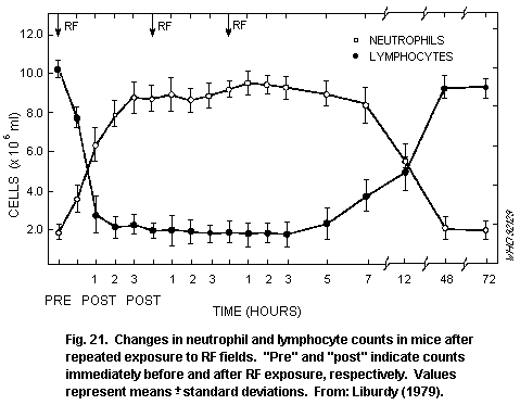 7.3.7 Cardiovascular system
The responses of the intact cardiovascular system to exposure to
RF, as shown in Table 24, are consistent with those associated with
conventional heating. Hence, depending on the ambient temperature, the
SAR, and duration of exposure, increases in heart rate (tachycardia),
no change, or decreases in heart rate (bradycardia) can be induced
during, and following, RF exposure.
Table 24. Cardiovascular system effects
Exposure conditions Effect on exposed group Reference
During exposure
2.45 GHz (CW); 100 W/m2, No heart rate effects in Birenbaum
SAR 2 W/kg, for 20 min rabbits et al.(1975)
applied to heads
2.45 GHz (CW), 50 or Increased heart rate only Chou et al.
800 W/m2 or 50 W/m2 at 800 W/m2 in (1980)
(pulsed) whole-body SAR rabbits; normal response
up to 15 W/kg to heating
2.45 GHz (CW); SAR No effect on arterial McRee et al.
2.3 W/kg for 6 h blood pressure, but heart (1988)
rate reduced by 10% in rats
4 GHz, CW or amplitude- Transient bradycardia during Mangel et al.
modulated at 16 Hz (70% CW or modulated RF in (1990)
mod.), for 30 min, SAR in anaesthetized rats
cortex 8.4, 16.8, or 42 W/kg
After exposure
2.45 GHz (CW), 30 min to At 6.5 W/kg, mild bradycardia Phillips
SARs up to 11.1 W/kg in in rats for up to 3 h; et al. (1975)
rats; environmental at 11.1 W/kg, pronounced
temperature 24 °C bradycardia for 2 h,
followed by mild tachycardia
and irregular heart rate for
short periods; threshold
between 4.5 and 6.5 W/kg
915 MHz (CW), SAR No change in heart weight D'Andrea
2.5 W/kg, for 8 h/day, in rats et al. (1980)
5 days/week, for 16 weeks
435 MHz (pulsed: 1 µs No change in resting Toler et al.
pulses at 1000 Hz) heart rate or mean (1988)
10 W/m2 whole-body arterial blood pressure
SAR approx 0.35 W/kg, for in rats
22 h/day over 6 months
An increase in cardiac output, heart rate, and blood pressure,
coupled with a decrease in peripheral resistance, has been reported in
rabbits exposed at SARs estimated at 10-15 W/kg (raising body
temperatures 0.5 °C) and in anaesthetized rats exposed at levels that
increased body temperature by about 3.5 °C. Following exposure, heart
rate decreased; the threshold for this effect was between 4.5 and 6.5
W/kg. Long-term exposure of rats at SARs of between 0.3 and 2.5 W/kg
did not affect heart rate or heart weight.
7.3.8 Reproduction and development
The assessment of the toxic effects of an agent on fertility and
the development of the embryo and fetus are of great importance. The
processes of meiosis, fertilization and implantation, and the high
rates of cell division and differentiation during development of the
embryo and fetus tend to be more susceptible to toxic insults than
many other processes in the tissues of the adult organism.
7.3.8.1 kHz studies
Studies in the kHz range are summarized in Table 25. The fields
used are generally of the type generated by clinical exposure systems
or by some types of visual display units. These studies have not shown
consistently reproducible effects. Exposure of developing chick
embryos to pulsed electromagnetic fields, including a signal of the
type used clinically for bone healing, for up to a week, had no effect
on malformation incidence (Sisken et al., 1986). Studies of effects on
mammals are of greater relevance to human health.
Two teratological studies (Tribukait et al., 1987; Stuchly et
al., 1988) on the effects of magnetic fields of the type used in VDUs
reported increased numbers of malformed fetuses in rodents, but, when
the results were analysed using the litter rather than the individual
fetus as the unit of observation, the increases were not significant
(Stuchly et al., 1988).
Table 25. Teratological studies in the kHz region
Exposure conditions Effect on exposed group Reference
Chicks
Pulsed magnetic fields Abnormal development, Delgado et al.
10, 100, or 1000 Hz; up particularly in cephalic (1982).
to 1.2 µT, for first region; effect most
48 h of development marked at 100 Hz
Pulsed electromagnetic No significant increase in Sisken et al.
fields, 3.8 kHz, 50 ms incidence of abnormalities (1986)
burst repeated at 2 Hz
(0.25 mT peak) or
4.4 kHz, 5 ms burst
repeated at 15 Hz
(1.6 mT) to embryos for
first 24 h or 7 days of
development
Table 25 (continued)
Exposure conditions Effect on exposed group Reference
20 kHz sawtooth magnetic No effect on incidence of Sandstrom et al.
fields 0.1-16 µT applied malformation (1987)
to embryos for first 42 or
47 h of development
Mammals
20 kHz sawtooth magnetic Significant increase in Tribukait et
fields 1 or 15 µT applied number of mouse fetuses al. (1987)
to embryos on days 0-14 of with external malformations
gestation at 15 µT (difference not
significant if analysed by
litter (Stuchly et al.,1988)
20 kHz sawtooth magnetic Increased number of implants Frolen et al.
fields 15 µT applied to and post-implantation (1987)
embryos on days 0-19 of deaths in mice; no effect on
gestation incidence of malformation
19 kHz sawtooth magnetic No effect on post-implantation Stuchly et al.
fields 5.7, 23, for or 66 survival in rats; increase (1988)
µT, for 7 h/day, before and in minor skeletal defects in
during gestation highest exposure group, but
only if analysed by individual
fetus and not by litter
7.3.8.2 MHz and GHz studies
(a) Fertility. Most of the studies on reproduction and development
in small mammals exposed to RF radiation have shown effects that can
be related to an increase in temperature, and can be produced by
thermal stress alone. It is well known that, in many species of
mammal, the development of male germ cells can be adversely affected
by increased testicular temperatures. The studies shown in Table 26
indicate that acute RF exposure of anaesthetized animals can, through
raising testicular temperature, affect the spermatogenic epithelium
and, thus, male fertility. However, the anaesthesia will have altered
the animals' abilities to regulate their testicular temperatures
(usually maintained 3-4 °C below body temperature). The exposure of
conscious animals has been found to have little effect on testicular
function, except after prolonged exposure at thermally significant
levels. Male rats, exposed long-term at about 6 W/kg, showed a slight
reduction in potential sperm production by the heat-sensitive
pachytene spermatocytes (Johnson et al., 1984) and were reported to be
temporarily less fertile (Berman et al., 1980).
Table 26. Effects on male fertility
Exposure conditions Effect on exposed group Reference
Anaesthetized
2.45 GHz (CW), or direct Depletion of primary Saunders &
heating of lower half of spermatocytes and spermatids Kowalczuk
body, for 30 min in mice; threshold temperature (1981);
for depletion 39 °C or SAR of Kowalczuk et
30 W/kg or greater; al. (1983)
increased number of abnormal
sperm at higher temperatures
1.3 GHz (pulsed), 8-10 Depletion of primary Lebovitz et
W/kg, for 60 min or more spermatocytes and spermatids al. (1987)
in rats; threshold temperature
39-41 °C
Conscious
2.45 GHz (CW), up to No effect on sperm count or Cairnie &
20 W/kg, 16 h/day, number of abnormal sperm in Harding
for up to 30 days conscious mice (1981)
2.45 GHz (CW), 5 W/kg, for No effect on conscious, Saunders
120 h over 8 weeks, then male mouse fertility, et al.(1988)
mice mated over next pregnancy rates 8 weeks
2.45 GHz (CW), 5.6 W/kg, Transient reduction in Berman et al.
for 80 h over 4 weeks conscious, male rat fertility, (1980)
50% of dams mated 3-9 days
after irradiation of males
showed pregnancies; rectal
temperature 41 °C
1.3 GHz (pulsed), 6.3 W/kg, No effect on sperm production, Lebovitz &
6 h/day for 9 days sperm morphology, testes mass, Johnson
etc. in rats; body temperature (1983);
rise of 1.5 °C; no effect Johnson et
on different stages al. (1984)
of spermatogenesis, except for
a reduction in heat sensitive
pachytene spermatocytes
1.3 GHz (CW), 9 W/kg, for No differences in testicular Lebovitz &
8 h, rectal temperature function of conscious rats Johnson (1987)
rise 4.5 °C
(b) Developmental (teratogenic) effects. Exposure to high levels of
RF will induce significant rises in maternal body temperature, and
result in deformities or defects in the offspring, as shown in Table
27. O'Connor (1980) concluded, from a review of the teratogenic
effects of exposure to RF, principally in mice and rats, that intense
exposures that result in significant maternal heating can result in
reduced fetal mass, specific abnormalities (especially exencephaly),
and in increased embryo and fetal losses. For rats, most of the
significant results were based on intense levels of exposure. The most
commonly reported defects were decreased fetal mass and increased
embryo and fetal losses.
RF teratogenesis has also been demonstrated in mice, though
generally at higher SARs. In one study, it was reported that RF
exposure at around 4-5 W/kg enhanced the effect of a chemical
teratogen.
In their review, Lary & Conover (1987) concluded that heat causes
birth defects and pre-natal mortality, when the temperature of the
pregnant mother exceeds 40 °C. Exposure that increases the core
temperature of pregnant dams to 39-41 °C does not usually result in
gross structural malformations, but may significantly increase the
incidence of pre-natal mortality, result in lower body weight, cause
histological or physiological changes, or alter the behaviour of the
exposed offspring. They suggest that only exposures that have an
appreciable heating effect are likely to affect the human embryo
adversely. In contrast, one study described teratological effects in
rats after exposure to 27.12 MHz at a whole-body SAR of 10-4 W/kg.
However, these results are difficult to reconcile with those of many
other studies carried out at the same frequency.
Table 27. Teratogenic effects in the MHz-GHz region
Exposure conditions Effect on exposed group Reference
Rats
27.12 MHz (CW), approx. Embryo and fetal deaths, and Lary et al.
11 W/kg, for 20-40 min; abnormalities at all stages (1982)
rectal temperature to 43 °C of development
27.12 MHz (CW), 33 kV/m, Various effects in offspring Brown-Woodman
0.8 A/m; mated rats related to temperature et al. (1988)
exposed on day 9 of increase and duration of
gestation; temperature exposure
increase maintained at
2.5-5 °C
27.12 MHz (CW), 1 W/m2, Decreased post-implantation Tofani et
0.1 mW/kg, fetuses exposed survival, reduced cranial al. (1986)
from day 0 to 20 of ossification in exposed rat
gestation fetuses
Table 27 (continued)
Exposure conditions Effect on exposed group Reference
6 GHz (CW), approx. Slight growth retardation in Jensh
7 W/kg, for 8 h/day, fetuses, no increased deaths (1984a,b)
throughout pregnancy or structural abnormalities
2.45 GHz (CW), 4 or Maternal temperature raised Berman et al.
6 W/kg, for 100 min/day, to 40 °C; no abnormalities (1981);
from day 6 to 15 of in fetuses; offspring exposed Berman &
gestation to higher levels had lower Carter (1984)
mean body weight
2.45 GHz (CW), 2-4 W/kg, No rectal temperature increase; Jensh et al.
for 6 h/day throughout no excess abnormalities in (1983a,b)
gestation fetuses; no altered performance
in neonatal reflex tests or
adult behaviour, except
increased activity in
exposed offspring
915 MHz (CW), 3.5 W/kg, No anatomical defects in Jensh et al.
for 6 h/day throughout fetuses or behavioural (1982a,b)
pregnancy alterations; maternal temperature
not increased
100 MHz (CW), 0.4 W/kg, No teratogenic or embryogenic Lary et al.
for 400 min/day, on days effects in offspring of rats (1983b)
6-11 of gestation
2.45 GHz (CW), 0.4 W/kg No effects on weight and DNA Merritt et
throughout gestation or RNA content of fetal rat al. (1984)
brain
Hamster
2.45 GHz (CW), 6 or 9 W/kg, Maternal rectal temperature Berman et al.
for 100 min/day, during days increase 0.4 and 1.6 °C; no (1982b)
6-14 of gestation of hamster effect in low-exposure group;
fetuses increased fetal deaths,
decreased fetal weight, and
decreased skeletal maturity
in high-exposure group
Mice
2.45 GHz (CW), 2.8 or Mean mass of live fetuses Berman et al.
22 W/kg, for 100 min/day, decreased in high-exposure (1978)
throughout gestation group
Table 27 (continued)
Exposure conditions Effect on exposed group Reference
2.45 GHz (CW), 7, 28, or At 40 W/kg: reduced no. Nawrot et
40 W/kg, 8 h/day, for implantation sites per al. (1981)
various times during litter and fetal weight,
gestation and increased malformations
2.45 GHz (CW), 16 W/kg, Lower fetal weight, delayed Berman et al.
for 100 min/day during days skeletal maturation, lower (1982a, 1984)
6-17 of gestation brain weight in exposed fetuses
2.45 GHz (CW), 4-5 W/kg, No teratogenic effects in Chazan et al.
for 2 h/day and 7 days per offspring of exposed (1983)
week from days 1 to 7, days animals
8 to 18, or days 1 to 18 of
gestation
2.45 GHz (CW), 1 or 10 At 4-5 W/kg: reduced fetal body Marcickiewicz
W/m2 (equal to 0.5, mass; exposure combined with et al. (1986)
4-5 W/kg) for 2 h/day, injection of cytosine arabinocide
from day 1 to 18 of enhanced incidence of
gestation abnormalities compared with
those on drug alone
7.3.9 Genetics and mutagenesis
Since the potential to induce heritable changes would be of
particular importance for protection standards, many studies designed
to examine the genetic consequences of exposure have been conducted.
Studies examining the possible hereditary consequences of RF exposure
are listed in Table 28, including those on germ cell chromosome
aberration frequencies and dominant lethal mutation frequencies
(assessed as the decreased survival of implanted embryos and fetuses).
Much experimental evidence suggests that acute or long-term RF
exposures do not result in an increase in chromosome aberration
frequency, when temperatures are maintained within physiological
limits. One study reported an increased frequency of cytogenetic
effects in mice exposed long-term at SARs between 0.05 and 20 W/kg.
However, this study was not successfully corroborated using a
different strain of mouse.
Table 28. Genetic and mutagenic effects
Exposure conditions Effect on exposed group Reference
Somatic cells
2.45 GHz (CW), up to 21 W/kg No increase in unstable chromo- Huang et al.
(in vivo), rectal temperature some aberrations in Chinese (1977)
rose by up to 1.6 0C hamster blood lymphocytes
2.45 GHz (CW), 21 W/kg, No sister chromatid exchanges McRee et al.
8 h/day for 28 days in mouse bone marrow cells (1981)
2.375 GHz (CW) and Partial hepatectomy in rats 5-6 Antipenko &
2.75 GHz (pulsed). days after exposure; cytological Koveshnikova
0.1, 0.5, 5.0 W/m2 study showed decreased rate of (1987)
7 h/day for 45 days chromosomal aberrations after
0.1 and 0.5 W/m2; increased
after 5.0 W/m2
Germ cells
2.45 GHz (CW), Increased chromosome ex- Manikowska -
0.05-20 W/kg, for 6 h changes and other cyto- Czerska et al.
over 2 weeks genetic abnormalities in germ (1985)
cells exposed as spermatocytes;
2.45 GHz (CW), No chromosome abnormalities Beechey et al.
0.05-20 W/kg, for 6 h in germ cells exposed as stem (1986)
over 2 weeks cells; rectal temperature in 20
W/kg group rose by up to 3 °C
1.7 GHz (CW), 25-45 W/kg, Induction of dominant lethal Varma &
for 30 min, or 5-9 W/kg, mutations in exposed mice; Traboulay
for 40 min over 2 weeks data inclusive (1977)
2.45 GHz (CW), 1.7 kW/m2, Increased dominant lethality Goud et al.
for 70 s reduced male fertility (1982)
2.45 GHz (CW), 43 W kg, No change in dominant Saunders et
for 30 min lethality, but reduced al. (1983)
pregnancy rate and pre-
implantation survival
2.45 GHz (CW), 5 W/kg, for No chromosomal abnormalities; Saunders et
120 h over 8 weeks no change in pregnancy rate al. (1988)
or dominant lethality
Table 28 (continued)
Exposure conditions Effect on exposed group Reference
2.45 GHz (CW) at 50 W/m2 No consistent pattern of Berman et al.
(0.9-4.7 W/kg) 4 h/day for responses, increased fetal (1980)
> 90 days mortality not related to
- at 100 W/m2, 5 h/day decreased live fetuses; no
for 5 day sperm cell mutagenesis
- at 280 W/m2, 4 h/day, 5
days/week over 4 weeks
In general, the data in Table 28 suggest that the only exposures
that are potentially mutagenic are those at high RF power densities,
which result in substantial increase in temperature.
7.3.10 Cancer-related studies
A summary of cancer-related animal studies is given in Table 29.
The number and types of studies are limited.
Exposure to RF levels sufficiently high to induce hyperthermia
has generally resulted in tumour regression following transplantation
of tumour cells (Preskorn et al., 1978; Roszkowski et al., 1980). In
contrast, an increase in tumour progression has been observed in mice
exposed long-term at lower, possibly thermogenic, SARs (Szmigielski et
al., 1982). This effect was related to a non-specific stress. The
authors suggested a transient shift in immune surveillance resulting
in a lowering of resistance to neoplastic growth, as a likely
explanation. Exposure at about 1 W/kg did not have any effect on
melanoma growth in mice (Santini et al., 1988).
The effects of exposure on spontaneous or chemically-induced
tumours have also been examined. In contrast to transplantation
studies, these can test for an effect on the process of
carcinogenesis. Two early studies (Prausnitz & Suskind, 1962; Skidmore
& Baum, 1974), relevant to cancer induction, but in which the
methodology was flawed in relation to an analysis of this end-point,
are described for completeness. An increased incidence of monocytic
leukosis (defined as a non-circulating neoplasm of white-blood cells)
and lymphatic or myeloid leukaemia (defined as a circulating
"leukosis") was reported in Swiss mice exposed to thermally
significant levels (half the acute LD50) of 9.27 GHz pulsed RF, for 5
days per week for 59 days (Prausnitz & Susskind, 1962). However, the
study suffered several deficiencies: leukosis and leukaemia were
inadequately defined, infection may well have confounded the results,
a large proportion of mice died without a cause of death being
identified, and statistical analysis was absent (Roberts 1983; Kirk
1984).
Table 29. Cancer-related studies
Exposure conditions Effect on exposed group Reference
Transplanted tumour cells
2.45 GHz (CW), 35 W/kg, for Retarded tumour growth and Preskorn et
20 min/day during days 11- tumour incidence in sarcoma- al. (1978)
14 of gestation; offspring injected offspring of exposed
injected with sarcoma pregnant mice; rectal temperature
cells at 16 days of age of dams rose over 2 °C; exposed
exposed for 36 days mice had increased longevity
2.45 GHz (CW), 25 W/kg, Temporary tumour regression Roszkowski et
2 h/day for 7 days; followed by renewed tumour al. (1980)
Injection of sarcoma growth 12 days later, when
cells in mice 14 days exposure 14 days after tumour
after, or just after, injection; accelerated tumour
RF exposure growth, if exposed before
implantation of tumour;
lung metastases increased
2.45 GHz (CW), 2-3 W/kg RF caused increase in Szmigielski
or 6-8 W/kg, 2 h/day, for sarcoma colonies in lungs in et al. (1982)
6 days/week; mice exposed mice injected intravenously
from 6 weeks of age to with these cells; chronic via
12 months of stress confinement caused similar
increase in lung tumours as
2-3 W/kg, but 6-8 W/kg produced
higher increase in tumours
2.45 GHz (CW and pulsed) No difference in mean tumour Santini et
10 W/m2, 1.2 W/kg prior surface area/animal, or in al. (1988)
to, and during, B16 melanoma mean survival time between
tumour transplantation and exposed or control mice
growth; exposed for
2.5 h/day, 6 times/week for
15 days, prior to injection of
melanoma cells, then exposed
to same schedule until death
Spontaneous or chemically-induced tumours
2.45 GHz (CW), 2-3 W/kg SAR-dependent acceleration of Szmigielski
or 6-8 W/kg, 2 h/day, for mammary tumours in mice et al. (1982)
6 days/week, mice exposed genetically predisposed to these
from 6 weeks of age to tumours, and acceleration of skin
12 months of stress tumours in mice painted with the
carcinogen 3,4-benzopyrene (BP)
Table 29 (continued)
Exposure conditions Effect on exposed group Reference
2.45 GHz (CW), 100 W/m2 Increased development of Szmigielski
4-5 W/kg, for 2 h/day, 5-6 chemically-induced hepatomas et al. (1988)
days/week for a few months and sarcomas in mice; survival
of exposed mice decreased;
increased frequency of skin
tumours in mice given
subcarcinogenic dose of BP
2.45 GHz (10 µs pulses at Total incidence of neoplasia Guy et al.
800 Hz) square wave- not significantly different (1985)
modulated at 8 Hz, from that in controls; however,
0.4 W/kg, continuous exposure increased number of primary
at 2-27 months of age malignancies (18) occurred early
(lifetime study of rats) in exposed group compared with
controls (5)
Skidmore & Baum (1974) reported that exposure for 5 days per week
for 33 weeks to very short pulses (5 ns rise time; 550 ms decay time)
of high field strength (447 kV/m) pulsed at 5 Hz, resulted in a
reduced incidence of leukaemia in AKR/J mice (which spontaneously
develop a high incidence of lymphatic leukaemia between 26 and 52
weeks of age) compared with controls at the end of the exposure.
However, the absence of a complete analysis of leukaemia incidence
(and other causes of death) precludes any conclusion being drawn from
this study. The authors also reported a zero incidence of mammary
tumours in 1-year-old female Sprague-Dawley rats that had been exposed
for 38 weeks; evaluation was probably premature for this end-point,
the tumours occur spontaneously mainly in older rats. A later study
(Baum et al., 1976) reported no effects on mammary tumour incidence
and other lesions in rats exposed for 94 weeks.
Two studies merit particular attention. The long-term exposure of
mice at SARs of between 2 and 8 W/kg resulted in an increase in the
number of sarcoma cell colonies in the lungs (following the injection
of sarcoma cells), as shown in Fig. 22, and in an SAR-dependent
increase in the rate of development of spontaneous mammary tumours and
chemically-induced skin tumours. Repeated microwave exposure, followed
by a "sub-carcinogenic" dose of carcinogen, resulted in an increased
number of skin tumours. A study of 100 rats exposed for most of their
lifetime at about 0.4 W/kg did not show any increased incidence of
non-neoplastic lesions compared with control animals; longevity was
very similar in both groups. However, the overall incidence of primary
malignancy in the exposed group (18) was significantly greater than
the control value (5), but was reported to be similar to the
spontaneous incidence given in the literature for the particular
strain of rat. Under these circumstances, it is difficult to draw any
firm conclusions.
7.3.7 Cardiovascular system
The responses of the intact cardiovascular system to exposure to
RF, as shown in Table 24, are consistent with those associated with
conventional heating. Hence, depending on the ambient temperature, the
SAR, and duration of exposure, increases in heart rate (tachycardia),
no change, or decreases in heart rate (bradycardia) can be induced
during, and following, RF exposure.
Table 24. Cardiovascular system effects
Exposure conditions Effect on exposed group Reference
During exposure
2.45 GHz (CW); 100 W/m2, No heart rate effects in Birenbaum
SAR 2 W/kg, for 20 min rabbits et al.(1975)
applied to heads
2.45 GHz (CW), 50 or Increased heart rate only Chou et al.
800 W/m2 or 50 W/m2 at 800 W/m2 in (1980)
(pulsed) whole-body SAR rabbits; normal response
up to 15 W/kg to heating
2.45 GHz (CW); SAR No effect on arterial McRee et al.
2.3 W/kg for 6 h blood pressure, but heart (1988)
rate reduced by 10% in rats
4 GHz, CW or amplitude- Transient bradycardia during Mangel et al.
modulated at 16 Hz (70% CW or modulated RF in (1990)
mod.), for 30 min, SAR in anaesthetized rats
cortex 8.4, 16.8, or 42 W/kg
After exposure
2.45 GHz (CW), 30 min to At 6.5 W/kg, mild bradycardia Phillips
SARs up to 11.1 W/kg in in rats for up to 3 h; et al. (1975)
rats; environmental at 11.1 W/kg, pronounced
temperature 24 °C bradycardia for 2 h,
followed by mild tachycardia
and irregular heart rate for
short periods; threshold
between 4.5 and 6.5 W/kg
915 MHz (CW), SAR No change in heart weight D'Andrea
2.5 W/kg, for 8 h/day, in rats et al. (1980)
5 days/week, for 16 weeks
435 MHz (pulsed: 1 µs No change in resting Toler et al.
pulses at 1000 Hz) heart rate or mean (1988)
10 W/m2 whole-body arterial blood pressure
SAR approx 0.35 W/kg, for in rats
22 h/day over 6 months
An increase in cardiac output, heart rate, and blood pressure,
coupled with a decrease in peripheral resistance, has been reported in
rabbits exposed at SARs estimated at 10-15 W/kg (raising body
temperatures 0.5 °C) and in anaesthetized rats exposed at levels that
increased body temperature by about 3.5 °C. Following exposure, heart
rate decreased; the threshold for this effect was between 4.5 and 6.5
W/kg. Long-term exposure of rats at SARs of between 0.3 and 2.5 W/kg
did not affect heart rate or heart weight.
7.3.8 Reproduction and development
The assessment of the toxic effects of an agent on fertility and
the development of the embryo and fetus are of great importance. The
processes of meiosis, fertilization and implantation, and the high
rates of cell division and differentiation during development of the
embryo and fetus tend to be more susceptible to toxic insults than
many other processes in the tissues of the adult organism.
7.3.8.1 kHz studies
Studies in the kHz range are summarized in Table 25. The fields
used are generally of the type generated by clinical exposure systems
or by some types of visual display units. These studies have not shown
consistently reproducible effects. Exposure of developing chick
embryos to pulsed electromagnetic fields, including a signal of the
type used clinically for bone healing, for up to a week, had no effect
on malformation incidence (Sisken et al., 1986). Studies of effects on
mammals are of greater relevance to human health.
Two teratological studies (Tribukait et al., 1987; Stuchly et
al., 1988) on the effects of magnetic fields of the type used in VDUs
reported increased numbers of malformed fetuses in rodents, but, when
the results were analysed using the litter rather than the individual
fetus as the unit of observation, the increases were not significant
(Stuchly et al., 1988).
Table 25. Teratological studies in the kHz region
Exposure conditions Effect on exposed group Reference
Chicks
Pulsed magnetic fields Abnormal development, Delgado et al.
10, 100, or 1000 Hz; up particularly in cephalic (1982).
to 1.2 µT, for first region; effect most
48 h of development marked at 100 Hz
Pulsed electromagnetic No significant increase in Sisken et al.
fields, 3.8 kHz, 50 ms incidence of abnormalities (1986)
burst repeated at 2 Hz
(0.25 mT peak) or
4.4 kHz, 5 ms burst
repeated at 15 Hz
(1.6 mT) to embryos for
first 24 h or 7 days of
development
Table 25 (continued)
Exposure conditions Effect on exposed group Reference
20 kHz sawtooth magnetic No effect on incidence of Sandstrom et al.
fields 0.1-16 µT applied malformation (1987)
to embryos for first 42 or
47 h of development
Mammals
20 kHz sawtooth magnetic Significant increase in Tribukait et
fields 1 or 15 µT applied number of mouse fetuses al. (1987)
to embryos on days 0-14 of with external malformations
gestation at 15 µT (difference not
significant if analysed by
litter (Stuchly et al.,1988)
20 kHz sawtooth magnetic Increased number of implants Frolen et al.
fields 15 µT applied to and post-implantation (1987)
embryos on days 0-19 of deaths in mice; no effect on
gestation incidence of malformation
19 kHz sawtooth magnetic No effect on post-implantation Stuchly et al.
fields 5.7, 23, for or 66 survival in rats; increase (1988)
µT, for 7 h/day, before and in minor skeletal defects in
during gestation highest exposure group, but
only if analysed by individual
fetus and not by litter
7.3.8.2 MHz and GHz studies
(a) Fertility. Most of the studies on reproduction and development
in small mammals exposed to RF radiation have shown effects that can
be related to an increase in temperature, and can be produced by
thermal stress alone. It is well known that, in many species of
mammal, the development of male germ cells can be adversely affected
by increased testicular temperatures. The studies shown in Table 26
indicate that acute RF exposure of anaesthetized animals can, through
raising testicular temperature, affect the spermatogenic epithelium
and, thus, male fertility. However, the anaesthesia will have altered
the animals' abilities to regulate their testicular temperatures
(usually maintained 3-4 °C below body temperature). The exposure of
conscious animals has been found to have little effect on testicular
function, except after prolonged exposure at thermally significant
levels. Male rats, exposed long-term at about 6 W/kg, showed a slight
reduction in potential sperm production by the heat-sensitive
pachytene spermatocytes (Johnson et al., 1984) and were reported to be
temporarily less fertile (Berman et al., 1980).
Table 26. Effects on male fertility
Exposure conditions Effect on exposed group Reference
Anaesthetized
2.45 GHz (CW), or direct Depletion of primary Saunders &
heating of lower half of spermatocytes and spermatids Kowalczuk
body, for 30 min in mice; threshold temperature (1981);
for depletion 39 °C or SAR of Kowalczuk et
30 W/kg or greater; al. (1983)
increased number of abnormal
sperm at higher temperatures
1.3 GHz (pulsed), 8-10 Depletion of primary Lebovitz et
W/kg, for 60 min or more spermatocytes and spermatids al. (1987)
in rats; threshold temperature
39-41 °C
Conscious
2.45 GHz (CW), up to No effect on sperm count or Cairnie &
20 W/kg, 16 h/day, number of abnormal sperm in Harding
for up to 30 days conscious mice (1981)
2.45 GHz (CW), 5 W/kg, for No effect on conscious, Saunders
120 h over 8 weeks, then male mouse fertility, et al.(1988)
mice mated over next pregnancy rates 8 weeks
2.45 GHz (CW), 5.6 W/kg, Transient reduction in Berman et al.
for 80 h over 4 weeks conscious, male rat fertility, (1980)
50% of dams mated 3-9 days
after irradiation of males
showed pregnancies; rectal
temperature 41 °C
1.3 GHz (pulsed), 6.3 W/kg, No effect on sperm production, Lebovitz &
6 h/day for 9 days sperm morphology, testes mass, Johnson
etc. in rats; body temperature (1983);
rise of 1.5 °C; no effect Johnson et
on different stages al. (1984)
of spermatogenesis, except for
a reduction in heat sensitive
pachytene spermatocytes
1.3 GHz (CW), 9 W/kg, for No differences in testicular Lebovitz &
8 h, rectal temperature function of conscious rats Johnson (1987)
rise 4.5 °C
(b) Developmental (teratogenic) effects. Exposure to high levels of
RF will induce significant rises in maternal body temperature, and
result in deformities or defects in the offspring, as shown in Table
27. O'Connor (1980) concluded, from a review of the teratogenic
effects of exposure to RF, principally in mice and rats, that intense
exposures that result in significant maternal heating can result in
reduced fetal mass, specific abnormalities (especially exencephaly),
and in increased embryo and fetal losses. For rats, most of the
significant results were based on intense levels of exposure. The most
commonly reported defects were decreased fetal mass and increased
embryo and fetal losses.
RF teratogenesis has also been demonstrated in mice, though
generally at higher SARs. In one study, it was reported that RF
exposure at around 4-5 W/kg enhanced the effect of a chemical
teratogen.
In their review, Lary & Conover (1987) concluded that heat causes
birth defects and pre-natal mortality, when the temperature of the
pregnant mother exceeds 40 °C. Exposure that increases the core
temperature of pregnant dams to 39-41 °C does not usually result in
gross structural malformations, but may significantly increase the
incidence of pre-natal mortality, result in lower body weight, cause
histological or physiological changes, or alter the behaviour of the
exposed offspring. They suggest that only exposures that have an
appreciable heating effect are likely to affect the human embryo
adversely. In contrast, one study described teratological effects in
rats after exposure to 27.12 MHz at a whole-body SAR of 10-4 W/kg.
However, these results are difficult to reconcile with those of many
other studies carried out at the same frequency.
Table 27. Teratogenic effects in the MHz-GHz region
Exposure conditions Effect on exposed group Reference
Rats
27.12 MHz (CW), approx. Embryo and fetal deaths, and Lary et al.
11 W/kg, for 20-40 min; abnormalities at all stages (1982)
rectal temperature to 43 °C of development
27.12 MHz (CW), 33 kV/m, Various effects in offspring Brown-Woodman
0.8 A/m; mated rats related to temperature et al. (1988)
exposed on day 9 of increase and duration of
gestation; temperature exposure
increase maintained at
2.5-5 °C
27.12 MHz (CW), 1 W/m2, Decreased post-implantation Tofani et
0.1 mW/kg, fetuses exposed survival, reduced cranial al. (1986)
from day 0 to 20 of ossification in exposed rat
gestation fetuses
Table 27 (continued)
Exposure conditions Effect on exposed group Reference
6 GHz (CW), approx. Slight growth retardation in Jensh
7 W/kg, for 8 h/day, fetuses, no increased deaths (1984a,b)
throughout pregnancy or structural abnormalities
2.45 GHz (CW), 4 or Maternal temperature raised Berman et al.
6 W/kg, for 100 min/day, to 40 °C; no abnormalities (1981);
from day 6 to 15 of in fetuses; offspring exposed Berman &
gestation to higher levels had lower Carter (1984)
mean body weight
2.45 GHz (CW), 2-4 W/kg, No rectal temperature increase; Jensh et al.
for 6 h/day throughout no excess abnormalities in (1983a,b)
gestation fetuses; no altered performance
in neonatal reflex tests or
adult behaviour, except
increased activity in
exposed offspring
915 MHz (CW), 3.5 W/kg, No anatomical defects in Jensh et al.
for 6 h/day throughout fetuses or behavioural (1982a,b)
pregnancy alterations; maternal temperature
not increased
100 MHz (CW), 0.4 W/kg, No teratogenic or embryogenic Lary et al.
for 400 min/day, on days effects in offspring of rats (1983b)
6-11 of gestation
2.45 GHz (CW), 0.4 W/kg No effects on weight and DNA Merritt et
throughout gestation or RNA content of fetal rat al. (1984)
brain
Hamster
2.45 GHz (CW), 6 or 9 W/kg, Maternal rectal temperature Berman et al.
for 100 min/day, during days increase 0.4 and 1.6 °C; no (1982b)
6-14 of gestation of hamster effect in low-exposure group;
fetuses increased fetal deaths,
decreased fetal weight, and
decreased skeletal maturity
in high-exposure group
Mice
2.45 GHz (CW), 2.8 or Mean mass of live fetuses Berman et al.
22 W/kg, for 100 min/day, decreased in high-exposure (1978)
throughout gestation group
Table 27 (continued)
Exposure conditions Effect on exposed group Reference
2.45 GHz (CW), 7, 28, or At 40 W/kg: reduced no. Nawrot et
40 W/kg, 8 h/day, for implantation sites per al. (1981)
various times during litter and fetal weight,
gestation and increased malformations
2.45 GHz (CW), 16 W/kg, Lower fetal weight, delayed Berman et al.
for 100 min/day during days skeletal maturation, lower (1982a, 1984)
6-17 of gestation brain weight in exposed fetuses
2.45 GHz (CW), 4-5 W/kg, No teratogenic effects in Chazan et al.
for 2 h/day and 7 days per offspring of exposed (1983)
week from days 1 to 7, days animals
8 to 18, or days 1 to 18 of
gestation
2.45 GHz (CW), 1 or 10 At 4-5 W/kg: reduced fetal body Marcickiewicz
W/m2 (equal to 0.5, mass; exposure combined with et al. (1986)
4-5 W/kg) for 2 h/day, injection of cytosine arabinocide
from day 1 to 18 of enhanced incidence of
gestation abnormalities compared with
those on drug alone
7.3.9 Genetics and mutagenesis
Since the potential to induce heritable changes would be of
particular importance for protection standards, many studies designed
to examine the genetic consequences of exposure have been conducted.
Studies examining the possible hereditary consequences of RF exposure
are listed in Table 28, including those on germ cell chromosome
aberration frequencies and dominant lethal mutation frequencies
(assessed as the decreased survival of implanted embryos and fetuses).
Much experimental evidence suggests that acute or long-term RF
exposures do not result in an increase in chromosome aberration
frequency, when temperatures are maintained within physiological
limits. One study reported an increased frequency of cytogenetic
effects in mice exposed long-term at SARs between 0.05 and 20 W/kg.
However, this study was not successfully corroborated using a
different strain of mouse.
Table 28. Genetic and mutagenic effects
Exposure conditions Effect on exposed group Reference
Somatic cells
2.45 GHz (CW), up to 21 W/kg No increase in unstable chromo- Huang et al.
(in vivo), rectal temperature some aberrations in Chinese (1977)
rose by up to 1.6 0C hamster blood lymphocytes
2.45 GHz (CW), 21 W/kg, No sister chromatid exchanges McRee et al.
8 h/day for 28 days in mouse bone marrow cells (1981)
2.375 GHz (CW) and Partial hepatectomy in rats 5-6 Antipenko &
2.75 GHz (pulsed). days after exposure; cytological Koveshnikova
0.1, 0.5, 5.0 W/m2 study showed decreased rate of (1987)
7 h/day for 45 days chromosomal aberrations after
0.1 and 0.5 W/m2; increased
after 5.0 W/m2
Germ cells
2.45 GHz (CW), Increased chromosome ex- Manikowska -
0.05-20 W/kg, for 6 h changes and other cyto- Czerska et al.
over 2 weeks genetic abnormalities in germ (1985)
cells exposed as spermatocytes;
2.45 GHz (CW), No chromosome abnormalities Beechey et al.
0.05-20 W/kg, for 6 h in germ cells exposed as stem (1986)
over 2 weeks cells; rectal temperature in 20
W/kg group rose by up to 3 °C
1.7 GHz (CW), 25-45 W/kg, Induction of dominant lethal Varma &
for 30 min, or 5-9 W/kg, mutations in exposed mice; Traboulay
for 40 min over 2 weeks data inclusive (1977)
2.45 GHz (CW), 1.7 kW/m2, Increased dominant lethality Goud et al.
for 70 s reduced male fertility (1982)
2.45 GHz (CW), 43 W kg, No change in dominant Saunders et
for 30 min lethality, but reduced al. (1983)
pregnancy rate and pre-
implantation survival
2.45 GHz (CW), 5 W/kg, for No chromosomal abnormalities; Saunders et
120 h over 8 weeks no change in pregnancy rate al. (1988)
or dominant lethality
Table 28 (continued)
Exposure conditions Effect on exposed group Reference
2.45 GHz (CW) at 50 W/m2 No consistent pattern of Berman et al.
(0.9-4.7 W/kg) 4 h/day for responses, increased fetal (1980)
> 90 days mortality not related to
- at 100 W/m2, 5 h/day decreased live fetuses; no
for 5 day sperm cell mutagenesis
- at 280 W/m2, 4 h/day, 5
days/week over 4 weeks
In general, the data in Table 28 suggest that the only exposures
that are potentially mutagenic are those at high RF power densities,
which result in substantial increase in temperature.
7.3.10 Cancer-related studies
A summary of cancer-related animal studies is given in Table 29.
The number and types of studies are limited.
Exposure to RF levels sufficiently high to induce hyperthermia
has generally resulted in tumour regression following transplantation
of tumour cells (Preskorn et al., 1978; Roszkowski et al., 1980). In
contrast, an increase in tumour progression has been observed in mice
exposed long-term at lower, possibly thermogenic, SARs (Szmigielski et
al., 1982). This effect was related to a non-specific stress. The
authors suggested a transient shift in immune surveillance resulting
in a lowering of resistance to neoplastic growth, as a likely
explanation. Exposure at about 1 W/kg did not have any effect on
melanoma growth in mice (Santini et al., 1988).
The effects of exposure on spontaneous or chemically-induced
tumours have also been examined. In contrast to transplantation
studies, these can test for an effect on the process of
carcinogenesis. Two early studies (Prausnitz & Suskind, 1962; Skidmore
& Baum, 1974), relevant to cancer induction, but in which the
methodology was flawed in relation to an analysis of this end-point,
are described for completeness. An increased incidence of monocytic
leukosis (defined as a non-circulating neoplasm of white-blood cells)
and lymphatic or myeloid leukaemia (defined as a circulating
"leukosis") was reported in Swiss mice exposed to thermally
significant levels (half the acute LD50) of 9.27 GHz pulsed RF, for 5
days per week for 59 days (Prausnitz & Susskind, 1962). However, the
study suffered several deficiencies: leukosis and leukaemia were
inadequately defined, infection may well have confounded the results,
a large proportion of mice died without a cause of death being
identified, and statistical analysis was absent (Roberts 1983; Kirk
1984).
Table 29. Cancer-related studies
Exposure conditions Effect on exposed group Reference
Transplanted tumour cells
2.45 GHz (CW), 35 W/kg, for Retarded tumour growth and Preskorn et
20 min/day during days 11- tumour incidence in sarcoma- al. (1978)
14 of gestation; offspring injected offspring of exposed
injected with sarcoma pregnant mice; rectal temperature
cells at 16 days of age of dams rose over 2 °C; exposed
exposed for 36 days mice had increased longevity
2.45 GHz (CW), 25 W/kg, Temporary tumour regression Roszkowski et
2 h/day for 7 days; followed by renewed tumour al. (1980)
Injection of sarcoma growth 12 days later, when
cells in mice 14 days exposure 14 days after tumour
after, or just after, injection; accelerated tumour
RF exposure growth, if exposed before
implantation of tumour;
lung metastases increased
2.45 GHz (CW), 2-3 W/kg RF caused increase in Szmigielski
or 6-8 W/kg, 2 h/day, for sarcoma colonies in lungs in et al. (1982)
6 days/week; mice exposed mice injected intravenously
from 6 weeks of age to with these cells; chronic via
12 months of stress confinement caused similar
increase in lung tumours as
2-3 W/kg, but 6-8 W/kg produced
higher increase in tumours
2.45 GHz (CW and pulsed) No difference in mean tumour Santini et
10 W/m2, 1.2 W/kg prior surface area/animal, or in al. (1988)
to, and during, B16 melanoma mean survival time between
tumour transplantation and exposed or control mice
growth; exposed for
2.5 h/day, 6 times/week for
15 days, prior to injection of
melanoma cells, then exposed
to same schedule until death
Spontaneous or chemically-induced tumours
2.45 GHz (CW), 2-3 W/kg SAR-dependent acceleration of Szmigielski
or 6-8 W/kg, 2 h/day, for mammary tumours in mice et al. (1982)
6 days/week, mice exposed genetically predisposed to these
from 6 weeks of age to tumours, and acceleration of skin
12 months of stress tumours in mice painted with the
carcinogen 3,4-benzopyrene (BP)
Table 29 (continued)
Exposure conditions Effect on exposed group Reference
2.45 GHz (CW), 100 W/m2 Increased development of Szmigielski
4-5 W/kg, for 2 h/day, 5-6 chemically-induced hepatomas et al. (1988)
days/week for a few months and sarcomas in mice; survival
of exposed mice decreased;
increased frequency of skin
tumours in mice given
subcarcinogenic dose of BP
2.45 GHz (10 µs pulses at Total incidence of neoplasia Guy et al.
800 Hz) square wave- not significantly different (1985)
modulated at 8 Hz, from that in controls; however,
0.4 W/kg, continuous exposure increased number of primary
at 2-27 months of age malignancies (18) occurred early
(lifetime study of rats) in exposed group compared with
controls (5)
Skidmore & Baum (1974) reported that exposure for 5 days per week
for 33 weeks to very short pulses (5 ns rise time; 550 ms decay time)
of high field strength (447 kV/m) pulsed at 5 Hz, resulted in a
reduced incidence of leukaemia in AKR/J mice (which spontaneously
develop a high incidence of lymphatic leukaemia between 26 and 52
weeks of age) compared with controls at the end of the exposure.
However, the absence of a complete analysis of leukaemia incidence
(and other causes of death) precludes any conclusion being drawn from
this study. The authors also reported a zero incidence of mammary
tumours in 1-year-old female Sprague-Dawley rats that had been exposed
for 38 weeks; evaluation was probably premature for this end-point,
the tumours occur spontaneously mainly in older rats. A later study
(Baum et al., 1976) reported no effects on mammary tumour incidence
and other lesions in rats exposed for 94 weeks.
Two studies merit particular attention. The long-term exposure of
mice at SARs of between 2 and 8 W/kg resulted in an increase in the
number of sarcoma cell colonies in the lungs (following the injection
of sarcoma cells), as shown in Fig. 22, and in an SAR-dependent
increase in the rate of development of spontaneous mammary tumours and
chemically-induced skin tumours. Repeated microwave exposure, followed
by a "sub-carcinogenic" dose of carcinogen, resulted in an increased
number of skin tumours. A study of 100 rats exposed for most of their
lifetime at about 0.4 W/kg did not show any increased incidence of
non-neoplastic lesions compared with control animals; longevity was
very similar in both groups. However, the overall incidence of primary
malignancy in the exposed group (18) was significantly greater than
the control value (5), but was reported to be similar to the
spontaneous incidence given in the literature for the particular
strain of rat. Under these circumstances, it is difficult to draw any
firm conclusions.
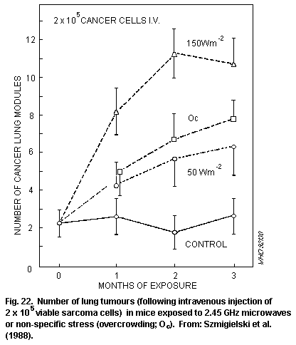 Tumour weights were not significantly different in rats implanted
with mammary adenocarcinoma tissue and either exposed 25 days later to
2 kHz magnetic fields of up to 2 mT for 1 h a day for 9 days or not
exposed (Baumann et al., 1989). Handling and restraint stress in
animals were identified as possible confounders for the detection of
subtle magnetic field effects.
7.3.11 Summary and conclusions
Most of the biological effects of acute exposure to RF fields are
consistent with responses to induced heating, resulting either in
rises in tissue or body temperature of about 1 °C or more, or in
responses for minimizing the total heat load. Most responses in
different animal species, exposed under various environmental
conditions, have been reported at SARs above about 1-2 W/kg.
These animal (particularly primate) data indicate the types of
response that are likely to occur in humans subject to a sufficient
heat load. However, direct quantitative extrapolation to humans is
difficult, given species differences in responses, in general, and in
thermoregulatory ability particularly.
The most sensitive animal responses to heat loads are
thermoregulatory adjustments, such as reduced metabolic heat
production and vasodilation, with thresholds ranging between about
0.05 and 5 W/kg, depending on environmental conditions. However, these
reactions form part of the natural repertoire of thermoregulatory
responses that serve to maintain normal body temperatures.
Transient effects seen in exposed animals that are consistent
with responses to increases in body temperature of 1 °C or more
(and/or SARs in excess of about 2 W/kg in primates and rats) include
the reduced performance of learned tasks and increased plasma
cortico-steroid levels. Other heat-related effects include temporary
haematopoietic and immune responses, possibly in conjunction with
elevated corticosteroid levels. The most consistent effects observed
are reduced levels of circulating lymphocytes and increased levels of
neutrophils, decreased natural killer cell function, and increased
macrophage activation; an increase in the primary antibody response of
B-lymphocytes has also been reported. Cardiovascular changes consonant
with increased heat load, such as increased heart rate and cardiac
output, have been observed, together with a reduction in the effects
of drugs, such as barbiturates, the action of which can be altered by
changes in circulation and clearance rates.
Most animal data indicate that implantation and the development
of the embryo and fetus are unlikely to be affected by exposures that
increase maternal body temperature by less than 1 °C. Above these
temperatures, adverse effects, such as losses in implantation, growth
retardation, and post-natal changes in behaviour, may occur, with more
severe effects occurring at higher maternal temperatures.
Most animal data suggest that low RF exposure that does not raise
body temperatures above the normal physiological range is not
mutagenic; thus, such exposure will not result in somatic mutation or
hereditary effects.
There is much less information describing the effects of
long-term, low-level exposure. So far, it is not apparent that any
long-term adverse effects can result from exposures below thermally
significant levels. The animal data indicate that male fertility is
unlikely to be affected by long-term exposure at levels insufficient
to raise body and testis temperatures. Cataracts have not been induced
in rabbits exposed at 100 W/m2 for 6 months, or in primates exposed
at 1.5 kW/m2 for 3 months.
A study of 100 rats, exposed for most of their lifetime at about
0.4 W/kg, did not show an increased incidence of non-neoplastic
lesions or total neoplasias compared with control animals; longevity
was very similar in both groups. There were differences in the overall
incidence of primary malignancies, but these could not necessarily be
attributed to the RF exposure. The possibility that exposure to RF
might influence the process of carcinogenesis is of particular
concern. So far, there is no definite evidence that RF exposure does
have an effect, but there is clearly a need for further studies to be
carried out. Overwhelmingly, the experimental data indicate that RF
fields are not mutagenic, and so they are unlikely to act as
initiators of carcinogenesis. In a few studies, evidence has been
sought of an enhancement of the effect of a known carcinogen. The
long-term exposure of mice at 2-8 W/kg resulted in an increase in the
progression of spontaneous mammary tumours and of skin tumours in mice
the skin of which was tested with a chemical carcinogen. Repeated RF
exposure followed by a "sub-carcinogenic" dose of carcinogen resulted
in an increased number of skin tumours; however, this study has been
reported only briefly, and the authors noted the need for experimental
confirmation.
In in vitro studies, enhanced cell transformation rates were
reported after RF exposure at 4.4 W/kg (alone or combined with
X-radiation) followed by treatment with a chemical promotor. The
latter data have not always been consistent between studies. It is
clear that studies relevant to carcinogenesis need replicating and
extending further, to reduce uncertainties in this area.
A substantial body of data exists describing in vitro
biological responses to amplitude-modulated RF radiation at SARs too
low to involve any response to heating. Some studies have reported
effects after exposure at SARs of less than 0.01 W/kg, occurring
within modulation frequency "windows" (usually between 1 and 100 Hz)
and sometimes within power density "windows".
Changes have been reported in the electroencephalograms of cats
and rabbits, in calcium ion mobility in the brain tissue in vitro
and in vivo, in lymphocyte cytotoxicity in vitro, and in the in
vitro activity of an enzyme involved in cell growth and division.
Some of these responses have been difficult to confirm, and their
physiological or pathological consequences are not clear. However, any
toxicological investigation should be based on tests carried out at
appropriate levels of exposure. It is important that these studies be
confirmed and extended to in vivo studies and that the health
implications, if any, for exposed people are determined. Of particular
importance, would be studies that link extremely low frequency,
amplitude-modulated RF interactions at the cell surface with changes
in DNA synthesis or transcription. It is worth noting that this
interaction implies a "demodulation" of the RF signal at the cell
membrane.
8. HUMAN RESPONSES
Epidemiology can be defined as the study of the occurrence of
illness; its main goals are to evaluate hypotheses about the causation
of illness and to relate disease occurrence to the characteristics of
people and their environment. Epidemiological studies of human
populations exposed to RF fields are few in number and are generally
limited in scope. The principal groups studied have been people
occupationally exposed in the military or in industry. Information
about worker health status has generally come from medical records,
questionnaires, and physical and laboratory examinations. Exposure
data have come from personnel records, questionnaires, environmental
measurements, and equipment-emission measurements. Determination of
actual exposure to RF fields and to other risk factors for the same
outcome is difficult in retrospective human studies.
Some studies of controlled exposures of volunteers have provided
valuable information on responses to RF exposure. These studies
include warming and pain thresholds for RF heating of the skin, RF
hearing, and RF shocks and burns. Clinical studies of accidental
overexposures provide information on acute-exposure responses.
8.1 Laboratory studies
8.1.1 Cutaneous perception
Exposure of the human body to RF fields can cause heating that is
detectable by the temperature-sensitive receptors in the skin. Several
investigators have determind experimentally the threshold intensities
that cause sensations of perceptible warmth, pain, and delay in
response to the stimulus in human subjects, as shown in
Table 30.
Adair (1983a) noted that RF exposures to frequencies of 30 GHz
and above would probably be similar to infrared in their perception
threshold values. However, over much of the RF spectrum, current
standards are set at levels that are below those that most would
consider detectable by sensation. Thus, cutaneous perception may be an
indicator of exposure only at RF frequencies of the order of several
gigahertz or more, which have wavelengths that are small in comparison
with the length of the exposed body, i.e., wavelengths comparable
with, or smaller than, the thickness of skin. Under these conditions,
most of the energy is absorbed in the outer tissue layers
Table 30. Cutaneous perception in humans
Exposure conditions Effects and thresholds Reference
3 GHz to inner forearm Threshold for pain Cook (1952)
Area 9.5 cm2 at 31 kW/m2: 20 s latency
at 8.3 kW/m2: 180 s latency
Area 53 cm2 at 5.6 kW/m2: 180 s latency
Pain at skin
temperature of 46 °C
3 GHz (pulsed) to inner Latency varied between Vendrik & Vos
forearm (area 13 cm2) less than 0.5 and 3.5 s (1958)
3-25 kW/m2
3 and 10 GHz (pulsed) Threshold for Hendler &
perception: Hardy (1960);
3 GHz, 1 s: 600 W/m2 Hendler et al.
3 GHz, 5 s: 320 W/m2 (1963);
10 GHz, 1 s: 190 W/m2 Hendler
10 GHz, 5 s: 130 W/m2 (1968)
Delay in response to
warming 2.4-6.6 s
2.88 GHz applied to fore- Delay in response: Schwan et al.
head area 38 cm2 (1966)
at 740 W/m2: 15-73 s
at 560 W/m2: 50-180 s
2.45 GHz (cw), 10 s to Threshold for Justesen et
forearm, area 100 cm2 perception of warmth al. (1982)
270 W/m2 (range 150-
440 W/m2); sensation of
warmth persisted for 0.7 s
after exposure ceased
2.88 GHz to forehead Reaction time to Schwan &
7 cm diameter warming not linearly Foster (1980)
proportional to reciprocal
of incident power density
containing thermal sensors. Cutaneous perception depends on the
frequency of the incident RF field. In the resonance region,
particularly, internal organs may suffer thermal damage (burns)
without any sensation of warmth during the exposure.
The studies that were conducted to determine the thresholds of
thermal pain and warmth sensations, were on human beings exposed to
frequencies predominantly in the approximate range of 3-10 GHz. These
data can be summarized as follows:
(a) There is a delay in response or reaction time, from the onset
of RF exposure to the sensation of warmth, which is variable,
from fractions of a second to many seconds, depending on the RF
frequency and power density;
(b) Reaction delay to the warming sensation of the RF field does
not appear to be linearly proportional to the reciprocal of the
incident power density;
(c) The threshold intensity for perception of warming or pain
from the RF field depends on incident RF frequency, and the area
and location of the exposed part of the body;
(d) The sensation of warmth can persist for a short time (part of
a second) after termination of exposure to the RF field.
It has been observed that pain thresholds are about two orders of
magnitude above the detection threshold, but the value is less
reliable and thermal damage can be produced at levels judged not
painful, especially with deeply penetrating microwaves (Justesen,
1988).
At lower frequencies, where the wavelengths are approximately
equal to, or longer than, the human body, modelling studies have shown
that much of the energy is absorbed within the body below the
superficial skin layers. Cutaneous perception of RF energy is not a
reliable sensory response that protects against potentially harmful
levels of RF over the broad frequency range of 300 kHz-300 GHz (US
EPA, 1984).
8.1.2 Other perception thresholds
Recently, Meister et al. (1989) reported effects on perception,
performance, and well-being in eight volunteers, exposed to a 2.45 GHz
field with power densities of up to 10 W/m2. Changes in visual
perception thresholds were reported at 5 and 10 W/m2, other effects
were also found at 10 W/m2. Although the health implication of these
results seems to be questionable, replication studies should be done
to validate the findings.
8.1.3 Auditory effects
Some people can perceive individual pulses of RF as audible
clicks, chirping, or buzzing sounds, depending on the pulsing regime
and intensity of the field. This phenomenon was first investigated by
Frey (1961). Since that time, there have been many studies on the
auditory responses of volunteers.
Other radiation parameters (peak power density, energy density
per pulse, and pulse width) are important in determining the threshold
for humans. The phenomenon depends on the energy in a single pulse and
not on the average power density. For instance, at 2.45 GHz and a
threshold energy density of 0.4 J/m2 per pulse, an energy absorption
per pulse of 16 mJ/kg, was calculated (Guy et al., 1975a).
Most experimental results indicate that the auditory perception
of RF pulses is due to the induction of thermoelastic waves in the
head, rather than to direct brain stimulation by the RF. For a more
extensive review see US EPA (1984) and NCRP (1986).
8.1.4 Induced-current effects
Currents can be induced in humans by RF fields in two ways: by
physical contact with metallic objects charged by RF fields (see
section 6.5), and by direct exposure to the electric and magnetic
field components of the RF field (see sections 5.2.1 and 5.2.2).
Currents induced in the body can be strong enough to exceed the
stimulation thresholds of certain excitable tissues, such as nerves
and muscles. At frequencies below about 100 kHz, biological effects
produced by induced currents can be more important than heating.
As is explained in section 5, results of experimental animal
studies and theoretical models can be used to identify frequency
dependent stimulation thresholds as a function of electric and
magnetic field strength. These are shown in Fig. 23 and 24,
respectively.
Fig. 23 illustrates the unperturbed electric field strength as a
function of frequency, which induces the indicated current density
(the dashed, straight lines) in the head or heart region for a person
exposed with the long axis of the body parallel to the orientation of
the E-field. Curve A represents the threshold for stimulation of nerve
or muscle cells and was derived from consideration of various data,
including threshold values for the stimulation of sensory receptors,
cardiac stimulation, stimulation of isolated neurons, stimulation
thresholds for nerve/muscle systems, and induction of membrane
potentials.
Fig. 24 represents the sinusoidal magnetic field as a function of
frequency for inducing current densities to the peripheral regions of
the head or heart. The curve A is the same as for Fig. 23. Curve B is
the threshold for diastole stimulation and represents a threshold
curve for injury (compare also with Fig. 12).
The data contained in Fig. 23 and 24 represent average values.
The uncertainties in these data extend over a factor of about 10.
Tumour weights were not significantly different in rats implanted
with mammary adenocarcinoma tissue and either exposed 25 days later to
2 kHz magnetic fields of up to 2 mT for 1 h a day for 9 days or not
exposed (Baumann et al., 1989). Handling and restraint stress in
animals were identified as possible confounders for the detection of
subtle magnetic field effects.
7.3.11 Summary and conclusions
Most of the biological effects of acute exposure to RF fields are
consistent with responses to induced heating, resulting either in
rises in tissue or body temperature of about 1 °C or more, or in
responses for minimizing the total heat load. Most responses in
different animal species, exposed under various environmental
conditions, have been reported at SARs above about 1-2 W/kg.
These animal (particularly primate) data indicate the types of
response that are likely to occur in humans subject to a sufficient
heat load. However, direct quantitative extrapolation to humans is
difficult, given species differences in responses, in general, and in
thermoregulatory ability particularly.
The most sensitive animal responses to heat loads are
thermoregulatory adjustments, such as reduced metabolic heat
production and vasodilation, with thresholds ranging between about
0.05 and 5 W/kg, depending on environmental conditions. However, these
reactions form part of the natural repertoire of thermoregulatory
responses that serve to maintain normal body temperatures.
Transient effects seen in exposed animals that are consistent
with responses to increases in body temperature of 1 °C or more
(and/or SARs in excess of about 2 W/kg in primates and rats) include
the reduced performance of learned tasks and increased plasma
cortico-steroid levels. Other heat-related effects include temporary
haematopoietic and immune responses, possibly in conjunction with
elevated corticosteroid levels. The most consistent effects observed
are reduced levels of circulating lymphocytes and increased levels of
neutrophils, decreased natural killer cell function, and increased
macrophage activation; an increase in the primary antibody response of
B-lymphocytes has also been reported. Cardiovascular changes consonant
with increased heat load, such as increased heart rate and cardiac
output, have been observed, together with a reduction in the effects
of drugs, such as barbiturates, the action of which can be altered by
changes in circulation and clearance rates.
Most animal data indicate that implantation and the development
of the embryo and fetus are unlikely to be affected by exposures that
increase maternal body temperature by less than 1 °C. Above these
temperatures, adverse effects, such as losses in implantation, growth
retardation, and post-natal changes in behaviour, may occur, with more
severe effects occurring at higher maternal temperatures.
Most animal data suggest that low RF exposure that does not raise
body temperatures above the normal physiological range is not
mutagenic; thus, such exposure will not result in somatic mutation or
hereditary effects.
There is much less information describing the effects of
long-term, low-level exposure. So far, it is not apparent that any
long-term adverse effects can result from exposures below thermally
significant levels. The animal data indicate that male fertility is
unlikely to be affected by long-term exposure at levels insufficient
to raise body and testis temperatures. Cataracts have not been induced
in rabbits exposed at 100 W/m2 for 6 months, or in primates exposed
at 1.5 kW/m2 for 3 months.
A study of 100 rats, exposed for most of their lifetime at about
0.4 W/kg, did not show an increased incidence of non-neoplastic
lesions or total neoplasias compared with control animals; longevity
was very similar in both groups. There were differences in the overall
incidence of primary malignancies, but these could not necessarily be
attributed to the RF exposure. The possibility that exposure to RF
might influence the process of carcinogenesis is of particular
concern. So far, there is no definite evidence that RF exposure does
have an effect, but there is clearly a need for further studies to be
carried out. Overwhelmingly, the experimental data indicate that RF
fields are not mutagenic, and so they are unlikely to act as
initiators of carcinogenesis. In a few studies, evidence has been
sought of an enhancement of the effect of a known carcinogen. The
long-term exposure of mice at 2-8 W/kg resulted in an increase in the
progression of spontaneous mammary tumours and of skin tumours in mice
the skin of which was tested with a chemical carcinogen. Repeated RF
exposure followed by a "sub-carcinogenic" dose of carcinogen resulted
in an increased number of skin tumours; however, this study has been
reported only briefly, and the authors noted the need for experimental
confirmation.
In in vitro studies, enhanced cell transformation rates were
reported after RF exposure at 4.4 W/kg (alone or combined with
X-radiation) followed by treatment with a chemical promotor. The
latter data have not always been consistent between studies. It is
clear that studies relevant to carcinogenesis need replicating and
extending further, to reduce uncertainties in this area.
A substantial body of data exists describing in vitro
biological responses to amplitude-modulated RF radiation at SARs too
low to involve any response to heating. Some studies have reported
effects after exposure at SARs of less than 0.01 W/kg, occurring
within modulation frequency "windows" (usually between 1 and 100 Hz)
and sometimes within power density "windows".
Changes have been reported in the electroencephalograms of cats
and rabbits, in calcium ion mobility in the brain tissue in vitro
and in vivo, in lymphocyte cytotoxicity in vitro, and in the in
vitro activity of an enzyme involved in cell growth and division.
Some of these responses have been difficult to confirm, and their
physiological or pathological consequences are not clear. However, any
toxicological investigation should be based on tests carried out at
appropriate levels of exposure. It is important that these studies be
confirmed and extended to in vivo studies and that the health
implications, if any, for exposed people are determined. Of particular
importance, would be studies that link extremely low frequency,
amplitude-modulated RF interactions at the cell surface with changes
in DNA synthesis or transcription. It is worth noting that this
interaction implies a "demodulation" of the RF signal at the cell
membrane.
8. HUMAN RESPONSES
Epidemiology can be defined as the study of the occurrence of
illness; its main goals are to evaluate hypotheses about the causation
of illness and to relate disease occurrence to the characteristics of
people and their environment. Epidemiological studies of human
populations exposed to RF fields are few in number and are generally
limited in scope. The principal groups studied have been people
occupationally exposed in the military or in industry. Information
about worker health status has generally come from medical records,
questionnaires, and physical and laboratory examinations. Exposure
data have come from personnel records, questionnaires, environmental
measurements, and equipment-emission measurements. Determination of
actual exposure to RF fields and to other risk factors for the same
outcome is difficult in retrospective human studies.
Some studies of controlled exposures of volunteers have provided
valuable information on responses to RF exposure. These studies
include warming and pain thresholds for RF heating of the skin, RF
hearing, and RF shocks and burns. Clinical studies of accidental
overexposures provide information on acute-exposure responses.
8.1 Laboratory studies
8.1.1 Cutaneous perception
Exposure of the human body to RF fields can cause heating that is
detectable by the temperature-sensitive receptors in the skin. Several
investigators have determind experimentally the threshold intensities
that cause sensations of perceptible warmth, pain, and delay in
response to the stimulus in human subjects, as shown in
Table 30.
Adair (1983a) noted that RF exposures to frequencies of 30 GHz
and above would probably be similar to infrared in their perception
threshold values. However, over much of the RF spectrum, current
standards are set at levels that are below those that most would
consider detectable by sensation. Thus, cutaneous perception may be an
indicator of exposure only at RF frequencies of the order of several
gigahertz or more, which have wavelengths that are small in comparison
with the length of the exposed body, i.e., wavelengths comparable
with, or smaller than, the thickness of skin. Under these conditions,
most of the energy is absorbed in the outer tissue layers
Table 30. Cutaneous perception in humans
Exposure conditions Effects and thresholds Reference
3 GHz to inner forearm Threshold for pain Cook (1952)
Area 9.5 cm2 at 31 kW/m2: 20 s latency
at 8.3 kW/m2: 180 s latency
Area 53 cm2 at 5.6 kW/m2: 180 s latency
Pain at skin
temperature of 46 °C
3 GHz (pulsed) to inner Latency varied between Vendrik & Vos
forearm (area 13 cm2) less than 0.5 and 3.5 s (1958)
3-25 kW/m2
3 and 10 GHz (pulsed) Threshold for Hendler &
perception: Hardy (1960);
3 GHz, 1 s: 600 W/m2 Hendler et al.
3 GHz, 5 s: 320 W/m2 (1963);
10 GHz, 1 s: 190 W/m2 Hendler
10 GHz, 5 s: 130 W/m2 (1968)
Delay in response to
warming 2.4-6.6 s
2.88 GHz applied to fore- Delay in response: Schwan et al.
head area 38 cm2 (1966)
at 740 W/m2: 15-73 s
at 560 W/m2: 50-180 s
2.45 GHz (cw), 10 s to Threshold for Justesen et
forearm, area 100 cm2 perception of warmth al. (1982)
270 W/m2 (range 150-
440 W/m2); sensation of
warmth persisted for 0.7 s
after exposure ceased
2.88 GHz to forehead Reaction time to Schwan &
7 cm diameter warming not linearly Foster (1980)
proportional to reciprocal
of incident power density
containing thermal sensors. Cutaneous perception depends on the
frequency of the incident RF field. In the resonance region,
particularly, internal organs may suffer thermal damage (burns)
without any sensation of warmth during the exposure.
The studies that were conducted to determine the thresholds of
thermal pain and warmth sensations, were on human beings exposed to
frequencies predominantly in the approximate range of 3-10 GHz. These
data can be summarized as follows:
(a) There is a delay in response or reaction time, from the onset
of RF exposure to the sensation of warmth, which is variable,
from fractions of a second to many seconds, depending on the RF
frequency and power density;
(b) Reaction delay to the warming sensation of the RF field does
not appear to be linearly proportional to the reciprocal of the
incident power density;
(c) The threshold intensity for perception of warming or pain
from the RF field depends on incident RF frequency, and the area
and location of the exposed part of the body;
(d) The sensation of warmth can persist for a short time (part of
a second) after termination of exposure to the RF field.
It has been observed that pain thresholds are about two orders of
magnitude above the detection threshold, but the value is less
reliable and thermal damage can be produced at levels judged not
painful, especially with deeply penetrating microwaves (Justesen,
1988).
At lower frequencies, where the wavelengths are approximately
equal to, or longer than, the human body, modelling studies have shown
that much of the energy is absorbed within the body below the
superficial skin layers. Cutaneous perception of RF energy is not a
reliable sensory response that protects against potentially harmful
levels of RF over the broad frequency range of 300 kHz-300 GHz (US
EPA, 1984).
8.1.2 Other perception thresholds
Recently, Meister et al. (1989) reported effects on perception,
performance, and well-being in eight volunteers, exposed to a 2.45 GHz
field with power densities of up to 10 W/m2. Changes in visual
perception thresholds were reported at 5 and 10 W/m2, other effects
were also found at 10 W/m2. Although the health implication of these
results seems to be questionable, replication studies should be done
to validate the findings.
8.1.3 Auditory effects
Some people can perceive individual pulses of RF as audible
clicks, chirping, or buzzing sounds, depending on the pulsing regime
and intensity of the field. This phenomenon was first investigated by
Frey (1961). Since that time, there have been many studies on the
auditory responses of volunteers.
Other radiation parameters (peak power density, energy density
per pulse, and pulse width) are important in determining the threshold
for humans. The phenomenon depends on the energy in a single pulse and
not on the average power density. For instance, at 2.45 GHz and a
threshold energy density of 0.4 J/m2 per pulse, an energy absorption
per pulse of 16 mJ/kg, was calculated (Guy et al., 1975a).
Most experimental results indicate that the auditory perception
of RF pulses is due to the induction of thermoelastic waves in the
head, rather than to direct brain stimulation by the RF. For a more
extensive review see US EPA (1984) and NCRP (1986).
8.1.4 Induced-current effects
Currents can be induced in humans by RF fields in two ways: by
physical contact with metallic objects charged by RF fields (see
section 6.5), and by direct exposure to the electric and magnetic
field components of the RF field (see sections 5.2.1 and 5.2.2).
Currents induced in the body can be strong enough to exceed the
stimulation thresholds of certain excitable tissues, such as nerves
and muscles. At frequencies below about 100 kHz, biological effects
produced by induced currents can be more important than heating.
As is explained in section 5, results of experimental animal
studies and theoretical models can be used to identify frequency
dependent stimulation thresholds as a function of electric and
magnetic field strength. These are shown in Fig. 23 and 24,
respectively.
Fig. 23 illustrates the unperturbed electric field strength as a
function of frequency, which induces the indicated current density
(the dashed, straight lines) in the head or heart region for a person
exposed with the long axis of the body parallel to the orientation of
the E-field. Curve A represents the threshold for stimulation of nerve
or muscle cells and was derived from consideration of various data,
including threshold values for the stimulation of sensory receptors,
cardiac stimulation, stimulation of isolated neurons, stimulation
thresholds for nerve/muscle systems, and induction of membrane
potentials.
Fig. 24 represents the sinusoidal magnetic field as a function of
frequency for inducing current densities to the peripheral regions of
the head or heart. The curve A is the same as for Fig. 23. Curve B is
the threshold for diastole stimulation and represents a threshold
curve for injury (compare also with Fig. 12).
The data contained in Fig. 23 and 24 represent average values.
The uncertainties in these data extend over a factor of about 10.
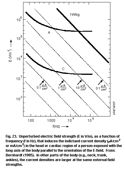
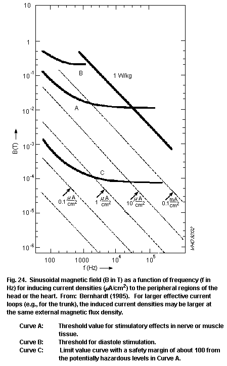 8.1.5 Thermoregulation
The need to understand and predict the thermal effects of
electromagnetic energy deposition arises from several perspectives: in
occupational and public health it is necessary to determine safe
limits of environmental exposure to RF fields, in medical therapeutic
applications there is a need to deposit electromagnetic energy in a
predetermined quantity in a specific location and volume, and,
finally, there is an RF energy deposition in diagnostic medical
applications, such as magnetic resonance imaging.
In all these instances, there is concern with the effects of
locally elevated temperatures resulting from the deposition of RF
energy, and the ability of the thermoregulatory system to dissipate
the thermal load without unduly stressing the physiological systems
involved.
In "thermally neutral" environments, with the body at rest, the
total heat production of the human body amounts to about 100 W, and
this heat production is offset by a heat loss of 100 W with 15-20 W of
evaporative heat loss from the skin and the respiratory tract; the
remainder of the heat loss is through radiation, convection, and
conduction to the surrounding environment. In strenuous exercise,
and/or in environments with elevated ambient temperatures and water
vapour pressure, the body temperature tends to increase. Healthy
individuals can sustain an increase in internal temperature from a
normal 37.0 °C to 39.0 °C with the latter temperature representing the
upper safe limit, even for young and healthy individuals. At 39.0 °C,
sweating at a rate of about one litre per hour is induced, and heart
rates become considerably elevated. From considerations of metabolism
and heat exchange, any metabolic heat production in a limited volume
of tissue does not result in a temperature rise exceeding 0.8 °C above
deep body temperature.
In normal, everyday life, thermal loads imposed by resting
metabolism, the thermal environment, or by muscular activity, vary
from a minimum of about 1 W/kg to 10 W/kg. Calculations relating
whole-body SAR to increases in body temperature are, in general,
supported by the limited results of studies of the responses of
patients and volunteers exposed to RF fields in magnetic resonance
imaging systems (Schaefer et al., 1985; Gordon et al., 1986; Kido et
al., 1987; Shellock & Crues, 1987, 1988; Shellock et al., 1989).
In these studies, the subjects were at rest and in controlled
environments. Exposure of healthy volunteers to up to 4 W/kg for 20-30
minutes resulted in body temperature increases in the range of 0.1-0.5
°C, confirming predictions derived from models of energy deposition
and thermoregulatory response. These exposures resulted in minimal
changes in blood pressure and respiration rate. At the higher SARs,
subjects felt warm during the procedure and each of them had visible
signs of sweating on their foreheads, chest, and abdomen.
Thermal stresses in the form of increased metabolic rates during
exercise, deposition of RF energy, or exposure to solar radiation,
tend to result in rises in body temperature and activation of
thermoregulatory responses, such as sweating and vasodilatation.
Different individuals have widely varying abilities to tolerate such
responses, depending on age, physical fitness, clothing, adaptation,
etc. Thermal stress from RF energy absorption is more severe when it
is combined with heavy clothing, or a very hot and humid environment.
The thermal effect of RF energy absorption could be beneficial and
stress reducing if it occurred in a cold or cool environment.
Thermal stresses for vulnerable populations, such as infants who
have an under-developed thermoregulatory system, or the elderly whose
thermoregulatory systems are no longer fully competent, must be
limited to less than that of an occupational population, but an
absolute level is difficult to define.
Mathematical models of the human thermal system make possible
reasonably accurate predictions of the steady state and the dynamics
of both the whole body thermal state, and local tissue temperatures,
under a variety of internal and external thermal stresses (Stolwijk &
Hardy, 1966, 1977; Wissler, 1964, 1981).
The development of models of RF energy deposition was initially
independent of the development of thermoregulatory models, though
similar simplifications had to be accepted. The models for human
thermoregulation and the models for RF energy deposition do not have
the same priorities or the same capabilities for spatial definition.
In addition, the level of knowledge of the parameters required for the
implementation of these models is different for the two types of
mathematical model. In human thermoregulation models, it is not of
crucial importance to describe in detail the local blood flow response
to tissue temperatures above 38 °C. However, in combined models, it is
very important that this characteristic is adequately incorporated,
particularly with respect to hyperthermia therapy.
Models that deal simultaneously with RF energy deposition in the
human body, and with the effects of the thermal environment on
thermoregulation and heat transfer in the human body have difficult
trade-offs between the degree of spatial definition that is pursued,
the degree of detail in the thermoregulatory response, and the cost of
computation required to produce and evaluate the predictions from such
combined models.
8.1.6 Contact currents
Persons coming in contact with ungrounded or poorly grounded
metallic objects in an RF field may experience perception, pain,
shock, burn, or even more severe reactions. Such phenomena occur for
sufficiently large objects and intense fields. These interactions are
described in section 6.5.
8.2 Epidemiological and clinical comparative studies
In studies on RF-exposed human populations, epidemiological
results are frequently based on estimates only of exposure
characteristics (RF frequency, power density, and exposure duration)
and some solely on a description of occupation. Despite these
limitations, they may provide useful information on the possible
effects of actual RF exposure in humans. In the assessment of RF-field
effects, comparative, clinical studies of a limited number of exposed
persons and controls may be useful.
Studies of health effects from exposure to RF fields have been
carried out since the 1940s, when man-made sources of RF energy led to
the increasing exposure of occupational groups and the general
population. These early studies have been reviewed (WHO, 1981). The
majority of reports in the literature concern people exposed in
military or industrial settings. Summaries of studies on the health of
humans exposed to RF fields are given in Tables 31-33. A wide variety
of conditions, symptoms, diseases, and clinical measurements have been
evaluated.
8.2.1 Mortality and morbidity studies
In the 1960s and 1970s, Soviet and Eastern European literature
described a collection of symptoms, reported to occur in personnel
industrially exposed to microwaves. These symptoms, which have been
variously called the "neurasthenic syndrome", the "chronic
overexposure syndrome", or "microwave sickness", are based on
subjective complaints, such as headaches, sleep disturbances,
weakness, decrease of sexual activity (lessened libido), impotence,
pains in the chest, and general poorly defined feelings of non-well-
being (Baranski & Czerski, 1976).
Table 31. Morbidity and mortality studies
Exposure conditions Effect on exposed group Reference
Radar (pulsed),two groups: No difference in health Czerski et al.
(i) <2 (ii) >2 up to status between 841 adult (1974b);
60 W/m2, for 1-10 years males in groups (i) and (ii) Siekierzynski
et al. (1974a,b)
Radar (pulsed), <50 W/m2 No effects in clinical Djordevic et al.
(<0.2 W/kg), for 5-10 evaluations in comparisons (1979)
years between 322 radar workers
and 220 non-radar workers;
however, more neurasthenic
symptoms in exposed group
Table 31 (continued)
Exposure conditions Effect on exposed group Reference
0.2-5 GHz (pulsed), approx. No effect on mortality Robinette &
10 W/m2, 0.05 W/kg in male military personnel Silverman (1977);
(max). Occasional exposure followed for over 20 years,
to 1 kW/m2 exposed for 2 years on Robinette et al.
average (1980)
(over 40 000 personnel)
Males: 2.56-4.1 GHz (CW), No effect on life span or Lilienfield et
0.05 W/m2 (max), cause of death of 1800 al. (1978)
0.0002 W/kg (max); employees and 3000
Females: 0.6-9.5 GHz dependents of US Embassy
(CW),0.018 W/m2 (max), personnel
0.0007 W/kg (max), for
0.5-4 years average
exposure
Long-term microwave Higher frequency of Friedman
exposure of military microwave exposure (1981)
personnel (interviews) in 14 polycythaemia cases
than in 17 age-matched
controls
Radar-exposed popula- Increased cancer mortality Lester & Moore
tions near air force bases compared with population- (1982); Lester
matched controls. (1985).
No increase in cancer mor- Polson & Merritt
tality compared with popu- (1985)
lation-matched controls
Children exposed to various Duration and severity of Shandala &
air pollutants and RF tonsilitis increased Zvinjatskovsky
(1988)
27 MHz shortwave dia- Association between heart Hamburger et al.
thermy (questionnaire to disease and work with (1983)
3004 physiotherapists) shortwave therapy (number
of treatments/week)
Work at 27 MHz plastic Upper limb paraesthesia Bini et al.
sealers (70% of measure- and eye irritation noted (1986)
ments at the head and among 30 exposed workers
hands >300 V/m) compared with 11 partially
exposed and 22 unexposed
workers
Table 31 (continued)
Exposure conditions Effect on exposed group Reference
Military personnel Increased risk of cancer Szmigielski et al.
exposed to RF/MW fields morbidity in a retrospective (1988)
<2 W/m2 with daily cohort study of military
incidental (minutes) personnel (study group size
exposures of 2-10 W/m2 not given)
(some times even
100-200 W/m2)
51 male/62 female Increase rates of paraesthesia Kolmodin-Hed-
operators of plastic in hands, neurasthenia, and man et al. (1988)
welding machines (27 MHz, eye complaints; diminished
50% of welders exceeded 2-point discrimination ability
250 W/m2) 23 female controls
(sewing machine operators)
Amateur radio operators Deaths from all causes less Milham (1985)
than expected from national
rates; increased risk of
leukaemia
1.3-10 GHz, 0.1 to 10-µs No differences in Nilsson et al.
pulses, RF exposure of neurological symptoms and (1989)
radar mechanics often findings between 17 exposed
exceeded 10 W/m2 and 12 controls; increased
protein band in CSF in
the exposed group
These early studies suffered from various deficiencies and their
results have not been replicated in later surveys. Some of the results
could have been attributed to other working conditions (e.g.,
Djordjevic et al., 1979), and, furthermore, it appears that the
working environments for exposed and control groups were not similar
in essential respects. Other factors could also have been operating to
produce more subjective complaints among the exposed workers, e.g., a
reporting bias because of enhanced awareness of the possible
"microwave sickness" syndrome.
Later studies on mortality and morbidity among US naval
personnel, occupationally exposed to radars, found no differences
between exposed and control groups (Robinette & Silverman, 1977;
Robinette et al., 1980).
In a study of US embassy personnel, with very low microwave
exposures, no significant effects were found (Lilienfield et al.,
1978). Studies on cancer mortality in populations around US Air Force
bases have given conflicting results, even contradictory findings,
when evaluating identical study groups (Lester & Moore, 1982; Polson
& Merritt, 1985; Lester, 1985). However, there are studies indicating
an increase in cancer in RF field-exposed populations. Friedman (1981)
reported a limited number of polycythemia cases with histories of
long-term exposure to microwaves, and, more recently, preliminary
reports from a retrospective cohort study of Polish military
personnel, occupationally exposed to RF, indicated an increased risk
of cancer (Szmigielski et al., 1988). Also a case study on a radar
mechanic, who developed acute myelogenous leukaemia, has been
published (Archimbaud et al., 1989).
Milham (1985), using records of licensed amateur radio operators
living on the west coast of the USA, derived standardized mortality
ratios (SMRs) and compared them with the mortility rates for the
population in the USA. Although the overall mortality rate was lower
for the radio amateurs, significantly raised SMRs were observed for
some types of leukaemias. However, it should be noted that around a
third of the radio amateurs were engaged in electrical/electronics
occupations. This may have involved exposure to solvents, PCBs, and
metal fumes. In general, studies on increased cancer risks in certain
"electrical" occupations (see, e.g., WHO, 1984, 1987) mainly refer to
exposure to 50/60 Hz magnetic and electric fields with little or no
contribution of 300 Hz-300 GHz radiation.
In studies on plastic welding machine operators, with RF exposure
levels sometimes exceeding existing national standards, upper limb
paraesthesias have been reported by Bini et al. (1986) and
Kolmodin-Hedman et al. (1988).
In a small study on radar mechanics, in which no differences were
found in neurological symptoms and signs compared with controls,
changes were reported in a protein band of the cerebral spinal fluid
(Nilsson et al., 1989). Because this study was small, its significance
with respect to health is unclear. The clinical observations of
Nilsson need to be confirmed.
Also described as part of the early "microwave sickness" syndrome
(see above) were effects on heart rate including bradycardia as well
as tachycardia, arterial hypertension (or hypotension), and changes in
cardiac conduction. With reference to this, the increased risk of
developing heart diseases found among physiotherapists working with
shortwave diathermy (Hamburger et al, 1983) calls for further studies.
The combined effects on children of various pollutants in the
environment (RF, noise, chemicals etc.) were studied by Shandala &
Zvinjatskovsky (1988), who found that the duration and severity of
tonsilitis were increased in the presence of RF.
8.2.2 Ocular effects
In health studies on RF field-exposed workers, general eye
irritation was described (Bini et al., 1986; Kolmodin-Hedman et al.,
1988). Lens opacities and cataracts have also been noted in some
studies, as shown in Table 32. In the most extensive study, however
(Appleton & McCrossan, 1972; Appleton et al., 1975), commented on by
Frey (1985) and Wike & Martin (1985), no differences were found
between exposed and unexposed military personnel. Where cases of
confirmed cataracts have been reported, exposures have exceeded 1
kW/m2.
8.2.3 Effects on reproduction
Only a limited number of studies, as shown in Table 33, have
investigated potential reproductive effects in humans exposed to RF in
the work environment. Sigler et al. (1965) found a higher incidence of
Downs syndrome in children whose fathers had worked with radars in the
military. From interviews of the fathers in the Sigler study and
additional information obtained from military records, Cohen et al.
(1977) could not confirm the result that the fathers had either an
excess of radar exposure or a larger proportion were exposed
personnel. The contradictory results probably reflect the difficulties
in exposure assessment in retrospective epidemiological studies.
Table 32. Lens opacities and cataracts in humans
Exposure conditions Effect on exposed group Reference
US Army and Air Force No difference in cataract Cleary et al.
veterans, radar incidence (1965)
personnel, 2644 exposed,
1956 controls
Microwave workers, More lens changes in Cleary &
736 exposed, 559 exposed group Pasternak (1966)
controls
Microwave workers, More lens changes in Majewska
60 MHz-10.7 GHz, exposed group (168 vs 148) (1968)
200 exposed, 200
controls
US military personnel, No differences in Appleton &
91 exposed, 135 controls incidence of lens McCrossan
opacities, vacuoles, (1972)
or subcapsular iridescence
US military personnel, Expanded study, same results Appleton et al.
1542 exposed, 801 (1975); Frey
controls (1985); Wike &
Martin (1985)
US military radar Lens abnormalities same Odland (1973)
personnel in exposed controls, except
377 exposed, 320 higher in exposed with pre-
controls existing visual defects
Table 32 (continued)
Exposure conditions Effect on exposed group Reference
Two groups of microwave No difference in lens Siekierzynski
workers: opacities between the et al. (1974a,b)
group 1: <2 W/m2 two groups
group 2: 2-60 W/m2
US Air Force and No difference in Shacklett et al.
civilian frequency of opacities, (1975)
personnel, 477 exposed, vacuoles or posterior
340 controls capsular iridescence
53 radio-linemen Increased incidence of Hollows &
installing and posterior subcapsular Douglas (1984)
maintaining radio, TV, cataracts
and repeater towers;
558 kHz-527 MHz,
0.8-39.6 kW/m2
Table 33. Reproductive effects in humans
Exposure conditions Effect on exposed group Reference
Work with radar in the Case-control study of the Sigler et al.
military fathers of 216 children (1965)
with Downs syndrome and
216 matched control
fathers: association between
radar exposure and Downs
syndrome
Work with radar in the Extended study from Sigler Cohen et al.
military et al. (1965) with additional (1977)
128 cases and 128 controls:
no association between
radar exposure of fathers
and Down's syndrome
3.6-10 GHz, hundreds to Decreased sperm number in Lancranjan
thousands of mW/m2, 31 males (70% of whom with et al. (1975)
0.003-0.04 W/kg neurasthenia) exposed for
1-17 years (8-year average)
compared with 30 healthy
controls
Table 33 (continued)
Exposure conditions Effect on exposed group Reference
Cohort study on pregnancy Physiotherapists had a better Kallen et al.
outcome of 2018 female than expected pregnancy (1982)
physiotherapists giving outcome; higher use of
birth to 2043 infants shortwave units among physio-
therapists giving birth to
malformed or still-born
infants
305 female RF welders No differences in pregnancy Kolmodin-
outcome compared with Hedman et al.
Swedish birth registers (1988)
Case-control study on 17% of "highly" exposed Larsen et al.
physiotherapists working were boys; exposure also (1991)
with shortwave diathermy associated with still-
birth/death within a
year, prematurity, and
low birth weight
Analysis of semen of 31 technicians with a very low-level
microwave exposure, revealed a reduced number of sperm compared with
a control group of 30 persons (Lancranjan et al., 1975). However, 70%
of the exposed group suffered from neurasthenia, which might wholly or
partly explain the results.
In a health study on operators of plastic welding machines
exposed to RF levels exceeding 250 W/m2 (Kolmodin-Hedman et al.,
1988), the pregnancy outcome for 305 female plastic welders during
1974-84 did not show any significant differences with the Swedish
average concerning malformation or prenatal mortality.
During the 1980s, two epidemiological studies indicated an
adverse pregnancy outcome among physiotherapists working with
shortwave diathermy (Kallen et al., 1982; Larsen et al., 1991). Kallen
et al. (1982), in Sweden, reported that physiotherapists as a group
had a slightly lower risk of perinatal deaths and major malformations
than the Swedish population for the same period. However, the
physiotherapists who gave birth to a malformed child, or who had a
perinatal death, had RF exposures (from microwave and shortwave
diathermy) higher than those recorded for the other physiotherapists.
In a Danish case-control study on physiotherapists working with
shortwave diathermy, Larsen et al. (1991) found that only 17% of the
"highly exposed" newborn infants were boys, and that exposure was
associated with stillbirth/death within a year, prematurity, and low
birth weight. The results suggest further study is necessary before
conclusions can be reached.
8.2.4 VDU studies
Concern about the effects of exposure to electromagnetic fields
and particularly about pregnancy outcome has been expressed with
regard to the use of VDUs. Work with such equipment may involve job
stress and ergonomic problems and these can be confounding factors in
studies of associated pregnancy outcomes. Studies have been reviewed
by Repacholi (1985), Bergqvist & Knave (1988), and Blackwell & Chang
(1988).
Blackwell & Chang (1988) pointed out that, in the USA and the
United Kingdom, about 10 million VDUs are in use. About 50% of these
are possibly used by women of childbearing age, and there are some 20
000 groups of women, in each of which at least 10 women could become
pregnant in one year. Since the naturally occurring pregnancy failure
rate is about 15%, there is a chance of about 29 "clusters" each year
in which more than half the pregnancies end in failure.
A large number of epidemiological studies have been conducted, in
order to elucidate whether VDU work during pregnancy increases the
risks of miscarriage or giving birth to a malformed child. While
Goldhaber et al. (1988) suggested there was some evidence of increased
spontaneous abortion rates among VDU operators, most studies have not
shown this (Bryant & Love, 1989; Goldhaber et al., 1988; McDonald et
al., 1988; Nielsen et al., 1989; Nurminen & Kurppa, 1988), or
threatened abortion, changes in placental weight, and maternal blood
pressure (Nurminen & Kurppa, 1988). Of these studies, just one
(Schnorr et al., 1991) included the measurement and assessment of the
emission of ELF and VLF electric and magnetic fields as exposure
factors. In this study, a cohort of female telephone operators, who
used VDUs at work, was compared with a cohort of operators who did not
use VDUs. Exposure was assessed by the number of hours worked per
week, from company records, and by measuring electric and magnetic
fields (45-60 Hz and 15 kHz) at the VDU work stations and at the
workstations without VDTs. Among 2430 women interviewed there were 882
pregnancies (366 exposed, 516 controls) that met the criteria for
inclusion in the study. No excess risk of spontaneous abortion was
found among women who used VDUs during the first trimester of
pregnancy (OR = 0.93, 95% CL, 0.63-1.38). There was no risk associated
with the use of VDUs when accounting for multiple pregnancies, early
and late abortions, and all fetal losses. No dose-response
relationship was apparent when examining the number of hours at the
VDU, or the measured electric and magnetic fields.
The study by McDonald et al. (1988) was designed around all women
who reported to 11 Montreal hospitals during 1982-84 for childbirths
or spontaneous abortion. They were interviewed on working conditions
during their current and previous pregnancies. Apart from an isolated
increase in renal urinary defects, the study showed no evidence of
increased malformation. However, the results are not so clear for
spontaneous abortion, especially among previous abortions. The design
of this study does, however, tend to exaggerate the odds ratio for VDU
exposed compared with non-exposed in previous pregnancies (Bergqvist,
1984; McDonald et al., 1988). By stratification, this systematic error
has been eliminated, and then the apparent increase in odds among VDU
exposed was absent (McDonald et al., 1988). A similar, but smaller,
error is also likely with regard to spontaneous abortion among current
pregnancies.
In a case-control study performed at three Kaiser Permanente
clinics in Northern California (Goldhaber et al., 1988), there was an
increase in spontaneous abortion among VDU operators compared with
referents. However, this significant increase was due to a trend in
one of the job categories (clerical workers), while a decrease in
relation to VDU work was reported for another job category (managers,
professionals). This contrary information from two job categories has
two ramifications: (1) the summary across job categories is not
justified; and (2) it makes the interpretation of magnetic fields as
a cause rather dubious, but does, instead, suggest job-specific
factors as possible causal factors.
Experimental studies, while showing a diverse outcome, have, as
a whole, failed to demonstrate an effect on reproductive processes in
magnetic field situations resembling those around VDUs.
Epidemiological studies have failed to show a difference between women
who worked and those who did not work at a VDU during pregnancy, and
interest has now turned to possible differences related to work
situations, e.g., stress, rather than physical emissions from the
VDUs.
8.2.5 Conclusions
In summary, the epidemiological and comparative clinical studies
do not provide clear evidence of detrimental health effects in humans
from exposure to RF fields. Some occupational groups, such as exposed
physiotherapists and industrial workers, should be studied further.
The question of whether RF might act as a carcinogen should be further
evaluated in epidemiological studies.
Occupational exposure to RF will be at higher levels than that
encountered by the general population, and, thus, there is less
likelihood of health effects in the general population as a whole.
8.3 Clinical case studies and accidental overexposures
In a survey of accidental overexposures to RF in the US Air Force
(Graham, 1985), 26 out of 58 individuals, with exposures exceeding 100
W/m2, reported that they had felt a warming sensation at the time of
overexposure. In clinical examinations, no abnormal findings were
recorded. Symptoms, such as headache, nausea, fatigue, malaise, and
heart palpitations, were often reported, however. Some high-level
exposures, e.g., at levels exceeding 5 kW/m2, resulted in anxiety
reactions so severe that hospitalization and sedation were necessary.
Similar symptoms were reported in a one-year, clinical, follow-up
study on two men who were accidentally, acutely irradiated with
600-900 W/m2 RF fields (Forman et al., 1982). Several months after
the incidents, hypertension was diagnosed in both patients. Exposures
to power densities of about 50 W/m2 for one or two hours were not
found to result in harmful health effects (Hocking et al., 1988).
In case reports, long-term neuropathies and chronic dysaesthesias
have been described after excessive microwave exposures from
malfunctioning microwave-ovens (Ciano et al., 1981; Tintially et al.,
1983; Fleck, 1983; Dickason & Barutt, 1984; Stein 1985). Also severe
burns have been reported at work with microwave ovens (Nicholson et
al., 1987). Similarly, Castillo & Quencer (1988) described the case of
a pilot who inadvertently stood in front of a functioning microwave
airfighter radar system for approximately five minutes. At that time
a moderate sensation of heat was perceived in the head and neck, and
after some time interstitial oedema and coagulation necrosis developed
in muscles of the neck. The pilot also noted a loss of recent memory
and extreme sleepiness.
9. HEALTH HAZARD ASSESSMENT
9.1 Introduction
The purpose of reviewing the scientific literature on effects of
exposure of various biological systems to RF fields is to assess its
possible impact on human health. Such an assessment is necessary for
the development of standards and guidelines limiting exposure to RF of
the general and working populations.
One of the problems encountered in assessing the possible health
effects of RF exposure over the whole range of frequencies covered in
this publication (i.e., 300 Hz-300 GHz) is that most studies have been
conducted at frequencies particularly in the low GHz region. Little
information is available from studies of human populations and only
limited data have been obtained on other biological systems,
particularly animals exposed to RF at frequencies below 10 MHz and
above 10 GHz.
The following categories of effects must be considered for risk
assessment. The first two of these are sufficiently well understood to
be used in risk assessment and the development of recommended limits
of exposure. The third category is reasonably well understood, but
quantitative data are sparse and any comprehensive recommendations to
protect workers and the general population have to be based on data at
other frequencies. The effects noted in the last two categories are
elaborately described and poorly understood. In view of their
importance in the possible promotion of cancer or of reproductive
failures, they must be considered. However, the lack of understanding
and the total absence of quantitative relationships between these
effects and either exposures or the outcomes in question makes it
impossible to derive recommended limits of exposure.
Points to consider for a health risk assessment of exposure to RF
fields are:
(a) Absorption of RF energy causes tissue heating. This is
recognized and has been well studied. This effect occurs from the
absorption of RF fields, especially at the higher end of the
frequency range (above about 1 MHz). RF heating is not directly
equivalent to heating by other forms of energy, because of the
very non-uniform energy deposition that occurs in biological
systems.
(b) At frequencies below about 100 MHz, currents can be induced in
humans by physical contact with ungrounded metallic objects (see
section 6.5). From 300 Hz to approximately 100 kHz, such currents
may result in the stimulation of electrically excitable tissues
above the threshold for perception or pain. At frequencies
between approximately 100 kHz and 100 MHz, contact currents of
sufficiently high density may cause burns.
(c) For frequencies below several hundred kHz, the predominant effect
is stimulation of excitable tissue resulting from currents
directly induced in the body by the RF fields. At these lower
frequencies, thermal interactions occur only at energy levels
much higher than interactions with excitable tissue.
(d) When RF energy is absorbed in the form of pulsed fields, the peak
power density in the pulse should be considered separately from
the average. Auditory perception is one example of a pulsed RF
field effect.
(e) When RF fields are amplitude modulated, effects in tissues have
been noted that do not manifest themselves with unmodulated RF
fields. Such effects are reported to have a complex dependence on
intensity and ELF modulation frequency. Too little information is
available to determine whether such effects occur in humans and,
thus, this effect cannot be used in a health risk assessment or
for setting human exposure limits.
9.2 Thermal effects
A number of factors in everyday life tend to increase the heat
load on the human body, such as high ambient temperatures, solar
radiation, and basal and exercise metabolism. Energy production can
reach levels of 3-6 W/kg in healthy people. In most individuals, the
thermoregulatory system can remove heat from the body at these rates
for extended periods of time. Limited experimental evidence and
theoretical calculations suggest that the exposure of resting humans
in moderate environmental conditions at whole body SARs of the order
of 1-4 W/kg for 30 minutes results in body temperature increases of
less than 1 °C. In addition, a review of the animal data (see section
7.3.4) indicates a threshold for behavioural responses in the same 1-4
W/kg range. Therefore, an occupational RF exposure guideline of 0.4
W/kg, based on thermal consideration, leaves a considerable margin of
safety for other limiting conditions, such as high ambient
temperature, humidity, or physical activity. Higher energy absorption
rates in extremities and limited body regions, do not appear to cause
adverse effects, for SAR values below thresholds that are dependent on
the body part and the volume.
In infants, the frail elderly, and in individuals taking certain
drugs, the thermoregulatory capacity may be much reduced and, as a
result, their tolerance for the combined effects of RF exposure,
exercise, solar radiation, and high ambient temperature, may be much
lower. Recognition that this tolerance is lower dictates that
guidelines for population exposure to RF fields be reduced. A
whole-body average SAR of 0.08 W/kg offers an additional safety
factor.
Significant overexposures at the higher frequencies that may
occur in occupational environments may result in very high SARs in
parts of the body, thus producing local burns. In such cases, the SAR
is so high that the normal avenues of heat transfer from the exposed
area are inadequate. The local tissue temperature quickly rises to
levels that denature proteins. Such burns may occur at depths much
greater than those usually associated with contact burns.
Thus, standards should be developed that, at a minimum, limit
exposure of the healthy and aware (occupational) population, so that
the whole-body average SAR does not exceed 0.4 W/kg. Additional
precautions must be exercised for situations that might cause large
peak values of the SAR, in order to eliminate rapid elevation of local
temperature by more than 1 °C. This requires that the peak (or local)
SARs should not exceed about 2 W/100 g in the extremities and 1 W/100
g in any other part of the body. The eye may need special
consideration, possibly by averaging over a mass of 10 g (i.e., 100
mW/10 g).
9.3 RF contact shocks and burns
At frequencies below a few hundred kilohertz, the electrical
stimulation of excitable membranes of nerves and muscle cells is a
well established phenomenon. These effects exist at very high
environmental field strengths, unlikely to occur in the general
environment. On the other hand, current densities sufficient for
stimulation and other potentially harmful effects can be produced, if
an individual makes contact with a conductive object energized by the
electric field component of an RF source.
For frequencies between 300 Hz and 100 kHz, perception, pain,
startle, or even inability to let go, may result from physical contact
with energized objects (see section 8.1.6). The thresholds are
expressed in terms of the current and are strongly frequency
dependent. Superficial and deep burns may occur as a result of contact
with metallic objects exposed to RF fields over a wide frequency
range. Sufficiently high current densities for contact burns can be
attained in RF fields that are too low to cause direct heating or
stimulation. Thresholds depend on the size and shape of the object,
field frequency, length and type of contact, and other parameters.
Field exposure guidelines should also contain RF limits to
eliminate hazards from shocks and burns. In this context, it should be
kept in mind whether the exposures occur under controlled or
uncontrolled conditions. Under uncontrolled exposure conditions, it is
not always possible to limit contact currents for some objects (e.g.,
vehicles) so that electric field strengths have to be reduced to
protect the general population. For workers, other measures, such as
protective clothing or contact avoidance, provide viable alternatives
for protection.
9.4 Induced current densities
At frequencies below approximately 1 MHz, interactions of RF
fields with biological systems and potential hazards can be considered
in terms of induced currents and their densities (see section 8.1.4).
The use of induced current densities, however, is only appropriate for
the assessment of acute, immediate effects, while it may have some
limitations for the complete evaluation of long-term effects. The
waveform of the RF field is an important factor to be considered in
the response of biological systems. However, peak instantaneous fields
strengths appear to be important in considering nerve and muscle cell
stimulation and for perturbing cell functions. Generally, for
frequencies above 300 Hz, the thresholds for effects increase with
frequency, up to frequencies where thermal effects dominate. For the
establishment of derived limits, the distribution of the current
densities within the body induced from RF fields have to be
considered. The treatment of this problem is restricted, at present,
to relatively simplified situations.
9.5 Pulsed RF fields
Experimental data suggest that thresholds for the biological
effects of absorbed energy at frequencies above hundreds of megahertz,
when in the form of short duration pulses (approx. 1-10 µs), are lower
than those for continuous fields at the same average energy level and
the same SAR. This indicates that the peak value of energy transfer to
the biological object can be an important determinant of the
biological effect. A well-investigated effect is the perception of
pulsed fields, such as from radar, as an audible sound described as a
click, chirp, or knocking sensation (see section 8.1.3).
Pulsed RF exposure effects observed in animals are suppression of
a startle response, stunning, ocular effects, and alterations in
responses to certain drugs. Thresholds in terms of the energy density
per pulse or the peak electric field strength for a given pulse
duration are known for these effects only at a limited number of
frequencies. Suppression of startle response was observed for pulse
durations of up to a few seconds. Shorter pulses with the same or
greater energy had a slightly enhanced effect on startle.
Since a single pulse, or a series of short pulses, of RF of very
high peak power density, but very low average power density, can
produce potentially harmful biological effects, it is necessary to
limit the maximum energy density per pulse. The available scientific
evidence is incomplete, and, therefore, the formulation of exposure
limits for pulsed fields presents difficulties.
9.6 RF fields amplitude modulated at ELF frequencies
Effects have been reported in in vitro systems and animals
exposed to RF fields of low intensities amplitude modulated at ELF.
Some of the same or similar effects have also been observed as a
result of exposure to ELF and VF fields. The effects usually exhibit
"window" characteristics, i.e., the effects occur only within
relatively narrow ranges, in both the modulation frequency and field
intensity. Even though the intensities of the fields in tissue at
which these effects occur are below the broadband thermal noise, there
are hypotheses that might account for such apparently aberrant
behaviour. The biological significance and possible adverse health
impact, if any, of the reported effects cannot be determined at this
time.
9.7 RF effects on tumour induction and progression
There have been isolated reports that, in certain cell lines and
in intact animals, RF exposures have been associated with increased
growth rates of cells and tumours and with increases in the incidence
of neoplastic transformations. Very few epidemiological studies have
been reported. The available evidence does not confirm that RF
exposure results in the induction of cancer, or causes existing
cancers to progress more rapidly. Because of incompleteness and
inconsistencies, the available scientific evidence is an entirely
inadequate basis for recommendations of health protection guidelines.
10. EXPOSURE STANDARDS
10.1 General considerations
The development of protection standards for any environmental
agent is a difficult and complex task. Setting exposure limits
requires an in-depth evaluation of the established scientific
literature, since to base standards on preliminary data or unproven
hypotheses means that the limit values may be either unprotective or
unduly restrictive. Using established scientific data allows exposure
limits to be determined with a higher degree of confidence about their
level of protection.
Certain criteria must be met, if claims of positive effects or
negative data are to be accepted within the body of scientifically
established effects (Michaelson, 1983; Repacholi, 1990):
(a) Experimental techniques, methods, and conditions should be as
completely described and objective as possible.
(b) All data analyses should be fully and completely objective, no
relevant data should be deleted from consideration, and uniform
analytical methods should be used.
(c) Results should demonstrate an effect of the relevant variable at
a high level of statistical significance using appropriate tests.
The effects of interest should ordinarily be shown by different
test organisms and the responses found be consistent.
(d) Results should be quantifiable and susceptible to confirmation by
independent researchers. Preferably, the studies should be
repeated and the data confirmed independently; or the claimed
effects should be consistent with results of similar studies,
where the biological systems involved were comparable.
From the body of established literature, a distinction must be
made between in vitro and in vivo studies. In vitro studies are
conducted to elucidate the mechanisms of interaction or to identify
biological effects or exposure parameters that need to be further
investigated to determine if they occur in vivo. Standard-setting
organizations can place only limited value on the results of in vitro
experiments.
An important part of the rationale for any exposure standard is
the definition of the population to be protected. Occupational health
standards are aimed at protecting healthy adults, exposed as a
necessary part of their work, who are aware of the occupational risk
and who are likely to be subject to medical surveillance. General
population standards must be based on broader considerations,
including widely different health status, special sensitivities,
possible effects on the course of various diseases, as well as
limitations in adaptation to environmental conditions and responses to
any kind of stress. Exposure limits for the general population must
include an adequate additional safety factor, also taking into account
the possibility of a 24-h exposure compared with 8-h occupational
exposure (or whatever the duration of the workday). Additionally, the
RF fields in the environment can be complex and may be affected by
reflections from buildings.
A distinction should be made between exposure limits and
equipment emission standards. The latter are based on safe operational
considerations, and should not allow exposure above the adopted
exposure limits.
Over the past decade, major advances in the study of RF fields have
come from the development of dosimetry as reviewed in section 5.
Methods of intercomparing the results of animal studies and relating
them to the human situation, have been developed to facilitate
standard-making. With increasing knowledge of RF dosimetry, standards
are becoming more specific.
10.2 Present trends
Many countries have now established health protection standards
or guidelines. There have been a number of in-depth reviews of current
RF standards (Czerski, 1985; Sliney, 1988; Grandolfo & Mild, 1989;
Repacholi, 1990; Szmigielski & Obara, 1989). Most of the early
standards addressed the microwave region only (300 MHz-300 GHz),
because of the introduction and proliferation of radars,
telecommunications, and radio and TV broadcasting. Later standards
recognised the vastly expanded use of the electromagnetic spectrum,
especially at lower frequencies, where concerns were raised about RF
exposures from induction heaters, heat sealers, and other industrial
applications.
RF exposure standards development is continuing, at present, and
with the availability of detailed reviews elsewhere, standards in
various countries and their rationales are not discussed here.
The maximum RF exposure levels permitted in some standards differ
by one to two orders of magnitude (factors between 20 and 100). It may
be speculated that these differences result from: (a) the physical and
biological effects data selected as the basis for the standards, (b)
the interpretation of these data, (c) the different purposes to be
served by the standards, (d) the compromises made between levels of
risk and degrees of conservatism, and (e) the influence of preceding
standards in each particular nation and in neighbouring areas having
allied socio-political outlooks. In recent years, an increasing number
of countries have adopted standards with limits identical, or very
close, to those of IRPA.
10.3 Recommendations by the IRPA
A joint WHO/IRPA Task Group on Radiofrequency and Microwaves
reviewed existing scientific literature (WHO, 1981). An evaluation of
the health risks of exposure to electromagnetic fields was made and
the rationale for the development of exposure limits was considered.
The Task Group suggested that RF exposure to power densities in the
range 1-10 W/m2 were acceptable for occupational exposure throughout
a complete working day and that higher exposures might be acceptable
for some frequency ranges and occasional exposure. For the general
population, it was suggested that lower, unspecified, exposure levels
were appropriate.
In 1984, IRPA issued recommendations based on the WHO publication
(WHO, 1981). These recommendations were more specific and provided
guidance on limits of exposure to electromagnetic fields in the
frequency range from 100 kHz to 300 GHz. The basic limits of exposure
formulated for the frequency region of 10 MHz and above were expressed
in terms of the specific absorption rate. In the frequency region
below 10 MHz, basic limits were expressed in terms of the electric and
magnetic field strengths.
The IRPA revision (1988a) of its 1984 guideline, shown in Tables
34 and 35, reaffirmed that research data, obtained over the past
years, did not alter the threshold whole-body exposure for health
effects on which the basic limit was derived: i.e., occupational
whole-body exposure to RF fields should not exceed 0.4 W/kg. The
revision was essentially a "fine tuning". Although the whole body
average SAR might not exceed 0.4 W/kg, several reports indicated that,
under certain conditions, local peak SARs in the extremities
(particularly wrists and ankles) could exceed the 0.4 W/kg value by a
factor of up to 300, at certain frequencies. Because of this, an
additional recommendation was introduced to limit the body-to-ground
current to 200 mA. It was also found that there was no adequate basis
for identifying SAR limits as averaged over any gram of tissue. IRPA
therefore recommended that the local SAR should not exceed 2W/100g in
the extremities (hands, wrists, ankles, and feet) and 1 W/100g in any
other part of the body.
Occupational exposure to frequencies up to 10 MHz should not
exceed the levels of unperturbed electric and magnetic field strengths
(rms), given in Table 34, when the squares of the electric and
magnetic field strengths are averaged over any 6-min period during the
working day, provided that the body-to-ground current does not exceed
200 mA, and the hazard for RF burns is eliminated. In general, RF
burns will not occur if the current at the point of contact does not
exceed 50 mA.
Table 34. IRPA occupational exposure limits for RF fieldsa
Frequency Unperturbed Unperturbed Equivalent plane-wave
range rms electric rms magnetic power density
field strength field strength
(MHz) (V/m)b (A/m)b (W/m2)b (mW/cm2)b
0.1-1 614 1.6/f - -
>1-10 614/f 1.6/f - -
>10-400 61 0.16 10 1
>400-2000 3f0.5 0.008f0.5 f/40 f/400
>2000-300 000 137 0.36 50 5
a From: IRPA (1988a).
b f = frequency in MHz.
Note: Hazards of RF burns should be eliminated by limiting currents
from contact with metal objects. In most situations, this may be
achieved by reducing the E values from 614 to 194 V/m in the range
from 0.1 to 1 MHz and from 614/f to 194/f0.5 in the range from >1
to 10 MHz.
The limits of occupational exposure given in Table 34 for the
frequencies between 10 MHz and 300 GHz are the working limits derived
from the SAR value of 0.4 W/kg. They apply to whole-body exposure from
one or more sources, averaged over any 6-min period during the working
day.
Exposure of the general population at frequencies up to 10 MHz
should not exceed the levels of unperturbed electric and magnetic
field strengths (rms) given in Table 35, provided that any hazard from
RF burns is eliminated.
Table 35. IRPA general population exposure limits for RF fieldsa
Frequency Unperturbed Unperturbed Equivalent plane-wave
range rms electric rms magnetic power density
field strength field strength
(MHz) (V/m)b (A/m)b (W/m2)b (mW/cm2)b
0.1-1 87 0.23/f0.5 - -
>1-10 87/f0.5 0.23/f0.5 - -
>10-400 27.5 0.073 2 0.2
>400-2000 1.375f0.5 0.0037f0.5 f/200 f/2000
>2000-300 000 61 0.61 10 1
a From: IRPA (1988a).
b f = frequency in MHz.
For RF-field exposure of the general population at frequencies
above 10 MHz, a SAR of 0.08 W/kg should not be exceeded when averaged
over any 6 min and over the whole body. The limits of RF exposure of
the general population given in Table 35 for the frequencies between
10 MHz and 300 GHz, are derived from the SAR value of 0.08 W/kg. These
limits apply to whole-body exposure from either continuous or
modulated electromagnetic fields from one or more sources, averaged
over any 6-min period during the 24-h day.
Although very little information is available at present on the
relation of biological effects with pulsed fields, a conservative
approach is to limit pulsed electric and magnetic field strengths, as
averaged over the pulse width, to 32 times the appropriate values
given in Tables 34 and 35 for workers and the public; or to limit the
equivalent plane-wave power density, as averaged over the pulse width,
to 1000 times the corresponding values in Tables 34 and 35. In
addition, the exposure as averaged over any 6 min should not exceed
the values indicated in these tables.
10.4 Concluding remarks
Various approaches have produced different philosophies of
protection guidelines and, thus, different exposure limits. It is
apparent that, in the light of the continuous advancement of
scientific results, the differences are decreasing and the revisions
of existing standards or the setting of new ones reflect, at least,
the tendency to merge to a common area.
The international cooperation in the development of more uniform
standards should be encouraged, because the lack of international
agreement on the protection standards to be used for non-ionizing
radiation constitutes a major drawback for the development of safety
regulations in countries where they do not yet exist (Duchêne &
Komarov, 1984). Efforts, outlined above, to achieve international
cooperation in the field of non-ionizing radiation together with
progress in knowledge on the biological effects will, hopefully, allow
protection against non-ionizing electromagnetic fields to develop in
a climate of international agreement.
11. PROTECTIVE MEASURES
In situations where recommended limits can be exceeded,
protective measures need to cover at least three types of potential
hazards.
- exposure to RF electric and magnetic fields;
- contact with ungrounded or poorly grounded metallic objects; and
- interference with implantable and other medical devices.
A programme of measurement surveys, inspections and education on
worker safety, is necessary for an effective protection programme.
Protective measures can be broadly divided into three categories:
engineering controls, administrative controls, and personal
protection.
11.1 Engineering measures
Engineering controls for limiting human exposure to RF fields
include design, siting, and installation of generating equipment.
These depend on the purpose of the equipment and its operational
characteristics. While strong fields around antennas of deliberate
radiators, such as broadcast transmitters or radars, are unavoidable,
appropriate design of the generating equipment can ensure negligibly
weak fields around cabinets housing generators and other electronic
circuits, and around transmission lines, such as cables and
waveguides. The limitation of leakage fields at the design and
manufacturing stages is more effective and less costly than later
remedies, such as additional shielding, barriers, etc. At the
frequency bands allocated for telecommunication, leakage (stray)
fields are frequently at such low levels that they are an
electromagnetic interference (EMI) problem rather than a health
problem.
However, at frequencies allocated for industrial, scientific, and
medical (ISM) uses, human exposure to strong stray fields is more
likely to occur, as exemplified by RF industrial heaters (West et al.,
1980; Stuchly et al., 1980; Eriksson & Mild, 1985; Joyner & Bangay,
1986b).
The siting and installation of deliberate transmitters must take
into account exposure standards, as well as other technical
considerations. It is important that an assessment of RF fields around
various antennas is made and particularly, in the near-field, is
verified by measurements. In siting deliberate radiators and
evaluating exposure fields, the existence of multiple RF sources has
to be taken into account where applicable. Often, broadcasting and
other communication or navigation transmitters are located on the same
tower. Furthermore, metal structures can cause reflections, and, thus,
produce local enhancement of the fields. However, depending on the
shape and location of the structure, it may also reduce the field. The
reduction usually occurs for fields of frequencies below approximately
10 MHz. If after the erection of a radio-transmitting structure, a
building is also to be erected, then it is recommended that planning
authorities seek guidance as to whether the new building could reflect
fields in such a way that exposure limits could be exceeded. This
would entail:
(a) obtaining assurances from the broadcasters that the field
intensities at the new site will not exceed relevant exposure limits,
and
(b) seeking assurances from the broadcasters and the builders that the
new building will not adversely affect broadcast coverage or
significantly increase fields in the vicinity, due to reflections,
such that the new levels exceed exposure limits.
Engineering controls against excessive contact currents include
the grounding of metal fences and other permanently located metal
objects, and the installation of special grounding straps on mobile
metal objects. Special techniques have to be used to ensure the
effective grounding of fences and other objects. Furthermore, the
contact currents should be measured after the grounding of the object.
RF hot spot - a special case
Tell (1990) conducted measurements and calculations directed to
applications in the VHF and UHF broadcasting bands, but the concepts
are also applicable to assessing RF hot spots near AM radio stations.
He summarized the problem of RF hot spots as shown below.
An RF hot spot may be defined as a point or small area in which
the local values of electric and/or magnetic field strengths are
significantly elevated above the typical ambient field levels and
often are confined near the surface of a conductive object. RF hot
spots usually complicate the process of evaluating compliance with
exposure standards, because it is often only at the small area of the
hot spots that fields exceed the exposure limits.
RF hot spots may be produced by an intersection of narrow beams
of RF energy (directional antennas), by the reflection of fields from
conductive surfaces (standing waves), or by induced currents flowing
in conductive objects exposed to ambient RF fields (re-radiation). RF
hot spots are characterized by very rapid spatial variation of the
fields and, typically, result in partial body exposures of individuals
near the hot spots. Uniform exposure of the body is essentially
impossible because of the high spatial gradient of the fields
associated with RF hot spots.
Several conclusions relevant to the exposure limit compliance
issue have been drawn from the results and experience of this
investigation:
(a) In the RF hot-spot situation, involving re-radiating objects, the
high, localized fields at the hot spot do not generally have the
capacity to deliver whole-body SARs to exposed individuals in
excess of exposure guidelines, where SARs are limited to 0.08
W/kg, regardless of the enhanced field magnitude. When the
ambient RF field strengths are already at, or above, the exposure
limits, the partial body exposure that accompanies proximity of
the body to the object will generally increase the whole-body SAR
only slightly.
(b) The high-intensity, electric and magnetic fields accompanying RF
hot spots are not good indicators of whole-body or spatial peak
SARs in the body, because of the high variability in coupling
between the body of an exposed person and the hot-spot source.
(c) A measurement of the contact current that flows between the
exposed person and a re-radiating object provides a meaningful
alternative to field measurements and makes possible the
evaluation of the peak SAR that may exist in a person touching
the hot-spot source.
(d) For most practical exposure situations, when hand contact is made
with a RF source, the greatest RF current will flow in the body,
resulting in the worst-case situation for peak SAR. The contact
case will result in significantly greater local SARs than for the
non-contact condition and should be assumed to be the exposure of
possible concern. This maximum SAR will be in the wrist, the
anatomical structure with the smallest cross-sectional area
through which the contact current can flow.
(e) Determining the wrist SAR for contact conditions requires a
measurement of the contact current, knowledge of the conductivity
of the tissues, and knowledge of the effective, conductive,
cross-sectional area.
(f) To determine whether a particular RF source meets absorption
criteria would be difficult and could be done only by a properly
qualified laboratory or by an appropriate scientific body for a
general class of equipment. In no case could a routine field
survey determine conformance with the SAR criteria. The
dosimetric procedures required for accurate SAR assessments
remain complex and are relegated, for many cases, to the
laboratory setting.
(g) Complex exposure environments, such as the interior of antenna
towers, that present highly localized RF fields on climbing
structures (e.g., ladders) are candidate locations where contact
current measurements may prove effective in evaluating compliance
with the exposure standards.
(h) Contact current measurements appear the only practical avenue of
evaluating RF hot spots found in public environments, where
ambient field levels are usually well within the standards, but
local fields are apparently excessive.
(i) Maximum contact currents are associated with the points on a
conducting object that generally exhibit the greatest surface
electric field strengths. Apparently this is because such points
have relatively low impedance and current is transferred when
contacted by the relatively low impedance of the human body.
11.2 Administrative controls
Administrative controls that can be used to reduce or prevent
exposure to RF fields are:
- access restriction, e.g., barrier fences, locked doors;
- occupancy restriction (only to authorized personnel);
- occupancy duration restriction (applicable only to workers);
- warning signs, and visible and audible alarms.
Protective measures should be applied also against ancillary
hazards such as the ignition of flammable gases and detonators or
blasting caps. Specific guidance on how to deal with these problems is
given elsewhere (Hall & Burstow, 1980; ANSI, 1985).
11.3 Personal protection
Protective clothing, such as conductive suits, gloves, and safety
shoes, can be used. However, very few are commercially available and
they are useful for RF shielding only over a specific frequency range.
The results of testing a few microwave suits have been published
recently (Guy et al., 1987; Joyner et al., 1989). Such suits should
not be used indiscriminantly. Their use should be confined to ensuring
compliance with exposure standards, when engineering and
administrative controls are insufficient to do so (Joyner et al.,
1989). Safety shoes have been proposed to reduce high local SARs for
people on the ground plane (Kanai et al., 1984). Safety glasses have
also been proposed for RF protection, but there is no convincing
evidence that any of them are effective. On the contrary, they may act
as receiving antennas and locally enhance the field.
11.4 Medical surveillance
Medical surveillance of workers should only be instituted if, in
the normal course of their work, they could be exposed to RF-field
intensities that would significantly exceed the general population
limits. Other than a pre-employment general medical examination to
determine baseline health status, a medical surveillance programme
would serve little purpose, unless workers could reasonably be exposed
to RF levels that approach or exceed occupational limits.
Medical surveillance of RF workers involves:
(a) The assessment of the health status of the worker before
commencing work (pre-employment assessment), during work, if
overexposures occur, and on termination of work involving RF
exposure.
(b) The detection and early treatment of signs of any adverse health
effects that might be due to RF exposure.
(c) The maintenance of precise and adequate medical records for
future epidemiological studies. The nature of the work and the
physical parameters of RF exposure (field strengths, exposure
durations, etc.) for each worker should be documented very
carefully.
In many countries, the initial and periodic medical examinations
of workers are a legal requirement; in others, industries and
governmental agencies may require pre-employment and periodic
examinations. Contraindications to employment involving RF exposure
should be identified by national authorities.
Over-exposures
When RF exposure exceeding occupational limits occurs, depending
on the circumstances, a medical examination may be required. It should
be noted that no unique syndrome for RF exposure has been identified
requiring highly specialized treatment. Treatment can be expected to
be symptomatic. From very high local exposures to RF of frequencies in
the GHz range, deep burns and local tissue necrosis may be observed
with a long-term and severe evolution. Very strong fields in the kHz
and low MHz range could result in symptoms due to involuntary muscle
contractions or stimulation of nervous tissue.
When RF over-exposure exceeds occupational limits, the following
is suggested (Hocking & Joyner 1988):
(a) The circumstances causing the over-exposure should be determined
and corrected.
(b) An investigation should determine the extent of over-exposure of
the worker(s).
(c) A medical examination should be conducted using data on the
over-exposure to direct the type of clinical examination.
11.5 Interference with medical devices and safety equipment
The susceptibility of electronic devices, particularly emergency
equipment, to interference from electromagnetic fields must be
evaluated in hospitals, clinics, and industry. Certain devices are
subject to interference at some frequencies at electric field
strengths below those permitted in many standards (Maskell, 1985).
Shielding of the devices or hospital rooms is a practical solution to
the problem.
A separate concern relates to electromagnetic interference with
implantable medical devices and, most prominently, cardiac pacemakers.
Improvements in pacemaker design have largely eliminated their
susceptibility, however, in some instances, interference may still
occur (Irnich, 1984; Sager, 1987). Cardiac pacemaker wearers need to
be informed by their physician about its susceptibility to
electromagnetic interference. RF workers who have implanted medical
devices should be evaluated prior to commencing (or resuming) work
(Hocking et al., 1991).
GLOSSARY
Wherever possible, this glossary gives terms and definitions
standardized by the International Electrotechnical Commission in the
International Electrotechnical Vocabulary (IEV) or by the
International Organization for Standardization (ISO). In such cases,
the IEV number, or the number of the ISO standard in which the
definition appears, is given in parentheses. This glossary was
compiled from WHO (1981) and US EPA (1984).
absorption. In radio wave propagation, attenuation of a radio wave
due to its energy being dissipated, i.e., converted into another form,
such as heat (IEV 60-20-105).
absorption cross-section effective area. Of an [antenna], oriented
for maximum power absorption unless otherwise stated, an area
determined by dividing the maximum power absorbed from a plane wave by
the incident power flux density, the load being matched to the
[antenna] (IEV 60-32-035).
antenna. The part of a radio system that is designed to radiate
electromagnetic waves into free space (or to receive them). This does
not include the transmission lines or waveguide to the radiator (IEV
60-30-005).
antenna, dipole. See dipole.
antenna directivity. See directivity.
antenna gain. See power gain of an antenna.
antenna, horn. See horn.
antenna isotropic. See isotropic radiator.
antenna pattern. See radiation pattern.
antenna regions. The distinction between electromagnetic fields far
from, and those near to, the antenna. The regions are usually
classified into three zones; near (static) zone, intermediate
(induction) zone and far zone, located by drawing spheres of different
radii around the antenna. The radii are approximately r < lambda for
the near zone, r approx.= lamda for the intermediate zone, and r >
lamda for the far zone. Note that lamda is the wavelength of the
electromagnetic field produced by the antenna. In the far zone, field
components (E and H) lie transverse to the direction of the
propagation, and the shape of the field pattern is independent of the
radius at which it is taken. In the near, and intermediate, zones, the
field patterns are quite complicated, and the shape is, in general, a
function of the radius and angular position (azimuth and elevation) in
front of the antenna.
antenna scanning. See scanning.
attenuation. The progressive diminution in space of certain
quantities characteristic of a propagation phenomenon (IEV 05-03-115).
athermal effect (nonthermal effect). Any effect of electromagnetic
energy on a body that is not a heat-related effect.
blood-brain barrier. A functional concept to explain the observation
that many substances transported by blood readily enter other tissues,
but do not enter the brain. The barrier functions as if it were a
continuous membrane lining the brain vasculature.
calcium efflux. The release of calcium ions from a sample into a
surrounding solution.
circularly polarized. If the electric field is viewed as a point in
space, the locus of the end point of the vector will rotate and trace
out an ellipse, once each cycle.
conductance. The reciprocal of resistance (IEV 05-20-170). Symbol:
G. Unit: siemens (S).
conductivity. The scalar or matrix quantity whose product by the
electric field strength is the conduction current density (IEV
121-02-1). It is the reciprocal of resistivity.
continuous wave. A wave whose successive oscillations are, under
steady-state conditions, identical.
current density. A vector of which the integral over a given surface
is equal to the current flowing through the surface. The mean density
in a linear conductor is equal to the current divided by the
cross-sectional area of the conductor (IEV 05-20-045.
cycle. The complete range of states or values through which a
phenomenon or periodic function passes before repeating itself
identically (IEV 05-02-050).
depth of penetration. For a plane wave electromagnetic field,
incident on the boundary of a good conductor, the depth of penetration
of the wave is the depth at which the field strength of the wave has
been reduced to 1/e, or approximately 37% of its original value.
dielectric constant. See permittivity.
dielectric material. A class of materials that act as electric
insulators. For this class, the conductivity is presumed to be zero,
or very small. The positive and negative charges in dielectrics are
tightly bound together so that there is no actual transport of charge
under the influence of a field. Such material alters electromagnetic
fields because of induced charges formed by the interaction of the
dielectric with the incident field.
dipole. A centre-fed open antenna excited in such a way that the
standing wave of current is symmetrical about the mid point of the
antenna (IEV 60-34-005).
directivity. That property of an antenna by virtue of which it
radiates more strongly in some directions than in others (IEV
60-32-130).
dosimetry. The measurement or the determination by calculations of
the internal electric field strength or induced current density, or of
the specific absorption (SA) or specific absorption rate (SAR)
distributions, in humans or animals exposed to electromagnetic fields
and waves.
duty factor. The ratio of (1) the sum of pulse durations to (2) a
stated averaging time. For repetitive phenomena, the averaging time is
the pulse repetition period (IEV 531-18-15).
duty ratio. The ratio, for a given time interval, of the on-load
duration to the total time (IEV 151-4-13).
effective radiated power in a given direction. The power supplied to
the antenna multiplied by the gain of the antenna in that direction
relative to a half-wave dipole (IEV 60-32-095).
electric field strength. The force on a stationary unit positive
charge at a point in an electric field. This quantity may be measured
in volts per metre (V/m).
electromagnetic energy. The energy stored in an electromagnetic
field (IEV 121-01-39).
electromagnetic wave. A wave characterized by variation of the
electric and magnetic fields (IEV 121-01-38).
exposure, intermittent. This term refers to alternating periods of
exposure and absence of exposure varying from a few seconds to several
hours. If exposure lasting a few minutes to a few hours alternates
with periods of absence of exposure lasting 18-24 hours (exposure
repeated on successive days), "repeated exposure" might be a more
appropriate term.
exposure, long-term. This term indicates exposure during a major
part of the lifetime of the biological system involved; it may,
therefore, vary from a few weeks to many years in duration.
far-field or far-zone. See radiation zone and antenna regions.
field strength. In radio wave propagation, the magnitude of a
component of specified polarization of the electric or magnetic field.
The term normally refers to the root-mean-square value of the electric
field (IEV 60-20-070).
Fraunhofer region. Of a transmitting [antenna] system, the region
which is sufficiently remote from the [antenna] system for the
wavelets arriving from the various parts of the system to be
considered to follow parallel paths (IEV 60-32-60).
free space. An ideal, perfectly homogeneous medium possessing a
relative dielectric constant of unity, in which there is nothing to
reflect, refract, or absorb energy. A perfect vacuum possesses these
qualities.
Fresnel region. Of a transmitting [antenna] system, the region near
the [antenna] system where the wavelets arriving from the various
parts of the system cannot be considered to follow parallel paths (IEV
60-32-065).
frequency. The number of sinusoidal cycles made by electromagnetic
waves in one second; usually expressed in units of hertz.
gain. The increase in power between two points 1 and 2 at which the
power is respectively P1 and P2, expressed by the ratio P2/P1
in transmission units (IEV 55-05-185).
gigahertz (GHz). One billion (1 000 000 000) hertz.
hertz (Hz). One cycle per second.
horn. An elementary [antenna] consisting of a waveguide in which one
or more transverse dimensions increase towards the open end (IEV
60-36-055).
hyperthermia. The condition of a temperature-regulating animal when
the core temperature is more than one standard deviation above the
mean core temperature of the species in resting conditions in a
thermoneutral environment.
hypothermia. The condition of a temperature-regulating animal when
the core temperature is more than one standard deviation below the
mean core temperature of the species in resting conditions in a
thermoneutral environment.
impedance, wave (at a given frequency). The ratio of the complex
number (vector) representing the transverse electric field at a point,
to that representing the transverse magnetic field at that point. The
sign is so chosen that the real part is positive (IEV 62-05-095).
induction zone; near zone. The region surrounding a transmitting
antenna in which there is a significant pulsation of energy to and fro
between the antenna and the medium. Note: The magnetic field strength
(multiplied by the impedance of space) and the electric field strength
are unequal and, at distances less than one tenth of a wavelength from
an antenna, vary inversely as the square or cube of the distance, if
the antenna is small compared with this distance (IEV 60-32-055).
irradiation, partial body. Exposure of only part of the body to
incident electromagnetic energy.
irradiation, whole body. Exposure of the entire body to incident
electromagnetic energy.
isotropic. Having the same properties in all directions.
isotropic radiator. An [antenna] which radiates uniformly in all
directions. This is a hypothetical concept used as a standard in
connection with the gain function (IEV 60-32-110).
kilohertz (kHz). One thousand (1000) hertz.
magnetic field strength. An axial vector quantity which, together
with magnetic induction, specifies a magnetic field at any point in
space. It can be detected by a small magnetized needle, freely
suspended, which sets itself in the direction of the field. The free
suspension of the magnetized needle assumes,however, that the medium
is fluid or that a small gap is provided of such a shape and in such
a direction that free movement is possible. As long as the induction
is solenoidal, the magnetic field is irrotational outside the spaces
in which the current density is not zero, so that it derives a
potential (non-uniform) therefrom. On the other hand, in the interior
of currents, its curl, in the rationalised system, is equal to the
vector current density, including the displacement current. The
direction of the field is represented at every point by the axis of a
small elongated solenoid, its intensity and direction being such that
it counterbalances all magnetic effects in its interior, whilst the
field intensity is equal to the linear current density of the solenoid
(IEV 05-25-020). Symbol: H. Unit: ampere per metre (A/m).
megahertz (MHz). One million (1 000 000) hertz.
metabolic rate. See resting metabolic rate.
metastable. A state that is not stable, but will exist for a long
period of time.
microwaves. Electromagnetic waves of sufficiently short wavelength
that practical use can be made of waveguide and associated cavity
techniques in their transmission and reception (IEV 60-02-025). Note:
the term is taken to signify waves having a frequency range of 300
MHz-300 GHz.
modulation. The process of varying the amplitude, frequency, or
phase of an RF carrier wave.
near-field. See induction zone.
non-ionizing radiation (NIR). Non-ionizing electromagnetic radiation
incorporates all radiations and fields of the electromagnetic spectrum
that do not normally have enough energy to produce ionization in
matter. NIRs have an energy per photon less than about 12 eV,
wavelengths longer than 100 nm, and frequencies lower than 300 THz.
permeability. The scalar or matrix quantity whose product by the
magnetic field strength is the magnetic flux density. Note: For
isotropic media, the permeability is a scalar; for anisotropic media,
a matrix (IEV 121-01-37). Synonym: absolute permeability. If the
permeability of a material or medium is divided by the permeability of
vacuum (magnetic constant) m, the result is termed relative
permeability. Symbol: µ. Unit: henry per metre (H/m).
permittivity; dielectric constant. A constant giving the influence
of an isotropic medium on the forces of attraction or repulsion
between electrified bodies (IEV 05-15-120). Symbol: epsilon. Unit:
farad per metre (F/m).
permittivity; relative. The ratio of the permitivity of a dielectric
to that of a vacuum (IEV 05-15-140). Symbol: epsilonr.
phase. Of a periodic phenomenon, the fraction of a period through
which the time has advanced relative to an arbitrary time origin.
plane wave. An electromagnetic wave in which the electric and
magnetic field vectors lie in a plane perpendicular to the direction
of wave propagation.
polarization. A vector quantity representing the state of dielectric
polarization of a medium, and defined at each point of the medium by
the dipole moment of the volume element surrounding that point,
divided by the volume of that element (IEV 05-15-115).
polarization, plane of. In a linearly polarized wave, the fixed
plane parallel to the direction of polarization and the direction of
propagation. Note: In optics the plane of polarization is normal to
the plane defined above (IEV 60-20-010).
power flux density. In radio wave propagation, the power crossing
unit area normal to the direction of wave propagation (IEV 60-20-075).
Symbol: W. Unit: watts per square metre (W/m2).
power (surface) density. Radiant power incident on a small sphere,
divided by the cross-sectional area of that sphere.
power gain of an antenna (in a given direction). The ratio, usually
expressed in decibels, of the power that would have to be supplied to
a reference antenna to the power supplied to the antenna being
considered, so that they produce the same field strength at the same
distance in the same direction; unless otherwise specified, the gain
is for the direction of maximum radiation; in each case the reference
antenna and its direction of radiation must be specified. For example:
half-wave loss-free dipole (the specified direction being in the
equatorial plane), an isotropic radiator in space (IEV 60-32-115).
Symbol: G. Unit: decibel (dB).
Poynting vector. A vector, the flux of which through any surface
represents the instantaneous electromagnetic power transmitted through
this surface (IEV 05-03-85). Synonym: power flux density.
pulse amplitude. The peak value of a pulse (IEV 55-35-100).
pulse duration. The interval of time between the first and last
instant at which the instantaneous value of a pulse (or of its
envelope if a carrier frequency pulse is concerned) reaches a
specified fraction of the peak amplitude (IEV 55-35-105).
pulse output power. The ratio of (1) the average output power to (2)
the pulse duty factor (IEV 531-41-14).
pulse repetition rate. The averge number of pulses in unit time
during a specified period (IEV 55-35-125).
radar. The use of radiowaves, reflected or automatically
retransmitted, to gain information concerning a distant object. The
measurement of range is usually included (IEV 60-72-005).
radiation field. That part of the field of an [antenna] which is
associated with an outward flow of energy (IEV 60-32-040).
radiation pattern; radiation diagram; directivity pattern. A diagram
relating power flux density (or field strength) to direction relative
to the [antenna] at a constant large distance from the [antenna].
Note: Such diagrams usually refer to planes or the surface of a cone
containing the [antenna] and are usually normalized to the maximum
value of the power flux density or field strength (IEV 60-32-135).
radiation zone. The region sufficiently remote from a transmitting
antenna for the energy in the wave to be considered as outward
flowing. Note: In free space, the magnetic field strength
(multiplied by the impedance of space) and the electric field strength
are equal in this region and, beyond the Fresnel region, vary
inversely with distance from the antenna. The inner boundary of the
radiation zone can be taken as one wavelength from the antenna if the
antenna is small compared with the distance (IEV 60-32-050).
radiofrequency (RF). Any frequency at which electromagnetic
radiation is useful for telecommunication (IEV 55-05-060). Note: in
this publication RF refers to the frequency range 300 Hz-300 GHz.
reflected wave. A wave, produced by an incident wave, which returns
in the opposite direction to the incident wave after reflection at the
point of transition (IEV 25-50-065).
resonance. The change in amplitude as the frequency of the wave
approaches or coincides with a natural frequency of the medium. The
whole-body absorption of electromagnetic waves presents its highest
value, i.e., the resonance, for frequencies (in MHz) corresponding
approximately to 114/L, where L is the height of the individual in
metres.
resting metabolic rate (RMR). The metabolic rate of an animal that
is resting in a thermoneutral environment, but not in the
postabsorptive state. The relationship of RMR (W/kg) to body mass, M
(kg), is RMR = 3.86M-0.24 Basal metabolic rate (BMR) is the rate of
energy production of an animal in a rested, awake, fasting, and
thermoneutral state.
root mean square (RMS). Certain electrical effects are proportional
to the square root of the mean value of the square of a periodic
function (over one period). This value is known as the effective value
or the root-mean-square (RMS) value, since it is derived by first
squaring the function, determining the mean value of this squared
value, and extracting the square root of the mean value to determine
the end result.
scanning. Of a radar [antenna], systematic variation of the beam
direction for search or angle tracking (IEV 60-72-095). The term is
also applied to periodic motion of a radiocommunication antenna.
scattering. The process by which the propagation of electromagnetic
waves is modified by one or more discontinuities in the medium which
have lengths of the order of the wave length (IEV 60-20-120); a
process in which a change in direction or energy of an incident
particle or incident radiation is caused by a collision with a
particle or a system of particles (ISO 921). The extent to which the
intensity of radiation is decreased in this manner is measured in
terms of the attenuation coefficient (scattering).
shield. A mechanical barrier or enclosure provided for protection
(IEV 151-01-18). The term is modified in accordance with the type of
protection afforded; e.g., a magnetic shield is a shield designed to
afford protection against magnetic fields.
specific absorption (SA). The energy absorbed per unit mass of
biological tissue, expressed in joules per kilogram (J/kg). SA is
defined as the quotient of the incremental energy absorbed by, or
dissipated in, an incremental mass contained in a volume element of a
given density. SA is the time integral of specific absorption rate
(SAR).
specific absorption rate (SAR). The rate at which energy is absorbed
in body tissues, in watts per kilogram (W/kg). SAR is defined as the
time derivative of the incremental energy absorbed by, or dissipated
in, an incremental mass contained in a volume element of a given
density. SAR is the dosimetric measure that has been widely adopted at
frequencies above about 100 kHz.
temperature regulation. The maintenance of the temperature or
temperatures of a body within a restricted range, under conditions
involving variable, internal and/or external heat loads. Biologically,
the existence of some degree of body temperature regulation by
autonomic or behavioural means.
temperature regulation, autonomic. The regulation of body
temperature by autonomic (i.e., involuntary) responses to heat and
cold, which modify the rates of heat production and heat loss (i.e.,
by sweating, thermal tachypnea, shivering, and variations in
peripheral vasomotor tone and basal metabolism).
temperature regulation, behavioural. The regulation of body
temperature by complex patterns of responses of the skeletal
musculature to heat and cold, which modify the rates of heat
production and/or heat loss (e.g., by exercise, change in body
conformation, and in the thermal insulation of bedding and, in humans,
of clothing, and by the selection of an environment that reduces
thermal stress).
thermal effect. In the biological tissue or system, an effect that
is related to heating of the tissue through the application of
electromagnetic fields, and that can occur through other forms of
heating.
thermogenic levels. Power densities of RF that produce a measurable
temperature increase in the exposed object.
thermoneutral zone. The range of ambient temperature within which
metabolic rate is at a minimum, and within which temperature
regulation is achieved by nonevaporative physical processes alone.
thermoregulation. See temperature regulation.
wave. A modification of the physical state of a medium which is
propagated as a result of a local disturbance (IEV 05-03-005).
waveguide. A system for the transmission of electromagnetic energy
by a wave not of TEM type. It may, for example, consist of a metal
tube, a dielectric rod or tube, or a single wire (IEV 62-10-005).
wavelength. The distance between two successive points of a periodic
wave in the direction of propagation, in which the oscillation has the
same phase (IEV 05-03-030). Symbol: lambda. Unit: metre (m).
wave, plane. A wave such that the corresponding physical quantities
are uniform in any plane perpendicular to a fixed direction (IEV
05-03-010).
wave, transmitted. A wave (or waves) produced by an incident wave
which continue(s) beyond the transition point (IEV 25-50-060).
wave, transverse. A wave characterised by a vector at right angles
to the direction of propagation (IEV 05-03-070).
whole-body exposure. Pertains to the case in which the entire body
is exposed to the incident electromagnetic energy or the case in which
the cross section (physical area) of the body is smaller than the
cross section of the incident radiation beam.
REFERENCES
ADAIR, E.R. (1983a) Sensation, subtleties, and standards: synopsis of
a panel discussion. In: Adair, E.R., ed. Microwaves and
thermoregulation. New York, Academic Press, pp. 231-238.
ADAIR, E.R. (1983b) Initiation of thermoregulatory sweating by
whole-body 2450 MHz microwave exposure. Fed. Proc., 42: 2658
(abstract).
ADAIR, E.R. (1988) Microwave challenges to the thermoregulatory
system. In: O'Connor, M.E. & Lovely, R.H., ed. Electromagnetic fields
and neurobehavioral function, New York, Alan R. Liss Inc., pp.
179-201.
ADAIR, E.R. & ADAMS, B.W. (1980a) Microwaves induce peripheral
vasodilation in squirrel monkey. Science, 207: 1381-1383.
ADAIR, E.R. & ADAMS, B.W. (1980b) Microwaves modify thermoregulatory
behaviour in squirrel monkey. Bioelectromagnetics, 1: 1-20.
ADAIR, E.R. & ADAMS, B.W. (1982) Adjustments in metabolic heat
production by squirrel monkeys exposed to microwaves. J. appl.
Physiol.: Respirat. Environ. Exercise Physiol., 52: 1049-1058.
ADAIR, E.R. & ADAMS, B.W. (1988) Microwave exposure at resonant
frequency alters behavioral thermoregulation. In: Abstracts, 10th
Annual Meeting of the Bioelectromagnetics Society, June 1988,
Stamford, Connecticut, p. 45.
ADEY, W.R. (1981) Tissue interactions with non-ionizing
electromagnetic fields. Physiol. Rev., 61: 435.
ADEY, W.R. (1983) Some fundamental aspects of biological effects of
extremely low frequency (ELF). In: Grandolfo, M., Michaelson, S.M., &
Rindi, A., ed. Biological effects and dosimetry of non-ionizing
radiation. New York, London, Plenum Press, pp. 561-580.
ADEY, W.R. (1988) Cell membranes: the electromagnetic environment and
cancer promotion. Neurochem. Res., 13: 671.
ADEY, W.R. (1989) The extracellular space and energetic hierarchies in
electrochemical signalling between cells. In: Allen, M.J., Cleary,
S.F., & Hawkridge, F.M., ed. Charge and field effects in biosystems-2.
New York, Plenum Publishing Corporation, p.263.
ADEY, W.R. (1990) Nonlinear electrodynamics in cell membrane
transductive coupling. In: Membrane transport and information storage.
New York, Alan Liss, Inc., pp. 1-27.
ADEY, W.R., BAWIN, S.M., & LAWRENCE, A.F. (1982) Effects of weak
amplitude-modulated microwave fields on calcium efflux from awake cat
cerebral cortex. Bioelectromagnetics, 3: 295-307.
AKOEV, I.G., ALEKSEEV, S.R., TJAZELOV, W.W., & FORMENKO, B.S. (1986)
[Primary mechanisms of action of radiofrequency radiation.] In: Akoev,
I.G., ed. [Biological effects of electromagnetic fields. Problems of
their use and safety.] Pushchino USSR Academy of Sciences (in
Russian).
ALBERT, E.N. (1977) Light and electron microscopic observations on the
blood-brain barrier after microwave irradiation. In: Hazzard, D.G.,
ed. Symposium on Biological Effects and Measurement of Radio
Frequency/Microwaves, Rockville, Maryland, US Department of Health,
Education and Welfare, pp. 294-304 (HEW Publication (FDA) 8026).
ALBERT, E.N., SLABY, F., ROCHE, J., & LOFTUN, J. (1987) Effect of
amplitude modulated 147 MHz radiofrequency radiation on calcium ion
efflux from avian brain tissue. Radiat. Res., 109:19-27.
ALLEN, S.G., BLACKWELL, R.P., & UNSWORTH, C. (1986) Field intensity
measurements, body currents, specific absorption rates and their
relevance to operators of dielectric PVC welding machines. In:
Proceedings of BNCE Conference on Heating and Processing 1-3000 MHz,
Cambridge, St John's College.
ALLEN, S.G., BLACKWELL, R.P., UNSWORTH, C., & DENNIS, J.A. (1988) The
measurement of body currents induced by radiofrequency fields. In: 7th
International Congress of IRPA, Radiation Protection Practice, Vol.
II, p. 607.
ALLIS, J.W. & SINHA, B.L. (1981) Fluorescence depolarisation of red
cell membrane fluidity. The effect of exposure to 1.0 GHz microwave
radiation. Bioelectromagnetics, 2: 13.
ALLIS, J.W. & SINHA-ROBINSON, B.L. (1987) Temperature-specific
inhibition of human red cell Na+/K+ ATPase by 2450-MHz microwave
radiation. Bioelectromagnetics, 8: 203-212.
ANDREUCCETTI, D., BINI, M., IGNESTI, A., OLMI, R., RUBINO, N., &
VANNI, R. (1988) Analysis of electric and magnetic fields leaking from
induction heaters. Bioelectromagnetics, 9: 373-379.
ANSI (1981) American National Standards Institute recommended practice
for the measurement of hazardous electromagnetic fields - RF and
microwave. New York, Institute of Electrical and Electronics
Engineers, (ANSI Committee C95.5-1981).
ANSI (1985) Safe distances from radiofrequency transmitting antennas
for electric blasting operations. New York, Institute of Electrical
and Electronics Engineers (ANSI C95.5-1985).
ANSI (1990) American National Standard safety level with respect to
human exposure to radiofrequency electromagnetic fields, 3kHz to 300
GHz. New York, Institute of Electrical and Electronics Engineers (ANSI
C95.1-1990).
ANTIPENKO, E.N. & KOVESHNIKOVA, I.V. (1987) [Cytogenetic effects of
microwaves of non-thermal intensity in mammals.] Dok. Akad. Nauk USSR,
296(3): 724-726 (in Russian).
APPLETON, B. & McCROSSAN, G.C. (1972) Microwave lens effects in
humans. Arch. Ophthal., 88: 259-262.
APPLETON, B., HIRSCH, S.E., & BROWN, P.V.K. (1975) Investigation of
single-exposure microwave ocular effects at 3000 MHz. Ann. N.Y. Acad.
Sci., 247: 125-134.
ARCANGELI, G., ARCANGEKI, G., GUERRA, A., LOVISOLO, G.A., CIVIDALLI,
A., MARINE, C., & MAURO, F. (1985) Tumour response to heat and
radiation: prognostic variables in the treatment of neck node
metastases from head and neck cancer. Int. J. Hypertherm., 1: 207-217.
ARCHIMBAUD, E., CHARRIN, C., GUYOTAT, D., & VIALA, J.J. (1989) Acute
myelogenous leukaemia following exposure to microwaves. Br. J.
Haematol., 73(2): 272-273.
BALCER-KUBICZEK, E.K. & HARRISON, G.H. (1985) Evidence for microwave
carcinogenesis in-vitro. Carcinogenesis, 6: 859-864.
BALCER-KUBICZEK, E.K. & HARRISON, G.H. (1989) Induction of neoplastic
transformation in C3H/10T+ cells by 2.45 GHz microwaves and phorbol
ester. Radiat. Res., 117: 531-537.
BARANSKI, S. & EDELWEJN, Z. (1974) Pharmacologic analysis of microwave
effects on the central nervous system in experimental animals. In:
Czerski, P., Ostrowski, K., Shore, M.L., Silverman, Ch., Suess, M.J.,
& Waldeskog, B., ed. Biological effects and health hazards of
microwave radiation. Warsaw, Polish Medical Publishers, pp. 119-127.
BARANSKI, S. & EDELWEJN, Z. (1975) Experimental morphologic and
electroencephalographic studies of microwave effects on the nervous
system. Ann. N.Y. Acad. Sci., 247: 109-116.
BARANSKI, S. & CZERSKI, P. (1976) Biological effects of microwaves.
Stroudsburg, Pennyslvania, Dowden, Hutchinson, and Ross, 234 pp.
BARANSKI, S, CZERSKI, P, & SZMIGIELSKI, S. (1971) The influence of
microwaves on the mitosis in vivo and in vitro. Postepy Fiz.
Medcznej, 6: 93-97.
BAUM, S.J., EKSTROM, M.E., SKIDMORE, W.D., WYANT, D.E., & ATKINSON,
J.L. (1976) Biological measurements in rodents exposed continuously
throughout their adult life to pulsed electromagnetic radiation.
Health Phys., 30: 161.
BAUMANN, S., COOPER, R., BERMAN, E., HOUSE, D., & JOINES, D. (1989)
Lack of effects from 2000 Hz magnetic fields on mammary adenocarcinoma
and reproductive hormones in rats. Bioelectromagnetics, 10 : 329-333.
BAWIN, S.M., GAVALAS-MEDICI, R.J., & ADEY, W.R. (1973) Effects of
modulated very high frequency fields on specific brain rhythms in
cats. Brain Res., 58: 365-384.
BAWIN, S.M., GAVALAS-MEDICI, R.J., & ADEY, W.R. (1974) Reinforcement
of transient brain rhythms by amplitude-modulated VHF fields. In:
Llaurado, J.G., Sances, A, & Battocletti, J.H., ed. Biological and
clinical effects of low frequency magnetic and electric fields.
Springfield, Charles C. Thomas, pp. 172-186.
BAWIN, S.M., KACZMAREK, L.K., & ADEY, W.R. (1975) Effects of modulated
VHF fields on the central nervous system. Ann. N.Y. Acad. Sci., 247:
74-81.
BEECHEY, C.V., BROOKER, D., DOWALCZUK, C.I., SAUNDERS, R.D., & SEARLE,
A.G. (1986) Cytogenetic effects of microwave irradiation on male germ
cells of the mouse. Int. J. radiat. Biol., 50: 909-918.
BERGQVIST, U. (1984) Video display terminals and health. Scand J. Work
Environ. Health, 10(Suppl. 2): 1-87.
BERGQVIST, U. & KNAVE, B.G. (1988) VDT work - An occupational health
hazard? In: Repacholi, M.H., ed. Non-ionizing radiations: physical
characteristics, biological effects and health hazard assessment.
London, IRPA Publications, pp. 395-409.
BERMAN, E. & CARTER, H.B. (1984) Decreased body weight in fetal rats
after irradiation with 2450-MHz (CW) microwaves. Health Phys., 46:
537-542.
BERMAN, E., KINN, J.B., & CARTER, H.B. (1978) Observations of mouse
fetuses after irradiation with 2.45 GHz microwaves. Health Phys., 35:
791-801.
BERMAN, E., CARTER, H.B., & HOUSE, D. (1980) Tests for mutagenesis and
reproduction in male rats exposed to 2450 MHz (CW) microwaves.
Bioelectromagnetics, 1: 65-76.
BERMAN, E., CARTER, H.B., & HOUSE, D. (1981) Observations of rat
fetuses after irradiation with 2450 MHz (CW) microwaves. J. microwave
Power, 16: 9-13.
BERMAN, E., CARTER, H.B., & HOUSE, D. (1982a) Reduced weight in mice
offspring after in utero exposure to 2450 MHz (CW) microwaves.
Bioelectromagnetics, 3: 285-291.
BERMAN, E., CARTER, H.B., & HOUSE, D. (1982b) Observations of Syrian
hamster fetuses after exposure to 2450 MHz microwaves. J. microwave
Power, 17: 107-112.
BERMAN, E., CARTER, H.B., & HOUSE, D. (1984) Growth and development of
mice offspring after irradiation in utero with 2450 MHz microwaves.
Teratology, 30: 402.
BERNARDI, P., MURA, A., & VEGNI, L. (1981) Field measurements in
proximity to medium frequency high power broadcast stations. IEEE
First Mediterranean Electrotechnical Conference, Tel Aviv (Paper
5.3.4).
BERNHARDT, J.H. (1979) The direct influence of electromagnetic fields
on nerve- and muscle cells of man within the frequency range of 1 Hz
to 30 MHz. Radiat. environ. Biophys., 16: 309-323.
BERNHARDT, J.H. (1985) Evaluation of human exposure to low frequency
fields. In: The impact of proposed radiofrequency radiation standards
on military operations. Neuilly sur Seine, France, NATO AGARD, pp.
8.1-8.18 (AGARD lecture series No 138).
BERNHARDT, J.H. (1986) Assessment of experimentally observed
bioeffects in view of their clinical relevance and the exposure at
work places. In: Bernhardt, J.H., ed. Biological effects of static and
extremely low frequency magnetic fields. Proceedings of Symposium,
Neuherberg, May 1985, Munich, MMV Medizin Verlag, pp. 157-168 (BGA
Schriften 3/86).
BERNHARDT, J.H. (1988) The establishment of frequency dependent limits
for electric and magnetic fields and evaluation of indirect effects.
Radiat. environ. Biophys., 27: 1-27.
BERNHARDT, J.H. & PAULY, H. (1973) On the generation of potential
differences across the membranes of ellipsoidal cells in an
alternating electrical field. Biophysik, 10: 89-98.
BICKMORE, R.W. & HANSEN, R.C. (1959) Antenna power densities in the
fresnel region, Proc. IRE, 47: 2119-2120.
BINI, M.G., IGNESTI, A., MILLANTA, L., RUBINO, N., & VANNI, R. (1980)
A comparative analysis of the various potentially polluting RF
sources. Alta Frequenza, XLIX: 76-84.
BINI, M., CHECCUCCI, A., IGNESTI, A., MILLANTA, L., OLMI, R., RUBINO,
N., & VANNI, R. (1986) Exposure of workers to intense RF electric
fields that leak from plastic sealers. J. microwave Power, 21: 33-40.
BIRENBAUM, L., KAPLAN, I.T., METLAY, W., ROSENTHAL, S.W., & ZARET,
M.M. (1975) Microwave and infra-red effects on heart rate, respiration
rate and subcutaneous temperature of the rabbit. J. microwave Power,
10: 3-18.
BLACKMAN, C.F., ELDER, J.A., WEIL, C.M., BENANE, S.G., EICHINGER,
D.C., & HOUSE, D.E. (1979) Induction of calcium-ion efflux from brain
tissue by radio-frequency radiation: Effects of modulation frequency
and field strength. Radio Sci., 14(6S): 93-98.
BLACKMAN, C.F., BENANE, S.G., ELDER, J.A., HOUSE, D.E., LAMPE, J.A.,
& FAULK, J.M. (1980a) Induction of calcium-ion efflux from brain
tissue by radiofrequency radiation: Effect of sample number and
modulation frequency on the power-density window. Bioelectromagnetics,
1: 35-43.
BLACKMAN, C.F., BENANE, S.G., JOINES, W.T., HOLLIS, M.A., & HOUSE,
D.E. (1980b) Calcium-ion efflux from brain tissue: Power-density vs
internal field-intensity dependencies at 50 MHz RF radiation.
Bioelectromagnetics, 1: 277-283.
BLACKMAN, C.F., BENANE, S.G., RABINOWITZ, J.R., HOUSE, D.E., & JOINES,
W.T. (1985) A role for the magnetic field in the radiation-induces
efflux of calcium ions from brain tissue, in vitro.
Bioelectromagnetics, 6: 327-337.
BLACKMAN, C.F., BENANE, S.G., HOUSE, D.E., JOINES, W.T., & SPIEGEL,
R.J. (1988) Effect of ambient levels of power-line-frequency electric
fields on a developing vertebrate. Bioelectromagnetics, 9(2): 129-140.
BLACKMAN, C.F., KINNEY, L.S., HOUSE, D.E., & JOINES, W.T. (1989)
Multiple power-density windows and their possible origin.
Bioelectromagnetics, 10: 115-128.
BLACKMAN, C.F., BENANE, S.G., HOUSE, D.E., & ELLIOTT, D.J. (1990)
Importance of alignment between local DC magnetic field and an
oscillating magnetic field in responses of brain tissue in vitro and
in vivo. Bioelectromagnetics, 11: 159-167.
BLACKMAN, C.F., BENANE, S.G., HOUSE, D.E. (1991) The influence of
temperature during electric- and magnetic-field-induced alteration of
calcium-ion release from in vitro brain tissue. Bioelectromagnetics,
12: 173-182.
BLACKWELL, R.P. (1980) Effects of microwave exposure on anaesthesia in
the mouse. In: Proceedings of the International Symposium on
Electromagnetic Waves and Biology, Jouy en Josas, June-July 1980.
Paris, URSI, CNFRS, pp. 71-73.
BLACKWELL, R.P. (1990) The personal current meter - A novel ankle
device for the measurement of RF body current in a mobile subject. J.
radiol. Prot., 10: 109-114.
BLACKWELL, R. & CHANG, A. (1988) Video display terminals and
pregnancy. A review. Br. J. Obstet. Gynaecol., 95: 446-453.
BLACKWELL, R.P. & SAUNDERS, R.D. (1986) The effects of low-level
radiofrequency and microwave radiation on brain tissue and animal
behaviour. Int. J. radiat. Biol., 50: 761-787.
BONKOWSKI, J. & MAKIEWICZ I. (1986) Very high& frequency
electromagnetic energy - a hazard to medical personnel. Ochr. Prac.,
40: 4-6.
BOTTOMLEY, P.A., REDINGTON R.W., EDELSTEIN, W.A., & SCHENCK, J.F.
(1985) Estimating radiofrequency deposition in body NMR imaging. Magn.
Res. Med., 2: 336-349.
BOTTREAU, A.M., CARISTAN, A., COSTA, O., DESCHAUX, P., DiGIUCOMO, E.,
GEFFARD, M., JOUSSOT-DUBIEN, J., LeDIRAISON, M., MOREAU, J.M., &
VEYRET, B. (1987) Effects of superimposed pulsed microwave and
magnetic fields on the immune system of mice. In: Abstracts, 9th
Annual Meeting of the Bioelectromagnetics Society, June 1987,
Portland, Oregon, p. 75.
BRH (1981) An evaluation of radiation emissions from video display
terminals, Rockville, Maryland, US Department of Health and Human
Services, Bureau of Radiological Health, FDA (Publication No. FDA
81-8153).
BROWN-WOODMAN, P.D., HADLEY, J.A., WATERHOUSE, J., & WEBSTER, W.S.
(1988) Teratogenic effects of exposure to radiofrequency (27.12 MHz)
from a shortwave diathermy unit. Ind. Health, 26(1): 1-10.
BRYANT, H.E. & LOVE, E.S. (1989) Video display terminal use and
spontaneous abortion risk. Int. J. Epidemiol., 18: 132-138.
BUDINGER, T.F. (1988) Safety of NMR in vivo imaging and
spectroscopy. In: Budinger, T.F. & Margulis, A.R., ed. Medical
magnetic resonance: a primer-1988. Berkeley, Society of Magnetic
Resonance in Medicine, Inc., pp. 327-343.
BYUS, C.V., LUNDAK, R.L., FLETCHER, R.M., & ADEY, W.R. (1984)
Alterations in protein kinase activity following exposure of cultured
human lymphocytes to modulated microwave fields. Bioelectromagnetics,
5: 341-351.
BYUS, C.V., KARTUN, K., PIEPER, S., & ADEY, W.R. (1988) Increased
ornithine decarboxylase activity in cultured cells exposed to low
energy modulated microwave fields and phorbol ester tumor promoters.
Cancer Res., 48: 4222-4226.
CAIRNIE, A.B. & HARDING, R.K. (1981) Cytological studies in mouse
testis irradiated with 2.45 GHz continuous-wave microwaves. Radiat.
Res., 87: 100-108.
CARPENTER, R.L. (1979) Ocular effects of microwave radiation. Bull.
N.Y. Acad. Med., 55: 1048-1057.
CARPENTER, R.L. & VAN UMMERSON, C.A. (1968) The action of microwave
power on the eye. J. microwave Power, 3: 3-19.
CARPENTER, R.L., BIDDLE, D.K., & VAN UMMERSON, C.A.(1960a) Opacities
in the lens of the eye experimentally induced by exposure to microwave
radiation. IRE Trans. med. Electronics, ME-7: 152-157.
CARPENTER, R.L., BIDDLE, D.K., & VAN UMMERSON, C.A. (1960b) Biological
effects of microwave radiation with particular reference to the eye.
In: Proceedings of the Third International Conference on Medical
Electronics, London, International Federation for Medical Electronics,
pp. 401-408.
CARPENTER, R.L., FERRI, E.S., & HAGAN, G.L. (1974) Assessing
microwaves as a hazard to the eye - Progress and problems, pp.
178-185. In: Czerski, P., Ostrowski, K., Silverman, C., Shore, M.L.,
Suess, M.J., & Waldeskog, B., ed. Biologic effects and health hazards
of microwave radiation. Warsaw, Polish Medical Publishers.
CASTILLO, M. & QUENCER R.M. (1988) Sublethal exposure to microwave
radar. J. Am. Med. Soc., 259 (3): 355.
CHATTERJEE, I., WU, D., & GANDHI, O.P. (1986) Human body impedance and
threshold currents for perception and pain for contact hazard analysis
in the VLF-MF Band. IEEE Trans. biomed. Eng., 33: 486-494.
CHAZAN, B., JANIAK, M., KOBUS, M., MARCICKIEWICZ, J., TROSZYNSKI, M.,
& SZMIGIELSKI, S. (1983) Effects of microwave exposure in utero on
embryonal, fetal and postnatal development of mice. Biol. Neonate, 44:
339-348.
CHIABRERA, A., GRATTAROLA, M., & VIVIANI, R. (1984) Interaction
between electromagnetic fields and cells: microelectrophoretic effect
on ligand and surface receptors. Bioelectromagnetics, 5: 173-191.
CHOU, C.-K. & GUY, A.W. (1973) Effect of 2450 MHz microwave fields on
peripheral nerves. In: Digest of technical papers, IEEE International
Microwave Symposium, Boulder, Colorado, June 1973, pp. 318-320.
CHOU, C.-K., YEE, K.-C., & GUY, A.W. (1980) Microwave radiation and
heart-beat rate of rabbits. J. microwave Power, 15: 87-93.
CHOU, C.-K., YEE, K.-C., & GUY, A.W. (1985) Auditory response in rats
exposed to 2.450 MHz electromagnetic fields in a circularly polarized
waveguide. Bioelectromagnetics, 6: 323-326.
CIANO, M. J., BURLIN, R., PARDIO, R., MILLS, R. L., & HENTZ, V. R.
(1981) High frequency electromagnetic radiation injury to the upper
extremity: local and systemic effects. A. plast. Surg., 7 (2):
128-135.
CLAPMAN, R.M. & CAIN, C.A. (1975) Absence of heart-rate effects in
isolated frog heart irradiated with pulse modulated microwave energy.
J. microwave Power, 10: 412-419.
CLEARY, S.F. (1989) Biological effects of radiofrequency radiation: An
overview. In: Franceschetti, G. Gandhi, O.P., & Grandolfo, M., ed.
Electromagnetic biointeraction. Mechanisms, safety standards
protection guides. New York, London, Plenum Press, pp.59-79.
CLEARY, S.F. & PASTERNACK, B.S. (1966) Lenticular changes in microwave
workers: A statistical study. Arch. environ. Health, 12: 23-29.
CLEARY, S.F. & WANGEMANN, R.T. (1976) Effect of microwave radiation on
pentobarbital-induced sleeping time. In: Johnson, C.C. & Shore, M.L.,
ed. Biological effects of electromagnetic fields. Selected papers of
the USNC/URSI Annual Meeting, Boulder, Colorado, October, 1975.
Rockville, Maryland, US Department of Health, Education, and Welfare,
Vol.1, pp. 311-323 (HEW Publication (FDA) 77-8010).
CLEARY, S.F., PASTERNACK, B.S., & BEEBE, G.W. (1965) Cataract
incidence in radar workers. Arch. environ. Health, 11: 179-182.
CLEARY, S.F., GARBER, F., & LIU, L.M. (1982) Effects of X-band
microwave exposure on rabbit erythrocytes. Bioelectromagnetics, 3:
453-466.
COHEN, B.H., LILIENFILED, A.M., KRAMER, A.M., & HYMAN, L.C.C. (1977)
Parental factors in Down's Syndrome: Results of the second Baltimore
case control study. In: Hook, E.B. & Porter, I.H., ed. Population
cytogenetics - Studies in humans. New York, Academic Press, pp.
301-352.
CONOVER, D.L., MURRAY, W.E., FOLEY, E.D., LARY, J.M., & PARR, W.H.
(1980) Measurement of electric- and magnetic-field strengths from
industrial radiofrequency (6-38 MHz). plastic sealers. Proc. IEEE, 68:
17-20.
CONOVER, D.L., MURRAY, W.E., LANG, J.M., & JOHNSON, P.H. (1986)
Magnetic field measurements near RF industrial heaters.
Bioelectromagnetics, 7: 83-90.
COOK, H.F. (1952) The pain threshold for microwave and infra-red
radiations. J. Physiol., 118: 1-11.
COURTNEY, K.R., LIN, J.C., GUY, A.W., & CHOU, C.K. (1975) Microwave
effect on rabbit superior cervical ganglion. IEEE Trans. microwave
Theory Tech., MTT-23: 809-813.
CZERSKI, P. (1985) Radiofrequency radiation exposure limits in Eastern
Europe. J. microwave Power, 20: 233.
CZERSKI, P., PAPROCKA-STONKA, E., & STOLANSKA, A. (1974a) Microwave
irradiation and the circadian rhythm of bone cell mitoses. J.
microwave Power, 9: 31-37.
CZERSKI, P., SIERKIERZYNSKI, M., & GIDYNSKI, A. (1974b) Health
surveillance of personnel occupationally exposed to microwaves. I.
Theoretical considerations and practical aspects. Aerospace Med., 45:
1137-1142.
DALZIEL, C.F. (1954a) The threshold of perception currents. IEEE Trans
Power Apparatus Syst., 73: 990-996.
DALZIEL, C.F. (1954b) The threshold of perception currents. Elec. Eng.
73: 625-630.
D'ANDREA, J.A., GANDHI, O.P., & KESNER, R.P. (1976) Behavioral effects
of resonant electromagnetic power absorption in rats. In: Johnson,
C.C. & Shore, M.L., ed. Biological effects of electromagnetic waves.
Rockville, Maryland, US Department of Health, Education, and Welfare,
FDA, Vol. I, pp. 257-273 (HEW Publication (FDA) 77-8010).
D'ANDREA, J.A., GANDHI, O.P., & LORDS, J.L. (1977) Behavioral and
thermal effects of microwave radiation at resonant and nonresonant
wavelengths. Radio. Sci., 12: 251-256.
D'ANDREA, J.A., GANDHI, O.P., LORDS, J.L., DURNEY, C.H., JOHNSON,
C.C., & ASTLE, L. (1979) Physiological and behavioral effects of
chronic exposure to 2450 MHz microwaves. J. microwave Power, 14:
351-362.
D'ANDREA, J.A., GANDHI, O.P., LORDS, J.L., DURNEY, C.H., ASTLE, L.,
STENSAAS, L.J., & SCHOENBERG, A.A. (1980) Physiological and behavioral
effects of prolonged exposure to 915 MHz microwaves. J. microwave
Power, 15: 123-135.
D'ANDREA, J.A., DEWITT, J.R., GANDHI, O.P., STENSAAS, S., LORDS, J.L.,
& NEILSON, H.C. (1986a) Behavioral and physiological effects of
chronic 2450 MHz microwave irradiation of the rat at 0.5 mW/cmi.
Bioelectromagnetics, 7: 45-56.
D'ANDREA, J.A., DEWITT, J.R., EMMERSON, R.Y., BAILEY, C., STENSAAS,
S., & GANDHI, O.P. (1986b) Intermittent exposure of rats to 2450 MHz
microwaves at 2.5 mW/cm2: Behavioral and physiological
DELGADO, J.M.R., LEAL, J., MONTEAGUDO, J.L., & GRACIA, M.G. (1982)
Embryological changes induced by weak, extremely low frequency
electromagnetic fields. J. Anat., 134: 533.
effects. Bioelectromagnetics, 7: 315-328.
DE LORGE, J.O. (1976) The effects of microwave radiation on behaviour
and temperature in Rhesus monkeys. In: Johnson, C.C. & Shore, M.L.,
ed. Biological effects of electromagnetic waves. Selected papers of
the USNC/URSI Annual Meeting, Boulder, Colorado, October, 1975.
Rockville, Maryland, US Department of Health, Education, and Welfare,
Vol.1, pp. 168-174 (HEW Publication (FDA) 77-8010).
DE LORGE, J.O. (1979) Operant behaviour and rectal temperature of
squirrel monkeys during 2.45-GHz microwave irradiation. Radio Sci.,
14: 217-225.
DE LORGE, J.O. (1984) Operant behaviour and colonic temperature of
Macaca mulatta exposed to radio frequency fields at above resonant
frequencies. Bioelectromagnetics, 5: 232-246.
DE LORGE, J.O. & EZELL, C.S. (1980) Observing responses of rats
exposed to 1.28-and 5.62-GHz microwaves. Bioelectromagnetics, 1:
183-198.
DELPIZZO, V. & JOYNER, K.J. (1987) On the safe use of microwave and
shortwave diathermy units. Austral. J. Physiother., 33: 152-161.
DENO, D.W. (1974) Calculating electrostatic effects of overhead
transmission lines. IEEE Trans. Power Appl. Syst., PAS-93: 1458-1471.
DENO, D.W. (1977) Current induced in the human body by high-voltage
transmission line electric field-measurement and calculation of
distribution and dose. IEEE Trans. Power Appar. Syst., PAS - 96:
1517-1527.
DEWITT, J.R., D'ANDREA, J.A., EMMERSON, R.Y., & GANDHI, O.P. (1987)
Behavioral effects of chronic exposure to 0.5 mW/cm2 of 2,450 MHz
microwaves. Bioelectromagnetics, 8: 149-157.
DICKASON, W.L. & BARUTT, J.P. (1984) Investigation of an acute
micro-wave-oven hand injury. J. hand Surg. [Am]., 9A (1): 132-135.
DIMBYLOW, P.J. (1987) Finite difference calculations of current
densities in a homogeneous model of a man exposed to extremely low
frequency electric fields. Bioelectromagnetics, 8: 355-375.
DIMBYLOW, P.J. (1988) The calculation of induced and absorbed power in
a realistic, heterogeneous model of the lower leg for applied electric
fields from 60 Hz to 30 MHz. Phys. Med. Biol., 33(12): 1453-1468.
DJORDJEVIC, Z. & KOLAK, A. (1973) Change in the peripheral blood of
the rat exposed to microwave radiation (2400 MHz) in conditions of
chronic exposure. Aerosp. Med., 44: 1051-1054.
DJORDJEVIC, Z., LAZAREVIC, N., & DJOKOVIC, V. (1977) Studies on the
haematologic effects of long-term, low-dose microwave exposure. Aviat.
space environ. Med., 48: 516-518.
DJORDJEVIC, Z., KOLAK, A., STOJKOVIC, M., RANKOVIC, N., & RISTIC, P.
(1979) A study of the health status of radar workers. Aviat. space
environ. Med., 50: 396-398.
DUCHENE, A. & KOMAROV, E. (1984) International Programmes and
Management of Non-ionizing Radiation Protection, Proceedings of the
IRPA 6th International Congress, Berlin. Cologne, TUV Rheinland, Vol.
3, pp. 1307-1310.
DUMANSKY, YU.D., KHOLYAVKO, F.R., & SOLDATCHENKOV, V.N. (1980)
[Methodical approaches to hygienic evaluation of radiolocation
devices.] Gig. i Sanit., 8: 42-44 (in Russian).
DUMANSKY, YU.D., KARACHEV, I., & IVANOV, D. (1985a) [Questions of
hygienic standard - setting of electromagnetic energy (EME).] Gig. i
Sanit., 3: 39-42 (in Russian).
DUMANSKY, YU.D., NIKITINA, N.G., SOLDATECHENKOV, V.N., & BITKIN, S.V.
(1985b) [Methods of sanitary defence zone construction and
construction limiting zone in radiolocation device location. In: Means
and methods of diminishing the adverse action of aviation upon the
environment under aviatransport processes.] Kiev, KIIGA, pp. 79-85 (in
Russian).
DUMANSKY, YU., IVANOV, D., & KARACHEV, I. (1986) [Evaluation of
electromagnetic situation in dwelling space and indoors.] Gig. i
Sanit., 3: 80-81 (in Russian).
DUMANSKY, YU., IVANOV, D., & NIKITINA, N.G. (1988) [Definition of
sanitary-defence zone and control for bichanel meteorologic
radiolocators.] Gig. i sanit., 5: 31-33 (in Russian).
DURNEY, C.H. (1980) Electromagnetic dosimetry for models of humans and
animals: a review of theoretical and numerical techniques, Proc. IEEE,
68: 33-40.
DURNEY, C.H., JOHNSON, C.C., BARBER, P.W., MASSOUDI, H., ISKANDER,
M.F., LORDS, J.L., RYSER, D.K., ALLEN, S.J., & MITCHELL, J.C. (1978)
Radiofrequency radiation dosimetry handbook, 2nd ed. Texas, Brooks Air
Force Base, USAF School of Aerospace Medicine (Report SAM-TR-78-22).
DURNEY, C.H., MASSOUDI, H., & ISKANDER, M.F. (1986) Radiofrequency
radiation dosimetry handbook, 4th ed. Texas, Brooks Air Force Base,
USAF School of Aerospace Medicine, pp. 286 (Report SAM-TR-85-73).
DUTTA, S.K., SUBRAMONIAN, A., GHOSH, B., & PARSHAD, R. (1984)
Microwave radiation-induced calcium ion efflux from human
neuroblastoma cells in culture. Bioelectromagnetics, 5: 71-78.
DUTTA, S.K., GHOSH, B., & BLACKMAN, C.F. (1989) Radiofrequency
radiation-induced calcium efflux enhancement from human and other
neuroblastoma cells in culture. Biolectromagnetics, 10: 197-202.
EDWARDS, G.S., DAVIS, C.C., SAFFER, J.D., & SWICORD, M.L. (1984)
Resonant microwave absorption of selected DNA molecules. Phys. Rev.
Lett., 53: 1284-1287.
EDWARDS, G.S., DAVIS, C.C., SAFFER, J.D., & SWICORD, M.L. (1985)
Microwave field driven acoustic modes in DNA. Biophys. J., 47:
799-807.
EHD (1980) Canada-wide survey of non-ionizing radiation emitting
medical devices. Part I. Short-wave and microwave devices. Ottawa,
Canada, Environmental Health Directorate, Health and Welfare Canada
(Publication 80-EHD-52).
ELDER, J.A. & CAHILL, D.F., ed. (1984) Biological effects of
radiofrequency radiation, Research Triangle Park, NC, US Environmental
Protection Agency (EPA-600/8-83-026).
ELLIOTT, G., GIES, P., JOYNER, K.H., & ROY, C.R. (1986)
Electromagnetic radiation emissions from video display terminals
(VDTs). Clin. exp. Optom., 69: 53-61.
EMERY, A.F., SHORT, R.E., GUY, A.W., & KRANING, K.K. (1976) The
numerical thermal simulation of the human body when undergoing
exercise or nonionizing electromagnetic irradiation. Trans. Am. Soc.
Mech. Eng., pp. 284-291.
EPRI (1979) Biological effects of high-voltage electric fields: An
update. Vol.1 and 2. Final report prepared by IIT Research Institute,
Chicago, Illinois. Palo Alto, California, Electric Power Research
Institute (EPRI EA-1123).
EPSTEIN, B.R. & FOSTER, K.R. (1983) Anisotropy in the dielectric
properties of skeletal muscle. Med. Biol. Eng. Comput., 21: 25-55.
ERIKSSON, A. & MILD, K. H. (1985) Radiofrequency electromagnetic
leakage fields from plastic welding machines. Measurements and
reducing measures. J. microwave Power, 20: 95-107.
FERRI, E.S. & HAGAN, G.J. (1976) Chronic low-level exposure of rabbits
to microwaves. In: Johnson, C.C. & Shore, M.L., ed. Biological effects
of electromagnetic waves. Selected papers of the USNC/URSI Annual
Meeting, Boulder, Colorado, October 1975. Rockville, Maryland, US
Department of Health, Education, and Welfare, Vol.1, pp. 129-142 (HEW
Publication (FDA) 77-8010).
FISHER, P.D., POZNANSKI, M.J., & VOSS, W.A.G. (1982) Effect of
microwave radiation 2450 MHz on the active and passive components of
efflux from human erythrocytes. Radiat. Res., 92: 441-422.
FLECK, H. (1983) Microwave oven burn. Bull. N. Y. Acad. Med., 59(3):
313-317.
FORMAN, S. A., HOLMES, C.K. McMANAMON, T.V., & WEDDING, W.R. (1982)
Psychological symptoms and intermittent hypertension following acute
microwave exposure. J. occup. Med., 24(11): 932-934.
FOSTER, K.R. & SCHWAN, H.P. (1986) Dielectric properties of tissues.
In: Polk, C. & Postow, E., ed. CRC handbook of biological effects of
electromagnetic fields. Boca Raton, Florida, CRC Press, pp. 27-96.
FOSTER, K.R. & SCHWAN, H.P. (1989) Dielectric properties of tissues
and biological materials. Crit. Rev. biomed. Eng., 17(1): 25-104.
FOSTER, K.R., STUCHLY, M.A., KRASZEWSKI, A., & STUCHLY, S.S. (1984)
Microwave dielectric absorption of DNA in aqueous solution.
Biopolymers, 23: 593-599.
FOSTER, K.R., EPSTEIN, B.R., & GEALT, M.A. (1987) "Resonances" in the
dielectric absorption of DNA? Biophys. J., 52: 421-425.
FRANCESCHETTI, G., GANDHI, O.P., & GRANDOLFO, M., ed. (1989)
Electromagnetic biointeraction. mechanisms, safety standards,
protection guides. New York, London, Plenum Press.
FREY, A. H. (1985) Data analysis reveals significant microwave-induced
eye damage in humans. J. microwave Power electromag. Energy, 20(1):
53-55.
FREY, A.H. & FELD, S.R. (1975) Avoidance by rats of illumination with
low power non-ionizing electromagnetic energy. J. comp. Physiol.
Psychol., 89: 183-188.
FREY, A.H. & MESSENGER, R. (1973) Human perception of illumination
with pulsed ultra-high-frequency electromagnetic radiation. Science,
181: 356-358.
FREY, A.H., FELD, S.R., & FREY, B., (1975) Neutral function and
behaviour: Defining the relationship. Ann. N. Y. Acad. Sci., 247:
433-439.
FREY, A.M. (1961) Auditory system response to radiofrequency energy.
Aerospace Med., 32: 1140-1142.
FRIEDMAN, H. L. (1981) Are chronic exposure to microwaves and
polycythemia associated [letter]. New England J. Med., 304(6):
357-358.
FROHLICH, H. (1968) Long-range coherence and energy storage in
biological systems. Int. J. quant. Chem., 2: 641-649.
FROHLICH, H. (1977) Possibilities of long-and short-range electric
interactions with biological systems. Neurosci. Res. prog. Bull., 15:
67-72.
FROLEN, H., SVEDENSTALL, B.M., BIERKE, P., & FELLNER-FELDEGG, H.
(1987) Repetition of a study of the effect of pulsed magnetic fields
on the development of fetuses in mice. English language version of
concluding report, June 1987. Sweden, National Institute of Radiation
Protection, pp. 86 (SSI Project 346).
GABRIEL, C., GRANT, E.H., TATA, R., BROWN, P.R., GESTBLOM, B., &
NORELAND, E. (1987) Microwave absorption in aqueous solutions of DNA.
Nature (Lond.), 328(9): 145-146.
GAGE, M.I. (1979a) Behaviour in rats after exposures to various power
densities of 2450 MHz microwaves. Neurobehav. Toxicol., 1: 137-143.
GAGE, M.I. (1979b) Microwave irradiation and ambient temperature
interact to alter rat behaviour following overnight exposure. J.
microwave Power, 14: 389-398.
GALVIN, M.J., ORTNER, M.J., & McREE, D.I. (1982) Studies on acute in
vivo exposure of rats to 2450-MHz microwave radiation. III.
Biochemical and haematologic effects. Radiat. Res., 90: 558-563.
GANDHI, O.P. & RIAZI, A. (1986) Absorption of millimetre waves by
human beings and its biological implications. IEEE Trans. microwave
Theory Tech., 34: 228-235.
GANDHI, O.P., DEFORD, J.F., & KANAI, H. (1984) Impedance method for
calculation of power deposition patterns in magnetically induced
hyperthermia. IEEE Trans. biomed. Eng., 31: 644-651.
GANDHI, O.P., CHATTERJEE, I., WU, D., D'ANDREA, J.A., & SAKAMOTO, K.
(1985a) Very low frequency (VLF) hazard study. Texas, Brooks Air Force
Base, USAF School of Aerospace Medicine (Report USAFSAM-TR-84).
GANDHI, O.P., CHATTERJEE, I., WU, D., & GU, Y.G. (1985b) Likelihood of
high rates of energy deposition in the human legs at the ANSI
recommended 3-30 MHz RF safety levels. Proc. IEEE, 73: 1145-1147.
GANDHI, O.P., CHEN, J.Y., & RIAZI, A. (1986) Currents induced in human
beings for plane-wave exposure conditions 0-50 MHz and for RF sealers.
IEEE Trans. biomed. Eng., 33: 757-767.
GOLDHABER, M.K., POLEN, M.R., & HIATT, R.A. (1988) The risk of
miscarriage and birth defects among women who use visual display
terminals during pregnancy. Am. J. ind. Med. 13: 695-706.
GOLDSTEIN, L. & SISKO, Z. (1974) A quantitative
electroencephalographic study of the acute effects of X-band
microwaves in rabbits. In: Czerski, P., Ostrowski, K., Shore, M.L.,
Silverman, Ch., Suess, M.J., & Waldeskog, B., ed. Biological effects
and health hazards of microwave radiation. Warsaw, Polish Medical
Publishers, pp. 128-133.
GORDON, C.J. (1983) Behavioral and autonomic thermoregulation in mice
exposed to microwave radiation. J. appl. Physiol.: Respirat. Environ.
Exercise Physiol., 55: 1242.
GORDON, C.J. (1987) Normalizing the thermal effects of radiofrequency
radiation: body mass versus total body surface area.
Bioelectromagnetics, 8: 111-118.
GORDON, C.J., SCHAEFER, D.J. ZIELONKA, J., & HECKER, J. (1986)
Thermoregulatory effects of magnetic resonance (MR) imaging. Fed.
Proc., 45: 1017.
GORDON, Z.A. (1974) [Biological effects of extremely high frequency
electromagnetic fields.] Moscow, Medicina (in Russian).
GOUD, G.N., USHA RANI, M.U., REDDY, P.P., REDDI, O.S., RAO, M.S., &
SAXENA, V.K. (1982) Genetic effects of microwave radiation in mice.
Mutat. Res., 103: 39-42.
GRAHAM, R.B. (1985) The medical results of human exposures to
radiofrequency radiation. In: The impact of proposed radio frequency
radiation standards on military operations. Neuilly-sur-Seine, France,
Advisory Group for Aerospace Research and Development (AGARD), pp.
6-1-6-8 (Lecture Series No. 138).
GRANDOLFO, M. & MILD, K. H. (1989) Worldwide public and occupational
radiofrequency and microwave protection guides. In: Franceschetti, G.,
Gandhi, O.P., & Grandolfo M., ed. Electromagnetic biointeraction
mechanisms, safety standards, protection guides. New York, London,
Plenum Press, pp. 99-134.
GRANDOLFO, M. & VECCHIA, P. (1988) Physical aspects of radiofrequency
electromagnetic field interactions. In: Repacholi, M.H., ed.
Non-ionizing radiations: physical characteristics, biological effects
and health hazard assessment. London, IRPA Publications, pp. 173-196.
GRANDOLFO, M., MARIUTTI, G., MONTELEONE, G., & GANGHIASCI, C. (1982)
Occupational exposure to radiofrequency and microwave electromagnetic
fields. G. Ital. Med. Lav., 4: 49-53.
GRANDOLFO, M., MICHAELSON, S.M., & RINDI, A., ed. (1983) Biological
effects and dosimetry of nonionizing radiation: radiofrequency and
microwave energies. New York, London, Plenum Press, p. 669.
GRANDOLFO, M., VECCHIA, P., & GANDHI O.P. (1990) Magnetic resonance
imaging calculation of radiofrequency power deposition in the human
torso model. Bioelectromagnetics, 11: 117-128.
GRUNDLER, W. & KEILMANN, F. (1983) Sharp resonances in yeast growth
prove nonthermal sensitivity to microwaves. Phys. Rev. Lett. 51(13):
1214-1216.
GRUNDLER, W. & KEILMANN, F. (1989) Resonant microwave effect on
locally fixed yeast microcolonies. Z. Naturforsch. (C), 44(9-10):
863-866.
GUY, A.W. (1985) Hazards of VLF electromagnetic fields. In: The impact
of proposed radiofrequency radiation standards on military operations.
Neuilly-sur-Seine, France, Advisory Group for Aerospace Research and
Development (AGARD), pp. 9.1-9.20 (Lecture Series No. 138).
GUY, A.W. (1987) Dosimetry associated with exposure to nonionizing
radiation: very low frequency to microwaves. Health Phys., 53:
569-584.
GUY, A.W. & CHOU, C.K. (1982) Hazard analysis: Very low frequency
through medium frequency range, Texas, Brooks Air Force Base, USAF
School of Aerospace Medicine, Aerospace Medical Division (Report
USAFSAM 33615-78-D-0617).
GUY, A.W., CHOU, C.K., LIN, J.C., & CHRISTENSEN, D. (1975a)
Microwave-induced effects in mammalian auditory systems and physical
materials. Ann. N.Y. Acad. Sci., 247: 194-218.
GUY, A.W., LIN, J.C., KRAMAR, P.O., & EMERY, A.F. (1975b) Effect of
2450 MHz radiation on the rabbit eye. IEEE Trans. microwave Theory
Tech., MTT-23: 492-498.
GUY, A.W., KRAMAR, P.O., HARRIS, C.A., & CHOU, C.K. (1980) Long-term
2450 MHz CW microwave irradiation of rabbits: Methodology and
evaluation of ocular and physiologic effects. J. microwave Power, 15:
37-44.
GUY, A.W., DAVIDOW, S., YUANG, G.Y., & CHOU, C.K. (1982) Determination
of electric current distributions in animals and humans exposed to a
uniform 60-Hz high-intensity electric field. Bioelectromagnetics, 3:
47-71.
GUY, A.W., CHOU, C.K., & NEUHAUS, B. (1984) Average SAR and SAR
distribution in man exposed to 450 MHz radiofrequency radiation. IEEE
Trans. microwave Theory Tech., MTT-32: 752-762.
GUY, A.W., CHOU, C-K, KUNZ, L.L., CROWLEY, J., & KRUPP, J. (1985)
Effects of long-term low-level radiofrequency radiation exposure on
rats. Volume 9. Summary. Texas, Brooks Air Force Base, USAF School of
Aerospace Medicine (USFSAM-TR-85-11).
GUY, A.W., CHOU, C.K., McDOUGALL, J.A., & SORENSEN, C. (1987)
Measurement of shielding effectiveness of microwave-protective suits.
IEEE Trans. microwave Theory Tech., 35: 984-993.
HAGMANN, M.J., LEVIN, R.L., & TURNER, P.F. (1985) A comparison of the
annular phased array to helical coil applicators for limb and torso
hyperthermia. IEEE Trans., BME-32: 916-927.
HALL, A. & BURSTOW, D.J. (1980) Risk of ignition of flammable gases
and vapours by radio transmission. Electrotechnology, Jan: 12-15.
HALLE, B. (1988) On the cyclotron resonance mechanism for magnetic
field effects on transmembrane ion conductivity. Bioelectromagnetics,
9(4): 381-385.
HAMRICK, P.E. & FOX, S.S. (1977) Rat lymphocytes in cell culture
exposed to 2450 MHz (CW) microwave radiation. J. microwave Power, 12:
125-132.
HAMRICK, P.E. & ZINKL, J.G. (1975) Exposure of rabbit erythrocytes to
microwave irradiation. Radiat. Res., 62: 164.
HAMBURGER, S., LOGUE, J.N., & STERNTHAL, P.M. (1983) Occupational
exposure to non-ionizing radiation and an association with heart
disease: an exploratory study. J. chronic Dis., 36: 791-802.
HANKIN, N.N. (1974) An evaluation of selected satellite communications
systems as sources of environmental microwave radiation. Silver
Springs, Maryland, US Environmental Protection Agency (Report
520/2-74-008).
HARVEY, S.M. (1984) Electric-field exposure of persons using video
display units. Bioelectromagnetics, 5: 1-12.
HENDLER, E. (1968) Cutaneous receptor response to microwave
irradiation. In: Hardy, J.D., ed. Thermal problems in aerospace
medicine. Maidenhead, England, Technivision Services, pp. 149-161.
HENDLER, E. & HARDY, J.D. (1960) Infrared and microwave effects on
skin heating and temperature sensation. IRE Trans med. Electron.,
Me-7: 143-152.
HENDLER, E., HARDY, J.D., & MURGATROYD, D. (1963) Skin heating and
temperature sensation produced by infra-red and microwave irradiation.
In: Herzfeld, C.M., ed. Temperature: Its measurement and control in
science and industry. Part 3. Biology and medicine. New York,
Reinhold, pp. 211-230.
HILL, D.A. (1984a) The effect of frequency and grounding on whole-body
absorption of human in E-polarized radiofrequency fields.
Bioelectromagnetics, 5: 131-146.
HILL, D.A. (1984b) Effect of separation from ground on human
whole-body RF absorption rates. IEEE Trans. microwave Theory Tech.,
MTT-32: 772-778.
HILL, D.A. (1984c) Application of human whole-body RF absorption
measurements to RFR safety standards. In: Mitchell, J.C., ed.
Proceedings of Radiofrequency Radiation Bioeffects. Texas, Brooks Air
Force Base, USAF School of Aerospace Medicine, 5301 pp.
HILL, D.A. & WALSH, J.A. (1985) Radiofrequency current through the
feet of a grounded man. IEEE Trans. Electromag. Compat., EMC-27:
18-23.
HO, H.S. & EDWARDS, W.P. (1977) Oxygen-consumption rate of mice under
differing dose rates of microwave radiation, Radio Sci., 12 (Suppl.):
131-138.
HOCKING, B. (1984) Microwave cataract in radiolinemen and controls
[letter]. Lancet, 2(8405): 760.
HOCKING, B & JOYNER, K. (1988) Health aspects of RFR accidents. III.
A protocol for assessment of health effects in RFR accidents. J.
microwave Power electromag. Energy, 23(2): 75-80.
HOCKING, B., JOYNER, K., & FLEMING, R. (1988) Health aspects of RFR
accidents. Part I. Assessment of health after a radiofrequency
radiation accident. J. microwave Power electromag. Energy, 23(2):
67-74.
HOCKING B., JOYNER, K.H., & FLEMING, A.J.J. (1991) Implanted medical
devices in workers exposed to radiofrequency radiation. Scan. J. Work
Environ. Health, 17: 1-6.
HOLLOWS, F.C. & DOUGLAS, J.B. (1984) Microwave cataract in
radiolinemen and controls. Lancet, 2(8399): 406-407.
HUANG, A.T-F & MOLD, N.G. (1980) Immunologic and haematopoietic
alterations by 2,450-MHz electromagnetic radiation.
Bioelectromagnetics, 1: 77-87.
HUANG, A.T., ENGLE, M.E., ELDER, J.A., KINN, J.B., & WARD, T.R. (1977)
The effect of microwave radiation (2450 MHz) on the morphology and
chromosomes of lymphocytes. Radio Sci., 12: 173-177.
HUNT, E.L., KING, N.W., & PHILLIPS, R.D. (1975) Behavioral effects of
pulsed microwave radiation. Ann. N.Y. Acad. Sci., 247: 440-453.
IEC PUBLICATION 479-1 (1984) Effects of current passing through the
human body. Part 1: General aspects, Chapter 1: Electrical impedance
of the human body, Chapter 2: Effects of alternating current in the
range of 15 Hz to 100 Hz, Chapter 3: Effects of direct current.
Geneva, Bureau Central de la Commission Electrotechnique
Internationale.
IEC PUBLICATIONS 479 (1987) Effects of current passing through the
human body, Part 2, Chapter 4: Effects of alternating current with
frequencies above 100 Hz. Geneva, Bureau Central de la Commission
Electrotechnique Internationale.
IEEE Committee Report (1978) Electric and magnetic field coupling from
high voltage power transmission lines - Classification of short-term
effects on people. New York, IEEE.
ILO (In press) Video display units - radiation protection guidance.
Geneva, International Labour Office.
IRNICH, W. (1984) Interference in pacemakers. Pace, 7: 1021-1048.
IRPA (1984) Interim guidelines on limits of exposure to radiofrequency
electromagnetic fields in the frequency range from 100 kHz to 300 GHz.
Health Phys., 46: 975-984.
IRPA (1988a) Guidelines on limits of exposure to radiofrequency
electromagnetic fields in the frequency range from 100 kHz to 300 GHz.
Health Phys., 54: 115-123.
IRPA (1988b) Alleged radiation risks form visual display units. Health
Phys., 54: 231-232.
IRPA (1991) Protection of patients undergoing a magnetic resonance
examination. Health Phys., 61(6): 923-928.
ITU (1981) Radio regulations. Geneva, General Secretariat of the
International Telecommunication Union.
JENSH, R.P. (1984a) Studies of the teratogenic potential of exposure
of rats to 600 MHz microwave radiation. I. Morphologic analysis at
term. Radiat. Res., 97: 272-281.
JENSH, R.P. (1984b) Studies of the teratogenic potential of exposure
of rats to 600 MHz microwave radiation. II. Postnatal
psychophysiologic evaluations. Radiat. Res., 97: 282-301.
JENSH, R.P., VOGEL, W.H., & BRENT, R.L. (1982a) Postnatal functional
analysis of prenatal exposure of rats to 915 MHz microwave radiation.
J. Am. Coll. Toxicol., 1: 73-90.
JENSH, R.P., WEINBERG, I., & BRENT, R.L. (1982b) Teratologic studies
of prenatal exposure of rats to 915 MHz microwave radiation. Radiat.
Res. 92: 160-171.
JENSH, R.P., VOGEL, W.H., & BRENT, R.L. (1983a) An evaluation of the
teratogenic potential of protracted exposure of pregnant rats to 2450
MHz microwave radiation. I. Morphologic analysis at term. J. Toxicol.
environ. Health, 11: 23-35.
JENSH, R.P., VOGEL, W.H., & BRENT, R.L. (1983b) An evaluation of the
teratogenic potential of protracted exposure of pregnant rats to 2450
MHz microwave radiation. II. Postnatal psychophysiologic analysis. J.
Toxicol. environ. Health, 11: 37-59.
JOHNSON, C.C. & GUY, A.W. (1972) Nonionizing electromagnetic wave
effects in biological materials and systems. Proc. IEEE, 60: 692-718.
JOHNSON, L., LEBOVITZ, R.M., & SAMSON, W.K. (1984) Germ cell
degeneration in normal and microwave-irradiated rats: Potential sperm
production rates at different developmental steps in spermatogenesis.
Anat. Rec., 209: 501-507.
JOHNSON, R.B, MYERS, D.E., GUY, A.W., & LOVELY, R.H. (1977)
Discriminative control of appetitative behaviour by pulsed microwave
radiation in rats. In: Johnson, C.C. & Shore, M.L., ed. Biological
effects of electromagnetic waves. Selected papers of the USNC/URSI
Annual Meeting, Boulder, Colorado, October 1975. Rockville, Maryland,
US Department of Health, Education, and Welfare, Vol. 1, pp.238-247
(HEW Publication (FDA) 77-8010).
JOHNSON, R.B., SPACKMAN, D., CROWLEY, J., THOMPSON, D., CHOU, C.K.,
KUNZ, L.L., & GUY, A.W. (1983) Effects of long-term low-level
radiofrequency radiation exposure on rats. Volume 4. Open-field
behaviour and corticosterone. Brooks Air Force Base, Texas, USAF
School of Aerospace Medicine (USAFSAM-TR-83-42).
JORDAN, E.C. & BALMAIN, K.G. (1968) Electromagnetic waves and
radiating system. New Jersey, Prentice Hall, pp. 317-338.
JOYNER, K.H. (1988) Measurement of electromagnetic radiation below 100
GHz. In: Repacholi, M.H., ed. Non-ionizing radiations. Physical
characteristics biological effects and health hazard assessment.
Proceedings of the International Non-ionizing Radiation Workshop,
Melbourne, 5-9 April 1988, pp. 373-393.
JOYNER, K.H. & BANGAY, M.J. (1986a) Exposure survey of civilian
airport radar workers in Australia. J. microwave Power, 21: 209-219.
JOYNER, K.H. & BANGAY, M.J. (1986b) Exposure survey of operators of
radiofrequency dielectric heaters in Australia. Health Phys., 50:
333-344.
JOYNER, K.H., COPELAND, P.R., & MACFARLANE, I.P. (1989) An evaluation
of a radiofrequency protective suit and electrically conductive
fabrics. IEEE Trans. EMC, 31(2): 129-137.
JUSTESEN, D.R. (1988) Microwave and infrared radiations as sensory,
motivational and reinforcing stimuli. In: O'Connor, M.E. & Lovely, R.
H., ed. Electromagnetic fields and neurobehavioral function. New York,
Alan R. Liss Inc., pp. 235-264.
JUSTESEN, D.R., ADAIR, E.R., STEVENS, J.C., & BRUCE-WOLFE, V. (1982)
A comparative study of human sensory thresholds: 2450 MHz microwaves
vs far-infra-red radiation. Bioelectromagnetics, 3: 117-125.
JUUTILAINEN, J. & SAALI, K. (1986) Development of chick embryos in 1
Hz to 100 kHz magnetic fields. Radiat. environ. Biophys., 25: 135.
KACZMAREK, L.K. & ADEY, W.R. (1973) The efflux of 45CA2+ and
O3HE-gamma-aminobutyric acid from cat cerebral cortex. Brain Res., 63:
331-342.
KALLEN, B., MALMQUIST, G., & MORITZ, U. (1982) Delivery outcome among
physiotherapists in Sweden: Is non-ionizing radiation a fetal hazard?
Arch. environ. Health, 37: 81-85.
KANAI, H., CHATTERJEE, I., & GANDHI, O.P. (1984) Human body impedance
for electromagnetic hazard analysis in the VLF to MF band. IEEE Trans.
microwave Theory Tech., 32: 763-771.
KARACHEV, I. & BITKIN, S. (1985) [Hygienic estimation of EMF tension
in the locations of TV stations.] Gig. naselyon. mest, Kiev, 24: 49-52
(in Russian).
KAUNE, W.T. & FORSYTHE, W.C. (1985) Current densities measured in
human models exposed to 60 Hz electric fields. Bioelectromagnetics, 6:
13-32.
KAUNE, W.T. & PHILLIPS, R.D. (1980) Comparison of the coupling of
grounded humans, swine and rats to vertical 60 Hz electric fields.
Bioelectromagnetics, 1: 117-129.
KIDO, D.K., MORRIS, J.W., ERICKSON, J.L., PLEWES, D.B., & SIMON, J.H.
(1987) Physiologic changes during high field strength MR imaging. Am.
J. Neuroradiol., 8, 263-2666.
KING, N.W., JUSTESEN, D.R., & CLARKE, R.L. (1971) Behavioral
sensitivity to microwave irradiation. Science, 172: 398-401.
KIRK, W.P. (1984) Life span and carcinogenesis. In: Elder, J.A. &
Cahill, D.F., ed. Biological effects of radiofrequency radiation.
Research Triangle Park, North Carolina, Health Effect Research
Laboratory, US Environmental Protection Agency, pp. 5-106-5-111
(EPA-600/8-83-026F).
KOLMODIN-HEDMAN, B., MILD, K.H., JONSSON, E., ANDERSSON, M-C., &
ERIKSSON, A. (1988) Health problems among operators of plastic welding
machines and exposure to radiofrequency electromagnetic fields. Ind.
Arch. occup. environ. Health, 60(4): 243-247.
KOWALCZUK, C.I., SAUNDERS, R.D., & STAPLETON, H.R. (1983) Sperm count
and sperm abnormality in male mice after exposure to 2.45 GHz
microwave radiation. Mutat. Res., 122: 155-161.
KRAMAR, P., HARRIS, C., EMERY, A.F., & GUY, A.W. (1978) Acute
microwave irradiation and cataract formation in rabbits and monkeys.
J. microwave Power, 13: 239-249.
KRASZEWSKI, A., STUCHLY, M.A., STUCHLY, S.S., HARTSGROVE, G., &
ADAMSKI, D. (1984) Specific absorption rate distribution in a
full-scale model of man at 350 MHz. IEEE Trans. microwave Theory
Tech., MTT-32: 779-782.
KUES, H.A., HIRST, L.W., LUTTY, G.A., D'ANNA, S.A., & DUNKELBERGER,
G.R. (1985) Effects of 2.45 GHz microwaves on primate corneal
endothelium. Bioelectromagnetics, 6: 177-188.
KUES, H.A., McLEOD, D.S., D'ANNA, S.A., LUTTY, G.A., & MONOHAN, J.C.
(1988) Histological evaluation of microwave-induced vascular leakage
in the iris. In: The Tenth Annual Bioelectromagnetics Society Meeting
Abstracts, June 1988. Stamford, Connecticut, p. 49.
LACOURSE, J.R., MILLER, W.T., VOGT, M., & SELIKOWITZ, S.M. (1985)
Effect of high-frequency current on nerve and muscle tissue. IEEE
Trans. biomed. Eng., 32: 82-86.
LAI, H., HORITA, A., & GUY, A.W. (1988) Acute low-level microwave
exposure and central cholinergic activity: studies of irradiation
parameters. Bioelectromagnetics, 9: 355-362.
LAI, H., CARINO, M.A., HORITA, A., & GUY, A.W. (1989) Low-level
microwave irradiation and central cholinergic activity: A
dose-response study. Bioelectromagnetics, 10: 203-208.
LAI, H., CARINO, M., HORITA, A., & GUY, A.W. (1990) Effects of acute
and repeated microwave exposures on benzodiazepine receptors in the
brain of the rat. In: Abstracts, 12th Annual Meeting of the
Bioelectromagnetics Society, June 1990, San Antonio, Texas, p. 41.
LANCRANJAN, I., MAICANESCU, M., RAFAILA, E., KLEPSCH, I., & POPESCU,
H.I. (1975) Gonadic function in workmen with long-term exposure to
microwaves. Health Phys., 29: 381-383.
LARSEN, A.I., OLSEN, J., & SVANE, O. (1991) Gender-specific
reproductive outcome and exposure to high-frequency electromagnetic
radiation among physiotherapists. Scand. J. Work Environ. Health, 17:
324-329.
LARY, J.M., CONOVER, D.L., FOLEY, E.D., & HANSER, P.L. (1982)
Teratogenic effects of 27.12 MHz radiofrequency radiation in rats.
Teratology, 26: 299-309.
LARY, J.M., CONOVER, D.L., JOHNSON, P.H., & BURG, J.R. (1983a)
Teratogenicity of 27.12 MHz radiation in rats is related to duration
of hyperthermic exposure. Bioelectromagnetics, 4: 249-255.
LARY, J.M., CONOVER, D.L., & JOHNSON, P.H. (1983b) Absence of
embryotoxic effects from low-level (non-thermal) exposure of rats to
100 MHz radiofrequency radiation. Scand. J. Work Environ. Health, 9:
120-127.
LARY, J.M. & CONOVER, D.L. (1987) Teratogenic effects of
radiofrequency radiation. IEEE Eng. Med. Biol. Mag., March: 42-46.
LEBOVITZ, R.M. & JOHNSON, L. (1983) Testicular function of rats
following exposure to microwave radiation. Bioelectromagnetics, 4:
107-114.
LEBOVITZ, R.M. & JOHNSON, L. (1987) Acute, whole body microwave
exposure and testicular function of rats. Bioelectromagnetics, 8:
37-43.
LEDNEV, V.D. (1990) Possible mechanism for influence of weak magnetic
fields on biosystems. Presented at the 12th Annual Meeting of
Bioelectromagnetic Society, San Antonio, Texas, June.
LEE, Q.P., GUY, A.W., LAI, H., & HORITA, A. (1987) The effects of
modulated radiofrequency radiation on the calcium efflux from the
chick brains in vitro. In: Ninth Annual Meeting of the
Bioelectromagnetics Society, Portland, Oregon, 21-25 June 1987.
Gaithersburg, Maryland, BEMS (Abstract D1).
LESTER, J.R. (1985) Reply to "Cancer mortality and Air Force bases: A
reevaluation." J. Bioelec., 4: 129-131.
LESTER, J.R. & MOORE, D.F. (1982) Cancer mortality and Air Force
bases. J. Bioelec., 1:77-82.
LIBOFF, A.R. (1985) Cyclotron resonance in membrane transport. In:
Chiabrera, A., Nicolini, C., & Schwan, H.P., ed. Interactions between
electromagnetic fields and cells. New York, London, Plenum Press, pp.
281-296.
LIBURDY, R.P. (1977) Effects of radio-frequency radiation on
inflammation. Radio Sci., 12: 179-183.
LIBURDY, R.P. (1979) Radiofrequency radiation alters the immune
system: Modification of T - and B-lymphocyte levels and cell-mediated
immunocompetence by hyperthermic radiation. Radiat. Res., 77: 34-46.
LIBURDY, R.P. (1980) Radiofrequency radiation alters the immune
system. II. Modulation of in vivo lymphocyte circulation. Radiat.
Res., 83: 63-73.
LIBURDY, R.P. & MAGIN, R.L. (1985) Microwave-stimulated drug release
from liposomes. Radiat. Res., 103: 266-275.
LIBURDY, R.P. & PENN, A. (1984) Microwave bioeffects in the
erythrocyte are temperature and pO2 dependent: Cation permeability
and protein shedding occur at the membrane phase transition.
Bioelectromagnetics, 5: 283-291.
LIBURDY, R.P. & VANEK, Jr, P.F. (1987) Microwaves and the cell
membrane. III. Protein shedding is oxygen and temperature dependent:
Evidence for cation bridge involvement. Radiat. Res., 109: 382.
LIDDLE, C.G. & BLACKMAN, C.F. (1984) Endocrine, physiological and
biochemical effects. In: Elder, J.A. & Cahill, D.F., ed. Biological
effects of radiofrequency radiation. Research Triangle Park, North
Carolina, Health Effect Research Laboratory, US Environmental
Protection Agency, pp. 5-79-5-93 (EPA-600/8-83-026F).
LIDDLE, C.G., PUTNAM, J.P., ALI, J.S., LEWIS, J.Y., BELL, B., WEST,
M., & LEWTER, O.H. (1980) Alteration of circulating antibody response
of mice exposed to 9-GHz pulsed microwaves. Bioelectromagnetics, l:
397-404.
LIDDLE, C.G., PUTNAM, J.P., LEWTER, O.H., WEST, M., & MORROW, G.
(1986) Circulating antibody response of mice to 9-GHz pulsed microwave
radiation. Bioelectromagnetics, 7(1): 91-94.
LILLIENFIELD, A.M., TONASCIA, J., TONASCIA, S., LIBAUER, C.A., &
CAUTHEN, G.M. (1978) Foreign service health status study - evaluation
of health status of foreign service and other employees from selected
eastern European posts. Final report. Washington, DC, Department of
State, pp. 436 (Contract No. 6025-619073) (NTIS PB-288163).
LIN, J.C. (1978) Microwave auditory effects and applications,
Springfield, Illinois, Charles C. Thomas.
LIN, J.C., OTTENBREIT, M.J., WANG, S-L., INOUE, S., BOLLINGER, R.O.,
& FRACASSA, M. (1979) Microwave effects on granulocytes and macrophage
precursor cells in mice in vitro. Radiat. Res., 80: 292-302.
LIN, J.C., SU, J.L., & WAN, Y. (1988) Microwave-induced thermoelastic
pressure wave propagated in the cat brain. Bioelectromagnetics, 9(2):
141-147.
LIN-LIU, S. & ADEY, W.R. (1982) Low frequency amplitude modulated
microwave fields change calcium efflux rates from synaptosomes.
Bioelectromagnetics, 3: 309-322.
LIU, L.M., NICKLESS, F.G., & CLEARY, S.F. (1979) Effects of microwave
radiation on erythrocyte membranes. Radio Sci., 14: 109.
LLOYD, D.C., SAUNDERS, R.D., FINNON, P., & KOWALCZUK, C.I. (1984) No
clastogenic effect from in vitro microwave irradiation of GO human
lymphocytes. Int. J. radiat. Biol., 46: 135-141.
LLOYD, D.C., SAUNDERS, R.D., MOQUET, J.E., & KOWALCZUK, C.I. (1986)
Absence of chromosomal damage in human lymphocytes exposed to
microwave radiation with hyperthermia. Bioelectromagnetics, 7:
235-237.
LOTZ, W.G. (1983) Influence of the circadian rhythm on body
temperature on the physiological response to microwaves: Day vs night
exposures. In: Adair, E.R., ed. Microwaves and thermoregulation. New
York, Academic Press, pp. 445-460.
LOTZ, W.G. (1985) Hyperthermia in radiofrequency-exposed Rhesus
monkeys: A comparison of frequency and orientation effects. Radiat.
Res., 102: 59-70.
LOTZ, W.G. & MICHAELSON, S.M. (1978) Temperature and corticosterone
relationships in microwave-exposed rats. J. appl. Physiol.: Respirat.
environ. Exercise Physiol., 44: 438-445.
LOTZ, W.G. & MICHAELSON, S.M. (1979) Effects of hypophysectomy and
dexamethasone on rat adrenal response to microwaves. J. appl.
Physiol.: Respirat. environ. Exercise Physiol., 47: 1284-1288.
LOTZ, W.G. & PODGORSKI, R.P. (1982) Temperature and adrenocortical
responses in Rhesus monkeys exposed to microwaves. J. appl. Physiol.:
Respirat. environ. Exercise Physiol., 53: 1565-1571.
LOTZ, W.G. & SAXTON, J.L. (1987) Metabolic and vasomotor responses of
Rhesus monkeys exposed to 225 MHz radiofrequency energy.
Bioelectromagnetics, 8: 73-89.
LOTZ, W.G. & SAXTON, J.L. (1988) Thermoregulatory responses in the
rhesus monkey during exposure at a frequency (255 MHz) near whole body
resonance. In: O'Connor, M.E. & Lovely, R.H., ed. Electromagnetic
fields and neurobehavioral function. New York, Alan R. Liss Inc., pp.
203-218.
LOVELY, R.H., MYERS, D.E., & GUY, A.W. (1977) Irradiation of rats by
918 MHz microwaves at 2.5 mW/cm2: Delineating the dose-response
relationship. Radio Sci., 12: 139-146.
LOVELY, R.H., MIZUMORI, S.J.Y., JOHNSON, R.B., & GUY, A.W. (1983)
Subtle consequences of exposure to weak microwave fields: Are there
nonthermal effects? In: Adair, E.R., ed. Microwaves and
thermoregulation. New York, Academic Press, pp. 401-429.
LOVISOLO, G.A., TOGNOLATTI, P., BENASSI, M., & MAURO, F. (1990)
[Methodological problems and prospectives of the control of high
quality of surface (low depth) electromagnetic field hyperthermia:
Situation in Italy with respect to that in Europe and
internationally.] In: [Quality control and optimization in the use of
radiation in medicine. Proceedings of Congress, Brescia, Italy.] pp.
103-112 (in Italian).
LU, S-T., LEBEDA, N., MICHAELSON, S.M., PETTIT, S., & RIVERA, D.
(1977) Thermal and endocrinological effects of protracted irradiation
of rats by 2450 MHz microwaves. Radio Sci., 12(S): 147-156.
LU, S-T., LOTZ, W.G., & MICHAELSON, S.M. (1980a) Advances in
microwave-induced neuroendocrine effects: The concept of stress. Proc.
IEEE, 68: 73-77.
LU, S-T., LOTZ, W.G., & MICHAELSON, S.M. (1980b) Delineating acute
neuroendocrine responses in microwave-exposed rats. J. appl. Physiol.:
Respirat. environ. Exercise Physiol., 48: 927-932.
LU, S-T, LEBDA, N., PETTIT, S., & MICHAELSON, S.M. (1981)
Microwave-induced temperature, corticosterone, and thyrotropin
interrelationships. J. appl. Physiol.: Respirat. environ. Exercise
Physiol., 50: 399-405.
LYLE, D.B., SCHECHTER, P., ADEY, W.R., & LUNDAK, R.L. (1983)
Suppression of T-lymphocyte cytotoxicity following exposure to
sinusoidally amplitude-modulated fields. Bioelectromagnetics, 4:
281-292.
MAGIN, R.L., LU, S-T., & MICHAELSON, S.M. (1977a) Microwave heating
effect on the dog thyroid gland. IEEE Trans. biomed. Eng., BME-24:
522-529.
MAGIN, R.L., LU, S-T., & MICHAELSON, S.M. (1977b) Stimulation of dog
thyroid by local application of high intensity microwaves. Am. J.
Physiol., 233: E363-E368.
MAJEWSKA, K. (1968) Investigations on the effect of microwaves on the
eye. Pol. med. J., 7: 989-994.
MALE, J.C. & EDMONDS, D. T. (1990) Ion vibrational procession, a model
for biological interactions with ELF magnetic fields. Presented at the
12th Annual Meeting of Biolectromagnetic Society, San Antonio, Texas,
June.
MALEEV, V.Y., KASHPUR, V.A., GLIBITSKY, G.M., KRASNITSKAYA, A.A., &
YERETELNIK, Y.V. (1987) Does DNA absorb microwave energy? Biopolymers,
26: 1965-1970.
MANGEL, G., HOLLAND, J., SZKLADANYI, A., THUROCZY, G., UNGER, E., &
SZABO, L.D. (1990) Effect of 2.45 GHz microwave irradiation on the
viability and metastatic ability of P388 lymphoid tumour cells. In:
Riklis, E., ed. Frontiers of radiation biology. VCH, Germany.
MANIKOWSKA-CZERSKA, E., CZERSKI, P., & LEACH, W.M. (1985) Effects of
2.45 GHz microwaves on meiotic chromosomes of male CBA/CAY mice. J.
Hered., 76: 71-73.
MARCICKIEWICZ, J., CHAZAN, B., NIEMIEC, T., SOKOLSKA, G., TROSZYNSKI,
N., LUCZAK, M., & SZMIGIELSKI, S. (1986) Microwave radiation enhances
teratogenic effect of cytosine arabinoside in mice. Biol. Neonate, 50:
75-82.
MASKELL, S.J. (1985) RF susceptibility of an EEG and consideration for
attenuating RFI in hospitals. IEEE Trans. Ind. Appl., 21: 876-881.
MAYERS, C.P. & HABERSHAW, J.A. (1973) Depression of phagocytosis: A
non-thermal effect of microwave radiation as a potential hazard to
health. Int. J. radiat. Biol., 24: 449-461.
McAFEE, R.D., LONGACRE, A., BISHOP, R.R., ELDER, S.T., MAY, J.G.,
HOLLAND, M.G., & GORDON, R. (1979) Absence of ocular pathology after
repeated exposure of unanaesthetised monkeys to 9.3-GHz microwaves. J.
microwave Power, 14: 41-44.
McDONALD, A.D., McDONALD, J.C., ARMSTRONG, B., CHERRY, N., NOLAN,
A.D., & ROBERTS, D. (1988) Work with visual display units in
pregnancy. Br. J. ind. Med., 45: 509-515.
McLEOD, B.R. & LIBOFF, A.R. (1986) Dynamic characteristics of membrane
ions in multifold configurations of low-frequency electromagnetic
radiation. Bioelectromagnetics, 7: 177-189.
McREE, D.I. & WACHTEL, H. (1980) The effects of microwave radiation on
the vitality of isolated frog sciatic nerves. Radiat. Res., 82:
536-546.
McREE, D.I. & WACHTEL, H. (1982) Pulse microwave effects on nerve
vitality. Radiat. Res., 91: 212-218.
McREE, D.I., FAITH, R., McCONNELL, E.E., & GUY, A.W. (1980) Long-term
2450-MHz CW microwave irradiation of rabbits: Evaluation of
haematological and immunological effects. J. microwave Power, 15:
45-52.
McREE, D.I., MACNICHOLS, G., & LIVINGSTON, G.K. (1981) Incidence of
sister chromatid exchange in bone marrow cells of the mouse following
microwave exposure. Radiat. Res., 85: 340-348.
McREE, D.I., GALVIN, M.J., & MITCHELL, C.L. (1988) Microwave effects
on the cardiovascular system: A model for studying the responsivity of
the automatic nervous system to microwaves. In: O'Connor, M.E. &
Lovely, R.H., ed. Electromagnetic fields and neurobehavioral function:
Progress in clinical and biological research. New York, Alan R. Liss
Inc., Vol. 257, pp. 153-177.
MEISTER, A., EGGERT, S., RICHTER, J., & RUPPE, I. (1989) [The effect
of a high frequency electromagnetic field (2.45 GHz) on the perception
process, mental performance and mental condition.] Z. gesamte Hyg.,
Berlin, 35(4): 203-205.
MERRITT, J.H., SHELTON, W.W., & CHAMNESS, A.F. (1982) Attempts to
alter Ca-452+ binding to brain tissue with pulse-modulated microwave
energy. Bioelectromagnetics, 3: 457-478.
MERRITT, J.H., HARDY, K.A., & CHAMNESS, A.F. (1984) In utero
exposure to microwave radiation and rat brain development.
Bioelectromagnetics, 5: 315-322.
METAXAS, A.C. & MEREDITH, R.J. (1983) Industrial microwave heating.
Exeter, Peter Peregrinus Ltd, pp. 281-282.
MICHAELSON, S.M. (1983) Microwave/radiofrequency protection guide and
standards. In: Grandolfo, M., Michaelson, S., & Rindi, A., ed.
Biological effects and dosimetry of non-ionizing radiation:
radiofrequency and microwave energies. New York, London, Plenum Press.
MICHAELSON, S.M., HOUK, W.M., LEBDA, N.J.A., LU, S.-T., & MAGIN, R.L.
(1975) Biochemical and neuroendocrine aspects of exposure to
microwaves. Ann. N.Y. Acad. Sci, 247: 21-45.
MIKOLAJCZYK, H. (1976) Microwave-induced shifts of gonadotropic
activity in anterior pituitary gland of rats. In: Johnson, C.C. &
Shore, M.L., ed. Biological effects of electromagnetic waves. Selected
papers of the USNC/URSI Annual Meeting, Boulder, Colorado, October
1975. Rockville, Maryland, US Department of Health, Education and
Welfare, Vol. 1, pp. 377-383 (HEW Publication (FDA) 77-8010).
MILD, K.H. & LOVSTRAND, K.G. (1990) Environmental and professionally
encountered electromagnetic fields. In: Gandi, O.P., ed. Biological
effects and medical applications of electromagnetic fields. Engelwood
Cliffs, New Jersey, Prentice Hall, Inc.
MILHAM, S. (1985) Silent Keys: leukaemia mortality in amateur radio
operators. Lancet, i: 8120.
MITCHELL, C.L., McREE, D.I., PETERSON, J., & TILSON, H.A. (1988) Some
behavioral effects of short-term exposure of rats to 2.45 GHz
microwave radiation. Bioelectromagnetics, 9: 259-268.
MOE, K.E., LOVELY, R.H., MYERS, D.E., & GUY, A.W. (1976) Physiological
and behavioral effects of chronic low level microwave radiation in
rats. In: Johnson, C. C. & Shore, M.L., ed. Biological effects of
electromagnetic waves. Selected papers of the USNC/URSI Annual
Meeting, Boulder, Colorado, October 1975. Rockville, Maryland, US
Department of Health, Education, and Welfare, Vol. 1, pp. 248-256 (HEW
Publication (FDA) 77-8010).
MONAHAN, J.C., KUES, H.A., McLEOD, D.S., D'ANNA, S.A., & LUTTY, G.A.
(1988) Lowering of microwave exposure threshold for induction of
primate ocular effects by timolol maleate. Abstract. Tenth Annual
Meeting, Bioelectromagnetic Society, Stamford, Connecticut, 19-23 June
(Abstract).
MYERSON, R.J., EMAMI, B.N., PILEPICH. M.V., FIELDS, J.N. PEREX, C.A.,
GERICHTEN VON, D., STRAUBE, W., NUSSBAUM, G., LEYBOVICH, L., &
SATHIASEELAN, V. (1989) Physical predictors of adequate hyperthermia
with the annular phased array. Int. J. Hyperther., 5: 749-755.
NAWROT, P.S., McREE, D.I., & STAPLES, R.E. (1981) Effects of 2.45 GHz
microwave radiation on embryofetal development in mice. Teratology,
24: 303-314.
NCRP (1981) Radiofrequency electromagnetic fields: properties,
quantities and units, biophysical interaction, and measurements.
Washington, DC, National Council on Radiation Protection and
Measurements, 134 pp. (NCRP Report No. 67).
NCRP (1986) Biological effects and exposure criteria for
radiofrequency electromagnetic fields. Bethesda, Maryland, National
Council on Radiation Protection and Measurements, 382 pp. (NCRP Report
No. 86).
NICHOLSON, C.P., GROTTING, J.C., & DIMICK, A.R., (1987) Acute
microwave injury to the hand. J. hand Surg. (Am.), 12(3): 446-449.
NIELSEN, C.V., BRANDT, L., HELSBORG, L., WALDSTROM, B., & NIELSEN,
L.T. (1989) [The effect of VDT work on the course of pregnancy.]
Report of the Department of Social Medicine, Aarhus, Denmark,
University of Aarhus.
NILSSON, R., HAMNERUIS, Y., MILD, K.H., HANSSON, H-A., HJELMQVIST, E.,
OLANDERS, S., & PERSSON, L.I. (1989) Microwave effects on the central
nervous system - a study of radar mechanics. Health Phys., 56(5):
777-779.
NORDESSEN, I., HANSSON-MILD, K., SANDSTROM, M., & MATTSSON, M.D.
(1989) [Effect of low frequency magnetic fields at a chromosomal level
in human amniotic cells.] Solna, Sweden, National Institute of
Occupational Health, p. 25 (in Swedish).
NURMINEN, T. & KURPPA, K. (1988) Office employment, work with video
display terminals, and the course of pregnancy. Scand. J. Work
Environ. Health, 14: 293-298.
O'CONNOR, M.E. (1980) Mammalian teratogenesis and radio-frequency
fields. Proc. IEEE, 68: 56-60.
ODLAND, L.T. (1973) Radiofrequency energy: A hazard to workers? Ind.
Med. Surg., 42: 23-26.
OLCERST, R.B., BELMAN, S., EISENBUD, M., MUMFORD, W.W., & RABINOWITZ,
J.R. (1980) The increased passive efflux of sodium and rubidium from
rabbit erythrocytes by microwave radiation. Radiat. Res., 82: 244-256.
OLSEN, R.G. (1982) Far-field dosimetric measurements in a full-sized
man model at 2.0 GHz. Bioelectromagnetics, 3: 433-441.
OLSEN, R.G. & GRINER, T.A. (1982) Electromagnetic dosimetry in a
sitting rhesus model at 225 MHz. Bioelectromagnetics, 3: 385-389.
ONORM (1986) [Microwave and radiofrequency electromagnetic fields;
definitions, limits of exposure, measurements.] Vienna,
Osterreichisches Normungsinstitut (Onorm S1120) (in German).
OSCAR, K.J. & HAWKINS, T.D. (1977) Microwave alteration of the
blood-brain barrier system of rats. Brain Res., 126: 281-293.
OSEPCHUK, J.M. (1979) A review of microwave oven safety. Microwave J.,
22: 25-37.
PARKER, L.N. (1973) Thyroid suppression and adrenomedullary activation
by low-intensity microwave radiation. Am. J. Physiol., 224: 1388-1390.
PENNES, H. H. (1948) Analysis of tissue and arterial blood
temperatures in the resting human forearm. J. appl. Physiol., 1:
93-122.
PEREZ, C.A., PAJAK, T.F., EMAMI, B.M., HORNBACK, N.B., TUPCHONG, L.,
& RUBIN, P. (1991) Randomized phase - III. Study comparing irradiation
and hyperthermia with irradiation alone in superficial measurable
tumours: final report by the Radiation Therapy Oncology Group. Am. J.
clin. Oncol. Cancer Clin. Trials (USA) 14(2): 133-141.
PETROVICK, Z., LANGHOLZ, B., GIBBS, F.A., SAPOZINK, M.D., KAPP, D.S.,
STEWART, R.J., EMAMI, B., OLESON, J., SENZER, N., SLATER, J., &
ASTRAHAN, M. (1989) Regional hyperthermia for advanced tumours: a
clinical study of 353 patients. Int. J. Radiat. Oncol. Biol. Phys.,
16: 601-607.
PHILLIPS, R.D., HUNT, E.L., CASTRO, R.D., & KING, N.W. (1975)
Thermoregulatory, metabolic and cardiovascular responses of rats to
microwaves. J. appl. Physiol., 38: 630-635.
POLK, C. & POSTOW, E., ed. (1986) CRC handbook of biological effects
of electromagnetic fields. Boca Raton, Florida, CRC Press.
POLSON, P. & MERRITT, J.H. (1985) Cancer mortality and Air Force
bases: A reevaluation. J. Bioelec., 4: 121-127.
PRATO, F.S., FRAPPIER, R.H., SHIVERS, R.R., KANKIERS,, M., ZABEL, P.,
DROST, D.J., & LEE, T.Y. (1990) Magnetic resonance imaging increases
the brain space of 153 gadolinium diethylene triaminopentascetic acid
in rats. In: Abstracts, 12th Annual Meeting of the Bioelectromagnetic
Society, June 1990, San Antonio, Texas, p. 46.
PRAUSNITZ, S. & SUSSKIND, C. (1962) Effects of chronic microwave
irradiation on mice. IRE Trans. Biomed. Electron., 9: 104-108.
PRESKORN, S.H., EDWARDS, W.D., & JUSTESEN, D.R. (1978) Retarded tumor
growth and greater longevity in mice after fetal irradiation by 2450
MHz microwaves. J. Surg. Oncol., 10: 483-492.
PRINCE, J. E., MORI, L.H., FRAZER, J.W., & MITCHELL, J.C. (1972)
Cytologic aspect of RF radiation in the monkey. Aerosp. Med., 43:
759-761.
RAGAN, H.A., PHILLIPS, R.D., BUSCHBOM, R.L., BUSCH, R.H., & MORRIS,
J.E. (1983) Haematologic and immunologic effects of pulsed microwaves
in mice. Bioelectromagnetics, 4: 383-396.
RAMA RAO, G., CAIN, C.A., LOCKWOOD, J., & TOMPKINS, W.A.F. (1983)
Effects of microwave exposure on the hamster immune system. II.
Peritoneal macrophage function. Bioelectromagnetics, 4: 141-155.
RAMA RAO, G., CAIN, C.A., & TOMPKINS, W.A.F. (1985) Effects of
microwave exposure on the hamster immune system. IV. Spleen cell IgM
haemolytic plaque formation. Bioelectromagnetics, 6: 41-52.
REILLY, J.P. (1988) Electrical models for neural excitation studies.
Johns Hopkins University, Applied Physics Laboratory, Tech. Digest, 9:
44-59.
REPACHOLI, M.H. (1983a) Sources and applications of radiofrequency and
microwave energy. In: Grandolfo, M., Michaelson, S.M., & Rindi, R.,
ed. Biological effects and dosimetry of nonionizing radiation:
radiofrequency and microwave energies. New York, London, Plenum Press,
pp. 19-41.
REPACHOLI, M.H. (1983b) Development of standards - Assessment of
health hazards and other factors. In: Grandolfo, M., Michaelson, S.M.,
& Rindi, A., ed. Biological effects and dosimetry of nonionizing
radiation: radiofrequency and microwave energies. New York, London,
Plenum Press, pp. 611-625.
REPACHOLI, M.H. (1985) Video display terminals - should operators be
concerned? Austral. phys. engin. Sci. Med., 8(2): 51-61.
REPACHOLI, M.H., ed. (1988) Non-ionizing radiations: physical
characteristics, biological effects and health hazard assessment.
London, IRPA Publications, 464 pp.
REPACHOLI, M.H. (1990) Radiofrequency field exposure standards:
Current limits and the relevant bioeffects data. In: Gandhi, O.P., ed.
Biological effects and medical applications of electromagnetic fields.
Englewood Cliffs, New Jersey, Prentice Hall, pp. 9-27.
RHEE, K.W., LEE, C.S., DAVIS, C.C., SAGRIPANTI, J.L., & SWICORD, M.L.
(1988) Further studies of the microwave absorption characteristics of
different forms of DNA in solution. (Abstract). 10th Annual Meeting of
Bioelectromagnetics Society, Stamford, Connecticut, p. 17.
ROBERTI, B., HEEBELS, G.H., HENDRICX, J.C.M., DE GREEF, A.H.A.M., &
WOLTHUIS, O.L. (1975) Preliminary investigations of the effects of
low-level microwave radiation on spontaneous motor activity in rats.
Ann. N.Y. Acad. Sci., 247: 417-424.
ROBERTS, N.J., Jr (1979) Temperature and host defence. Microbiol.
Rev., 43: 241-259.
ROBERTS, N.J., Jr (1983) Radiofrequency and microwave effects on
immunological and haematopoietic systems. In: Grandolfo, M.,
Michaelson, S.M., & Rindi, A., ed. Biological effects and dosimetry of
nonionizing radiation, radiofrequency and microwave energies. New
York, London, Plenum Press, pp. 429-459.
ROBERTS, N.J., Jr, LU, S.T., & MICHAELSON, S.M. (1983) Human leukocyte
functions and the US safety standard for exposure to radio-frequency
radiation. Science, 220: 318-320.
ROBERTS, N.J., Jr, MICHAELSON, S.M., & LU, S.T. (1984) Exposure of
human mononuclear leukocytes to microwave energy pulse modulated at 16
or 60 Hz. IEEE Trans. microwave Theory Tech., MTT-32: 803-807.
ROBERTS, N.J., Jr, MICHAELSON, S.M., & LU, S.T. (1986) The biological
effects of radiofrequency radiation: A critical review and
recommendations. Int. J. radiat. Biol., 50: 379-420.
ROBINETTE, C.D. & SILVERMAN, C. (1977) Causes of death following
occupational exposure to microwave radiation (radar) 1950-1974. In:
Hazzard, D.G., ed. Symposium on the Biological Effects and Measurement
of Radiofrequency/Microwaves. Washington, DC, Department of Health,
Education, and Welfare (HEW Publication No (FDA) 77-8026).
ROBINETTE, C.D., SILVERMAN, C., & JABLON, S. (1980) Effects upon
health of occupational exposure to microwave radiation (radar). Am. J.
Epidemiol., 112: 39-53.
ROGERS, S.J. (1981) Radiofrequency burn hazards in the MF/HF band.
(Aeromedical Review 3-81), pp. 76-89. In: Proceedings of a Workshop on
the Protection of Personnel Against Radiofrequency Electromagnetic
Radiation, Texas, Brooks Air Force Base, USAF/SAM Aerospace Medical
Division.
ROSENTHAL, S.W., BIRENBAUM, L., KAPLAN, I.T., METLAY, W., SNYDER,
W.Z., & ZARET, M.M. (1976) Effects of 35 and 107 GHz CW microwaves on
the rabbit eye. In: Johnson, C.C. & Shore, M.L., ed. Biological
effects of electromagnetic waves. Selected Papers of the USNC/URSI
Annual Meeting, Boulder, Colorado, October 1975. Rockville, Maryland,
US Department of Health, Education, and Welfare, Vol. 1, pp. 110-128
(HEW Publication (FDA) 77-8010).
ROSS, S.M., LIBURDY, R.P., BUDINGER, T.F., SALFORD, L.S., BRUN, A.,
PERSSON, B.R.R., ROOS, M.S., de MARINCOR, O.J., & BRENNAN, K.M. (1990)
Possibility that the blood-bone barrier (BBB) of the rat to albumin is
not significantly altered by nuclear magnetic resonance imaging
(NMRI)fields. In: Abstracts, 12th Annual Meeting of the
Bioelectromagnetics Society, June, 1990, San Antonio, Texas. p. 46.
ROSZKOWSKI, W., WREMBEL, J.K., ROSZKOWSKI, K., JANIAK, M., &
SZMIGIELSKI, S. (1980) Does whole-body hyperthermia therapy involve
participation of the immune system? Int. J. Cancer, 25: 289-292.
ROTKOVSKA, D., VACEK, A., & BARTONICKOVA, A. (1987) Effects of
microwaves on the colony-forming ability of haemopoietic stem cells in
mice. Acta oncol., 26: 233-236.
ROZZELL, T.C. (1985) West Germany EMF exposure standard (BEMS
Newsletter, 55).
RUGGERA, P.S. (1980) Measurements of emission levels during microwave
and short wave diathermy treatments. Rockville, Maryland, US
Department of Health and Human Services, FDA (Publication No. FDA
80-8119).
SAA (1988) Radio-frequency radiation - principles and methods of
measurement. Sydney, Standards Association of Australia.
SAGER, D.P. (1987) Current facts on pacemaker electromagnetic
interference and their application to clinical care. Heart Lung, 16:
211-221.
SANDSTROM, M., HANSSON-MILD, K., & LOVTRUP, S. (1987) Effects of weak
pulsed magnetic fields on chick embryogenesis. In: Knave, B. &
Wideback, P.G., ed. Work with display units 86. Amsterdam, Elsevier,
p. 135.
SANTINI, R., HOSNI, M., DESCHAUX, P., & PACKECO, H. (1988) B16
melanoma development in black mice exposed to low-level microwave
radiation. Bioelectromagnetics, 9(1): 105-107.
SANZA, J.N. & DE LORGE, J. (1977) Fixed interval behaviour of rats
exposed to microwaves at low power densities. Radio Sci., 12: 273-277.
SAUNDERS, R.D., & KOWALCZUK, C.I. (1981) Effects of 2.45 GHz microwave
radiation and heat on mouse spermatogenic epithelium. Int. J. radiat.
Biol., 40: 623-632.
SAUNDERS, R.D., DARBY, S.C., & KOWALCZUK, C.I. (1983) Dominant lethal
studies in male mice after exposure to 2.45 GHz microwave radiation.
Mutat. Res., 117: 345-356.
SAUNDERS, R.D., KOWALCZUK, C.I., BEECHEY, C.V., & DUNFORD, R. (1988)
Studies of the induction of dominant lethals and translocations in
male mice after chronic exposure to microwave radiation. Int. J.
radiat. Biol., 53: 983-992.
SAUNDERS, R.D., KOWALCZUK, C.I., & SIENKIEWICZ, Z.J. (1991) The
biological effects of non-ionizing electromagnetic fields and
radiation: III. Radiofrequency and microwave radiation. Oxfordshire,
England, National Radiological Protection Board (NRPB R 240).
SAVIN, B.M. (1986) Safety regulations for non-ionizing radiation. In:
Hygienic standardization of NIR. Moscow, Medicina, pp. 115-146.
SAVIN, B.M., NIKONOVA, K.W., LOBANOVA, E.A., SADCZIKOVA, M.N., &
LOBED, E.K. (1983) [Novelties in safety standards of EM radiation of
the microwave range.] Gig. Truda, 3: 1 (in Russian).
SCHAEFER, D.J., BARBER, B.J., GORDON, C.J., ZIELONKA, J. & HECKER, J.
(1985) Thermal effects of magnetic resonance imaging (MRI). In:
Abstracts, Meeting of the Society of Magnetic Resonance in Medicine,
Vol. 2, pp. 925-926, Berkeley, California, Society of Magnetic
Resonance in Medicine.
SCHLAGEL, C.J. & AHMED, A. (1982) Evidence for genetic control of
microwave-induced augmentation of complement receptor-bearing B
lymphocytes. J. Immunol., 129(4): 1530-1533.
SCHLAGEL, C.J., SULEK, K., HO, H.S., LEACH, W.M., AHMED, A., & WOODY,
J.N. (1980) Biological effects of microwave exposure. II Studies on
the mechanisms controlling susceptibility to microwave-induced
increases in complement receptor-positive spleen cells.
Bioelectromagnetics, 1: 405-414.
SCHNORR, T.M., GRAJEWSKI, B.A., HORNUNG, R.W., THUN, M.J., EGELAND,
G.M., MURRAY, W.E., CONOVER, D.L., & HALPERIN, W.E. (1991) Video
display terminals and the risk of spontaneous abortion. New England J.
Med., 324: 727-733.
SCHOLL, D.J. & ALLEN, S.J., (1979) Skilled visual-motor performance by
monkeys in a 1.2-GHz microwave field. Radio Sci., 12: 247-252.
SCHROT, J., THOMAS, J.R., & BANVARD, R.A. (1980) Modification of the
repeated acquisition of response sequences in rats by low-level
microwave exposure. Bioelectromagnetics, 1: 89-99.
SCHWAN, H.P. (1984) Frequency selective propagation of extracellular
electrical stimuli to intracellular compartments. In: Adey, W.R. &
Lawrence, A.F., ed. Nonlinear electrodynamics in biological systems.
New York, London, Plenum Press, pp. 327-338.
SCHWAN, H.P. (1985) Biophysical principles of interactions and forces.
In: Grandolfo, M., Michaelson, S.M., & Rindi, A., ed. Biological
effects and dosimetry of static and ELF electromagnetic fields. New
York, London, Plenum Press, p. 243-271.
SCHWAN, H.P. & FOSTER, K.R. (1980) RF field interactions with
biological systems: Electrical properties and biophysical mechanisms.
Proc. IEEE, 68: 104-113.
SCHWAN, H.P., ANNE, A., & SHER, L. (1966) Heating in living tissues.
Philadelphia, US Naval Air Engineering Center (NAEC-ACEL-534).
SCHWARTZ, J.L., HOUSE, D.E., & MEALING, S.A.R. (1990) Exposure of frog
hearts to CW or amplitude-modulated VHF fields: selective efflux of
calcium ions at 16 Hz. Bioelectromagnetics, 11: 349-358.
SCOTT, A.C. (1985) Soliton oscillations in DNA. Phys. Rev. A., 31:
3518-3519.
SCOTT, R.S., CLAY, L., STOREY, K.V., & JOHNSON, R.J. (1985) Transient
microwave induced neurosensory reactions during superficial
hyperthermia treatment. Int. J. radiat. Oncol. Biol. Phys., 11(3):
561-566.
SEAMAN, R.L. (1977) Effects of microwave radiation on Aplysian
ganglion cells. In: Adey, W.R. & Bawin, S.M., ed. Brain interactions
with weak electric and magnetic fields. pp. 45-48 (Neurosciences
Research Programme Bulletin, 15(1)).
SERVANTIE, A.M. & ETIENNE, J. (1975) Synchronization of cortical
neurons by a pulsed microwave field as evidenced by spectral analysis
of electrocorticograms from the white rat. Ann. N.Y. Acad. Sci., 247:
82-86.
SERVANTIE, B., BERTHARION, G., JOLY, R., SERVANTIE, A.M., ETIENNE, J.,
DREYFUS, P., & ESCOUBET, P. (1974) Pharmacologic effects of a pulsed
microwave field. In: Czerski, P., Ostrowski, K., Shore, M.L.,
Silverman, Ch., Suess, M.J., & Waldeskog, B., ed. Biological effects
and health hazards of microwave radiation. Warsaw, Polish Medical
Publishers, pp. 119-127.
SHACKLETT, D.E., TREDICI, T.J., & EPSTEIN, D.L. (1975) Evaluation of
possible microwave-induced lens changes in the Unites States Air
Force. Aviat. space environ. Med., 46: 1403-1406.
SHANDALA, M. & ZVINYATSKOVSKY, YA. (1988) [Environment and health of
the population.] Kiev, Zdorovja, p. 150 (in Russian).
SHANDALA, M., DUMANSKY, YU., & SERDYIUK, A. (1983) [Environmental
electromagnetic factors and questions of their regulations.] In:
[Experimental and practical problems in the biology of electromagnetic
radiation.] Pushchino, Sbornik nauchnykh, pp. 113-122 (in Russian).
SHANDALA, M.G. & VIROGNODOV, G.I. (1990) Non-ionizing microwave
radiation as autoimmune indicator. In: Abstracts, 12th Annual Meeting
of the Bioelectromagnetics Society, June 1990, San Antonio, Texas, p.
102.
SHANDALA, M.G., RUDNEV, M.I., & NAVAKATIAN, M.A. (1977) Patterns of
change in behavioral reactions to low power densities of microwaves
(Abstract). International Symposium on the Biological Effects of
Electromagnetic Waves (URSI), Airlie, Virginia, p. 88.
SHANDALA, M.S. & VINOGRADOV, G.I. (1982) [Autoallergic effects of
electromagnetic energy (EHF) and their influence on the fetus and
offspring.] Vestn. Akad. Med. USSR, 10: 13-16 (in Russian).
SHELLOCK, F.G. & CRUES, J.V. (1987) Temperature, heart rate and blood
pressure changes associated with clinical MR imaging at 1.5 T.
Radiology, 163: 259-262.
SHELLOCK, F. G. & CRUES, J.V. (1988) Temperature changes caused by MR
imaging of the brain with a head coil. Am. J. Neuroradiol. 9: 287-291.
SHELLOCK, F.G., SHAEFER, D.J., & CRUES, J.V. (1989) Alterations in
body and skin temperatures caused by MR imaging: is the recommended
exposure for radiofrequency radiation too conservative? Br. J.
Radiol., 61: 904.
SHELTON, W.W. & MERRITT, J.H. (1981) In vitro study of microwave
effects on calcium efflux in rat brain tissue. Bioelectromagnetics, 2:
161-167.
SHEPPARD, A.R., BAWIN, S.M., & ADEY, W.R. (1979) Models of long-range
order in cerebral macromolecules: Effects of sub-ELF and of modulated
VHF and UHF fields. Radio Sci., 14(S): 141-145.
SHEPPARD, A.R., FRENCH, E., & ADEY, W.R. (1980) Extracellular
alternating currents change firing rate in Aplysia pacemaker neurons.
Soc. Neurosci. Abstr., 6: 197.
SIEKIERZYNSKI, M. (1972) The influence of microwave radiation on iron
metabolism in rabbits. Med. Lotnic., 39: 53-77.
SIEKIERZYNSKI, M., CZERSKI, P., MILCZAREK, H., GIDYNSKI, A.,
CZARNECKI, C., DZIUK, E., & JEDRZEJCZAK, W. (1974a) Health
surveillance of personnel occupationally exposed to microwaves. II.
Functional disturbances. Aerospace Med. 45: 1143-1145.
SIEKIERZYNSKI, M., CZERSKI, P., GIDYNSKI, A., ZYDECKI, S., CZARNECKI,
C., DZIUK, E., & JEDRZEJCZAK, W. (1974b) Health surveillance of
personnel occupationally exposed to microwaves III. Lens translucency.
Aerospace Med., 45: 1146-1148.
SIGLER, A.T., LILIENFIELD, A.M., COHEN, B.H., & WESTLAKE, J.E. (1965)
Radiation exposure in parents of children with mongolism (Down's
Syndrome). Bull. J. Hopkins Hosp., 117: 374-399.
SISKEN, B.F., FOWLER, I., MAYAUD, C., RYABY, J.P., RYABY, J., & PILLA,
A.A. (1986) Pulsed electromagnetic fields and normal chick
development. J. Bioelec., 5: 25.
SKIDMORE, W.D. & BAUM, S.J. (1974) Biological effects in rodents
exposed to 108 pulses of electromagnetic radiation. Health Phys.,
26: 391.
SLINEY, D.H. (1988) Current RF safety standards. In: Repacholi, M.H.,
ed. Non-ionizing radiations: physical characteristics, biological
effects and health hazard assessment. London, IRPA Publications, pp.
219-233.
SMIALOWICZ, R.J. (1976) The effect of microwaves (2450 MHz) on
lymphocyte blast transformation in vitro. In: Johnson, C.C. & Shore,
M.L., ed. Biological effects of electromagnetic waves. Selected papers
of the USNC/URSI Annual Meeting, Boulder, Colorado, October 1975.
Rockville, Maryland, US Department of Health, Education and Welfare,
Vol.1, pp. 472-483 (HEW Publication (FDA) 77-8010).
SMIALOWICZ, R.J. (1984) Haematologic and immunologic effects. In:
Elder, J.A. & Cahill, D.F., ed. Biological effects of radiofrequency
radiation. Research Triangle Park, North Carolina, Health Effect
Research Laboratory, US Environmental Protection Agency, pp. 5-13-5-28
(EPA-600/8-83-026F).
SMIALOWICZ, R.J., RIDDLE, M.M., BRUGNOLOTTI, P.L., SPERRAZZA, J.M., &
KINN, J.B. (1979a) Evaluation of lymphocyte function in mice exposed
to 2450 MHz (CW) microwaves. In: Stuchly, S.S., ed. Electromagnetic
fields in biological systems. Edmonton, Canada, International
Microwave Power Institute, pp. 122-152.
SMIALOWICZ, R.J., KINN, J.B., & ELDER, J.A. (1979b) Perinatal exposure
of rats to 2450 MHz CW microwave radiation: Effects on lymphocytes.
Radio Sci., 14: 147-153.
SMIALOWICZ, R.J., WEIL, C.M., MARSH, P., RIDDLE, M.M., ROGERS, R.R.,
& REHNBERG, B.F. (1981a) Biological effects of long-term exposure of
rats to 970 MHz radiofrequency radiation. Bioelectromagnetics, 2:
279-284.
SMIALOWICZ, R.J., ALI, J.S., BERMAN, E., BURSIAN, S.J., KINN, J.B.,
LIDDLE, C.G., REITER, L.W., & WEIL, C.M. (1981b) Chronic exposure of
rats to 100-MHz (CW) radiofrequency radiation: Assessment of
biological effects. Radiat. Res., 86: 488-505.
SMIALOWICZ, R.J., BRUGNOLOTTI, P.L., & RIDDLE, M.M. (1981c) Complement
receptor positive spleen cells in microwave (2450 MHz) irradiated
mice. J. microwave Power, 16: 73-77.
SMIALOWICZ, R.J., WEIL, C.M., KINN, J.B., & ELDER, J.A. (1982)
Exposure of rats to 425-MHz (CW) radiofrequency radiation: Effects on
lymphocytes. J. microwave Power, 17: 211-221.
SMIALOWICZ, R.J., ROGERS, R.R., GARNER, R.J., RIDDLE, M.M., LUEBKE,
R.W., & TOWE, D.G. (1983) Microwaves (2,450 MHz) suppress murine
natural killer cell activity. Bioelectromagnetics, 4: 371-381.
SPALDING, J.F., FREYMAN, R.W., & HOLLAND, L.M. (1971) Effects of 800
MHz electromagnetic radiation on body weight, activity, haematopoiesis
and life span in mice. Health Phys., 20: 421.
SPIEGEL, R.J. (1976) ELF coupling to spherical models of man and
animals. IEEE Trans. biomed. Eng., 23: 387-391.
SPIEGEL, R.J. (1981) Numerical determination of induced currents in
humans and baboons exposed to 60 Hz electric fields. IEEE Trans. EMC,
23: 382-390.
SPIEGEL, R.J. (1982) The thermal response of a human in the near-zone
of a resonant thin-wire antenna. IEEE Trans. microwave Theory Tech.,
30: 177-184.
SPIEGEL, R.J., DEFFENBAUGH, D.M., & MANN, J.E. (1980) A thermal model
of the human body exposed to an electromagnetic field.
Bioelectromagnetics, 1: 253-270.
STEIN, J. M. (1985) Hand exposure to microwaves [letter]. Ann. emerg.
Med., 14 (3): 278-279.
STERN, S., MARGOLIN, L., WEISS, B., & MICHAELSON, S.M. (1979)
Microwaves: Effects on thermoregulatory behaviour in rats. Science,
206: 1198-1201.
STOLWIJK, J.A.J., & HARDY, J.D. (1966) Temperature regulation in man
- A theoretical study. Pflugers Archiv., 291: 129-162.
STOLWIJK, J.A.J. & HARDY, J.D. (1977) Control of body temperature. In:
Lee, D.H.K., ed. Handbook of physiology - Reactions to environmental
agents. Baltimore, Williams and Wilkins, Chapter 4.
STORM, F.K., ELLIOTT, R.S., HARRISON, W.H., KAISER, L.R., & MORTON,
D.L. (1981) Radiofrequency hyperthermia of advanced human sarcomas. J.
surg. Oncol., 17: 94-98.
STRUZAK, R.G. (1982) Terrestrial electromagnetic environment. In:
Rotkiewicz, W., ed. Electromagnetic compatibility in radio
engineering. Amsterdam, Elsevier Science Publishers, and Warsaw,
Widawnictwa komunikacji i Lacznosci, pp. 3-56.
STRUZAK, R.G. (1985) Vestigial radiation from industrial, scientific,
and medical radiofrequency equipment. In: Kikuchi, H., ed. Nonlinear
and environmental electromagnetics. Amsterdam, Elsevier Science
Publishers, pp. 223-252.
STUCHLY, M.A. (1977) Potentially hazardous microwave radiation
sources: a review. J. microwave Power, 12: 370-381.
STUCHLY, M.A. (1983) Fundamentals of the interactions of
radiofrequency and microwave energies with health. In: Grandolfo, M.,
Michaelson, S. M., & Rindi, A., ed. Biological effects and dosimetry
of nonionizing radiation: radiofrequency and microwave energies. New
York, London, Plenum Press, pp. 75-93.
STUCHLY, M.A. (1986) Human exposure to static and time-varying
magnetic fields. Health Phys., 51: 215-225.
STUCHLY, M.A. & LECUYER, D.W. (1985) Induction heating and operator
exposure to electromagnetic fields. Health Phys., 49: 693-700.
STUCHLY, M.A. & LECUYER, D.W. (1987) Electromagnetic fields around
induction heating stoves. J. microwave Power, 22: 63-69.
STUCHLY, M.A. & LECUYER, D.W. (1989) Exposure to electromagnetic
fields in arc welding. Health Phys., 56: 297-302.
STUCHLY, M. A. & MILD, K. H. (1987) Environmental and occupational
exposure to electromagnetic fields. IEEE Eng. Med. Biol. Mag., 6:
15-17.
STUCHLY, M.A. & STUCHLY, S.S. (1986) Experimental radio and microwave
dosimetry. In: Polk, C. & Postow, E., ed. Handbook of biological
effects of electromagnetic fields. Boca Raton, Florida, CRC Press, pp.
229-272.
STUCHLY, M.A. & STUCHLY, S.S. (1987) Measurements of electromagnetic
fields in biomedical applications. CRC crit. Rev. biomed. Eng., 14:
241-288.
STUCHLY, M.A. & STUCHLY, S.S. (1990) Electrical properties of
biological substances. In: Gandhi, O.P., ed. Biological effects and
medical applications of electromagnetic fields, Englewood Cliffs, New
Jersey, Prentice Hall Inc., pp. 75-112.
STUCHLY, M.A., REPACHOLI, M.H., LECUYER, D.W., & MANN, R. (1980)
Radiation survey of dielectric (RF) heaters in Canada. J. microwave
Power, 15: 113-121.
STUCHLY, M.A., REPACHOLI, M.H., LECUYER, D.W., & MANN, R.D. (1982)
Exposure to the operator and patient during short wave diathermy
treatments. Health Phys., 42: 341-366.
STUCHLY, M.A., REPACHOLI, M.H., & LECUYER, D.W. (1983a) Operator
exposure to radiofrequency fields near a hyperthermia device. Health
Phys., 45: 101-107.
STUCHLY, M.A., REPACHOLI, M.H., LECUYER, D.W., & MANN, R.D. (1983b)
Radiofrequency emissions from video display terminals. Health Phys.,
45: 772-775.
STUCHLY, M.A., KRASZEWSKI, A., & STUCHLY, S.S. (1985) Exposure of
human body models in the near- and far-field. A comparison. IEEE
Trans. Biomed. Eng., BME-32: 609-616.
STUCHLY, M.A., SPIEGEL, R.J., STUCHLY, S.S., & KRASZEWSKI, A. (1986)
Exposure of man in the near-field of a resonant dipole: comparison
between theory and measurements. IEEE Trans. microwave Theory Tech.,
MTT-34: 26-30.
STUCHLY, M.A., KRASZEWSKI, A., STUCHLY, S.S., HARTSGROVE, G.W., &
SPIEGEL, R.J. (1987) Energy deposition in a heterogeneous model of
man: near-field exposures. IEEE Trans. Biomed. Eng., BME-34: 944-950.
STUCHLY, M.A., RUDDICK, J., VILLENEUVE, D., ROBINSON, K., REED, B.,
LECUYER, D.W., TAN, K., & WONG, J. (1988) Teratological assessment of
exposure to time-varying magnetic field. Teratology, 38: 461.
STUCHLY, S.S., KRASZEWSKI, A., STUCHLY, M.A., HARTSGROVE, G., &
ADAMSKI, D. (1985) Energy deposition in a model of man in the
near-field. Bioelectromagnetics, 6: 115-129.
STUCHLY, S.S., STUCHLY, M.A., KRASZEWSKI, A., & HARTSGROVE, G. (1986)
Energy deposition in a model of man; frequency effects. IEEE Trans.
biomed. Eng., BME-33: 702-711.
STUCHLY, S.S., KRASZEWSKI, A., STUCHLY, M.A., HARTSGROVE, G., &
SPIEGEL, R.J. (1987) RF energy deposition in a heterogeneous model of
man: far-field exposures. IEEE Trans. biomed. Eng., BME-34: 951-957.
SUESS, M.J. & BENWELL-MORISON, D.A., ed. (1989) Non-ionizing radiation
protection, 2nd ed. Copenhagen, World Health Organization Regional
Office for Europe, 346 pp. (European Series No. 25).
SULTAN, M.F., CAIN, C.A., & TOMPKINS, W.A.F. (1983a) Effects of
microwaves and hyperthermia on capping of antigen-antibody complexes
on the surface of normal mouse B lymphocytes. Bioelectromagnetics, 4:
115-122.
SULTAN, M.F., CAIN, C.A., & TOMPKINS, W.A.F. (1983b) Immunological
effects of amplitude-modulated radiofrequency radiation: B lymphocyte
capping. Bioelectromagnetics, 4: 157-166.
SZMIGIELSKI, S. & OBARA, T. (1989) The rationale for the Eastern
European radiofrequency and microwave protection guides. In:
Franceschetti, G., Gandhi O.P., & Grandolfo, M., ed. Electromagnetic
biointeraction - Mechanisms, safety standards, protection guides. New
York, London, Plenum Press, pp. 135-151.
SZMIGIELSKI, S., SZUDZINSKI, A., PIETRASZEK, A., BIELEC, M., &
WREMBEL, J.K. (1982) Accelerated development of spontaneous and
benzopyrene-induced skin cancer in mice exposed to 2450 MHz microwave
radiation. Bioelectromagnetics, 3: 179-191.
SZMIGIELSKI, S., BIELEC, M., LIPSKI, S., & SOKOLSKA, G. (1988)
Immunologic and cancer-related aspects of exposure to low-level
microwave and radiofrequency fields. In: Marino, A.A., ed. Modern
bioelectricity. New York, Marcel Dekker, Inc., pp. 861-925.
SZUDZINSKI, A., PIETRASZEK, A., JANIAK, M., WREMBEL, J., KALCZEK, M.,
& SZMIGIELSKI, S. (1982) Acceleration of the development of
benzopyrene-induced skin cancer in mice by microwave radiation. Arch.
dermatol. Res., 274: 303-312.
TAKASHIMA, S., ONARAL, B., & SCHWAN, H.P. (1979) Effects of modulated
RF energy on the EEG of mammalian brains. Radiat. environ. Biophys.,
16: 15-27.
TAKASHIMA, S., GABRIEL, C., SHEPPARD, R.J., & GRANT, E.H. (1984)
Dielectric behaviour of DNA solutions at radio and microwave
frequencies (at 20 °C). Biophys. J., 46: 29-34.
TELL, R.A. (1983) Instrumentation and measurement of electromagnetic
fields: Advanced Study Institute, series A. Life Sci., 49: 95-162.
TELL, R.A. (1990) RF hot spot fields: The problem of determining
compliance with the ANSI radiofrequency protection guide. NAB
Engineering Conference Proceedings, pp. 419-431.
TELL, R. A. & MANTIPLY, E. D. (1980) Population exposure to VHF and
UHF broadcast radiation in the United States. Proc. IEEE, 68: 6-12.
TELL, R.A., MANTIPLY, E.D., DURNEY, C.H., & MASSOUDI, H. (1982)
Electric and magnetic field intensities and associated induced body
currents in man in close proximity to a 50 kW AM standard broadcast
station. Las Vegas, Nevada, US Environmental Protection Agency,
Electromagnetic Radiation Analysis Branch, and Salt Lake City, Utah,
Departments of Electrical Engineering and Bioengineering, University
of Utah.
TENFORDE, T.S. & BUDINGER, T.F. (1986) Biological effects and physical
safety aspects of NMR imaging and in vivo spectroscopy. In: Thomas,
S.R. & Dixon, R.L., ed. NMR in medicine: Instrumentation and clinical
applications. New York, American Association of Physicists in Medicine
(Medical Monograph No. 14).
TENFORDE, T.S. & KAUNE, W.T. (1987) Interaction of extremely low
frequency electric and magnetic fields with humans. Health Phys., 53:
595-606.
THOMAS, J.R., YEANDLE, S.S., & BURCH, L.S. (1976) Modification of
internal discriminative stimulus control of behaviour by low levels of
pulsed microwave radiation. In: Johnson, C.C. & Shore, M.L., ed.
Biological effects of electromagnetic waves. Selected papers of the
USNC/URSI Annual Meeting, Boulder, Colorado, October 1975. Rockville,
Maryland, US Department of Health, Education, and Welfare, Vol. 1, pp.
201-214 (HEW Publication (FDA) 77-8010).
THOMAS, J.R., BURCH, L.S., & YEANDLE, S.S. (1979) Microwave radiation
and chlordiazepoxide: Synergistic effects on fixed-interval behaviour.
Science, 203: 1357-1358.
TINTINALLI, J. E., KRAUSE, G., & GURSEL, E. (1983) Microwave radiation
injury. Ann. emerg. Med., 12(10): 645-647.
TOFANI, S., AGNESOD, G., OSSOLA, P., FERRINI, S., & BUSSI, R. (1986)
Effects of continuous low-level exposure to radiofrequency radiation
on intrauterine development in rats. Health Phys., 51: 489-499.
TOLER, J., POPOVIC, V., BONASERA, S., POPOVIC, P., HONEYCUTT, C., &
SGOUTAS, D. (1988) Long-term study of 435 MHz radio-frequency
radiation on blood-borne end points in cannulated rats. Part II:
Methods, results, and summary. J. microwave Power, 23: 105-136.
TRIBUKAIT, B., CEKAN, E., & PAULSSON, L.E. (1987) Effects of pulsed
magnetic fields on embryonic development in mice. In: Knave, B. &
Wideback, P.G., ed. Work with display units 86. Amsterdam, Elsevier,
p. 129.
US EPA (1986) Federal radiation protection guidance; Proposed
alternatives for controlling public exposure to radiofrequency
radiation; Notice of proposed recommendations. Fed. Reg., Part II,
51(146): 27318-27339 (July 30, 1986).
VAN ZANDT, L.L. (1986) Resonant microwave absorption by dissolved DNA.
Phys. Rev. Lett., 57: 2085-2087.
VENDRIK, A.J.H. & VOS, J.J. (1958) Comparison of the stimulation of
the warmth sense organ by microwave and infrared. J. appl. Physiol.,
13: 435-444.
WACHTEL, H. (1985) Synchronization of neural firing patterns by
relatively weak ELF fields. In: Grandolfo, M., Michaelson, S.M., &
Rindi, A. ed. Biological effects and dosimetry of static and ELF
electromagnetic fields. New York, London, Plenum Press, pp.313-328.
WACHTEL, H., SEAMAN, R., & JOINES, W. (1975) Effects of low-intensity
microwaves on isolated neurons. Ann. N. Y. Acad. Sci., 247: 46-62.
WACHTEL, H., BEBIO, D., VARGAS, C., BASSEN, H., & BROWN, D. (1989)
Comparison of the efficacy of pulsed versus CW microwave fields in
evoking body movements. In: The Eleventh Annual International IEEE
Engineering in Medicine and Biology Society Conference Proceedings.
WAY, W.I., KRITIKOS, H., & SCHWAN, H. (1981) Thermoregulatory
physiologic responses in the human body exposed to microwave
radiation. Bioelectromagnetics, 2: 341-356.
WEAVER, J.C. & ASTUMIAN, R.D. (1990) The response of living cells to
very weak electric fields: The thermal noise limit. Science, 247:
459-461.
WEST, D., GLASER, Z., THOMAS, A., ALEXANDER, V., CONOVER, D., MURRAY,
W., CURTIS. R., MALLINGER, S., ROBBINS, A., & BINGHAM, E. (1980)
Radiofrequency (RF) heaters and sealers: potential health hazards and
their prevention. Am. Ind. Hyg. Assoc. J., 41: A22-A38.
WHITE, D.R.J. (1980) A handbook on electromagnetic shielding material
and performance. Gainesville, Don White Consultants, p. 164.
WHO (1981) Environmental health criteria 16: Radiofrequency and
microwaves. Geneva, World Health Organization, 134 pp.
WHO (1984) Environmental health criteria 35: Extremely low frequency
(ELF) fields. Geneva, World Health Organization, 131 pp.
WHO (1987) Environmental health criteria 69: Magnetic fields. Geneva,
World Health Organization, 197 pp.
WIKE, E.L. & MARTIN, E.J. (1985) Comments on Frey's "Data analysis
reveals significant microwave-induced eye damage in humans". J.
microwave Power electromag. Eng., 20(3): 181-184.
WIKTOR-JEDRZEJCZAK, W., AHMED, A. CZERSKI, P., LEACH, W.M., & SELL,
K.W. (1977a) Immune response of mice to 2450-MHz radiation: Overview
of immunology and empirical studies of lymphoid splenic cells. Radio
Sci., 12(S): 209-219.
WIKTOR-JEDRZEJCZAK, W., AHMED, SELL, K.W., CZERSKI, P., & LEACH, W.M.
(1977b) Microwaves induce an increase in the frequency of complement
receptor-bearing lymphoid spleen cells in mice. J. Immunol., 118:
1499-1502.
WIKTOR-JEDRZEJCZAK, W., AHMED, A., CZERSKI, P., LEACH, W.M., & SELL,
K.W. (1980) Effect of microwaves (2450-MHz) on the immune system in
mice: Studies of nucleic acid and protein synthesis.
Bioelectromagnetics, 1: 161-170.
WILLIAMS, R.J., MCKEE, A., & FINCH, E.D. (1975) Ultrastructural
changes in the rabbit lens induced by microwave radiation. Ann. N.Y.
Acad. Sci., 247: 166-174.
WILLIAMS, W.M., HOSS, W., FORMANIAK, M., & MICHAELSON, S.M. (1984a)
Effect of 2450 MHz microwave energy on the blood-brain barrier to
hydrophilic molecules. A. Effect on the permeability to sodium
fluorescein. Brain Res. Rev., 7: 165-170.
WILLIAMS, W.M., DEL CERRO, M., & MICHAELSON, S.M. (1984b) Effect of
2450 MHz microwave energy on the blood-brain barrier to hydrophilic
molecules. B. Effect on the permeability to HRP. Brain Res. Rev., 7:
171-181.
WILLIAMS, W.M., PLATNER, J., & MICHAELSON, S.M., (1984c), Effect of
2450 MHz microwave energy on the blood-brain barrier to hydrophilic
molecules. C. Effect on the permeability to (14C) sucrose. Brain Res.
Rev., 7: 183-190.
WILLIAMS, W.M., LU, S-T., DEL CERRO, M., & MICHAELSON, S.M. (1984d)
Effect of 2450 MHz microwave energy on the blood-brain barrier to
hydrophillic tracers. Brain Res. Rev., 7: 191-212.
WISSLER, E.H. (1964) A mathematical model of the human thermal system.
Bull. Math. Biophys. 26: 147-166.
WISSLER, E.H.(1981) Mathematical simulation of thermoregulatory
behaviour. Houston, Texas, American Society of Mechanical Engineers.
WONG, L.S., MERRIT, J.H., & KIEL, J.L. (1985) Effects of 20-MHz
radiofrequency radiation on rat haematology, splenic function, and
serum chemistry. Radiat. Res., 103: 186-195.
YANG, H.K., CAIN, C.A., LOCKWOOD, J., & TOMPKINS, W.A.F., (1983)
Effects of microwave exposure on the hamster immune system. I. Natural
killer cell activity. Bioelectromagnetics, 4: 123-139.
YAO, K.T.S. (1982). Cytogenetic consequences of microwave irradiation
on mammalian cells incubated in vitro. J. Hered., 73: 133-138.
YEE, K-C., CHOU, C.K., & GUY, A.W. (1984) Effect of microwave
radiation on the beating rate of isolated frog hearts.
Bioelectromagnetics, 5: 263-270.
YEE, K-C., CHOU, C-K., & GUY, A.W. (1988) Influence of microwaves on
the beating rate of isolated rat hearts. Bioelectromagnetics, 9(2):
175-181.
RESUME ET RECOMMANDATIONS EN VUE D'ETUDES FUTURES
1 Résumé
1.1 Propriétés physiques et effets biologiques correspondants
Le présent document porte sur les effets biologiques des champs
électromagnétiques dans la gamme de fréquence de 300 Hz à 300 GHz,
gamme qui comprend les radiofréquences (RF) (100 kHz à 300 GHz),
traitées dans une précédente publication (OMS, 1981). Pour simplifier,
on utilise dans la suite du document l'abréviation RF pour désigner
les champs électromagnétiques de fréquences comprises entre 300 Hz et
300 GHz. Ce domaine de fréquences comprend notamment les micro-ondes
dont les fréquences se situent entre 300 MHz et 300 GHz.
Pour définir le niveau d'exposition à des champs
électromagnétiques qui se situent dans le domaine des micro-ondes
(appelées également hyperfréquences) on utilise en général la "densité
de puissance" qui s'exprime normalement en watts par mètre carré
(W/m2) ou encore en milliwatts ou microwatts par mètre carré
(mW/m2 ou µW/m2). Toutefois, à proximité des sources RF de plus
grande longueur d'onde, il est nécessaire de préciser l'intensité du
champ électrique (V/m) et du champ magnétique (A/m) pour décrire le
champ électromagnétique.
Les conditions d'exposition peuvent être fortement modifiées par
la présence d'objets, le degré de perturbation dépendant de la taille,
de la forme, de l'orientation dans le champ et des propriétés
électriques de ces objets. La distribution du champ résultant peut
donc être très complexe tant à l'intérieur qu'à l'extérieur des
systèmes biologiques exposés aux champs électromagnétiques. La
réfraction des ondes à l'intérieur de ces systèmes peut focaliser
l'énergie transmise ce qui entraîne une hétérogénéité importante du
champ et de pl'énergie cédée à la matière. Des différences dans les
taux d'absorbtion de l'énergie peuvent entraîner l'apparition de
gradients thermiques générateurs d'effets biologiques locaux,
difficiles à prévoir et probablement spécifiques. La géométrie et les
propriétés électriques des systèmes biologiques jouent également un
rôle déterminant dans l'intensité et la répartition des courants
induits à des fréquences inférieures à celles des micro-ondes.
Lorsqu'un champ électromagnétique passe d'un milieu à un autre,
il peut être réfléchi, réfracté, transmis ou absorbé en fonction de sa
fréquence et de la conductivité de l'objet placé dans ce champ.
L'énergie RF absorbée peut être transformée en d'autres formes
d'énergie et perturber le fonctionnement du système biologique. Pour
l'essentiel, cette énergie est transformée en chaleur. Toutefois on ne
peut pas expliquer tous les effets des champs électromagnétiques par
un simple mécanisme biophysique où l'énergie est absorbée puis
transformée en chaleur. On a montré qu'aux fréquences inférieures à
environ 100 kHz, il y a induction de champs électriques qui peuvent
stimuler les tissus nerveux. Au niveau microscopique, on pense qu'il
peut y avoir d'autres interactions susceptibles de perturber les
systèmes biologiques macromoléculaires complexes (membranes
cellulaires, structures infracellulaires).
1.2 Sources de l'exposition
1.2.1 Collectivité
Des études approfondies menées aux Etats-Unis au sein de la
collectivité afin de déterminer la valeur de fond du champ
électromagnétique ambiant a fait ressortir une exposition médiane de
l'ordre de 50 µW/m2. Ce sont principalement les émissions de radio
à haute fréquence qui sont à l'origine des champs électromagnétiques
ambiants. Les enquêtes ont montré que moins de 1 % de la population
était exposé à des densités de puissance dépassant 10 mW/m2. A
proximité immédiate des émetteurs (c'est-à-dire à une distance de
l'ordre d'une demi-longueur d'onde du champ incident) l'exposition
peut être plus importante et être accrue par la présence de
conducteurs avoisinants. Une étude s'impose dans chaque cas
particulier.
1.2.2 Environnement domestique
Dans l'environnement domestique, les sources de radiofréquences
sont constituées de divers appareils: fours à micro-ondes, réchauds à
induction, alarmes électroniques, installations vidéo et téléviseurs.
Les fuites provenant des fours à micro-ondes peuvent atteindre 1,5
W/m2 à 0,3 m et 0,15 W/m2 à 1 m. Le meilleur moyen de limiter
l'exposition au rayonnement émis par les appareils domestiques
consiste à améliorer leur conception et à effectuer des contrôles à la
production.
1.2.3 Lieu de travail
Sur divers lieux de travail on utilise des corps de chauffe
diélectriques pour le formage du bois et le soudage des plastique, des
chauffages à induction pour le travail des métaux ainsi que des
installations vidéo. Les installations vidéo produisent des champs
électriques et magnétiques dont la fréquence se situe entre 15 et 35
kHz ainsi que des champs modulés de très basse fréquence. Les
personnels qui travaillent sur ou près des tours ou des antennes de
radio-télévision peuvent être exposés à des champs importants allant
respectivement jusqu'à 1 kV/m et 5 A/m. A proximité des installations
de radar, les travailleurs peuvent être exposés à des densités
importantes de puissance de crête s'ils se trouvent dans le faisceau
de radio fréquence à quelques mètres de l'antenne émettrice (jusqu'à
10 mW/m2). En général, la densité moyenne de puissance à proximité
des radars de contrôle du trafic aérien est de l'ordre de 0,03-0,8
W/m2.
Dans les ambiances de travail, le meilleur moyen d'assurer la
protection du personnel est de s'en tenir aux spécifications
d'émission relatives aux différents équipements et le cas échéant,
d'assurer un contrôle continu au moyen d'instruments de mesure
appropriés.
Des risques d'exposition particuliers existent en milieu
médicalisé lorsqu'on utilise des appareils de diathermie pour traiter
la douleur et l'inflammation des tissus. Les personnes qui manipulent
ces appareils risquent une exposition professionnelle relativement
importante au rayonnement parasite que l'on peut réduire au moyen
d'écrans appropriés ou par une conception convenable de l'appareil. On
a mesuré des champs de 300 V/m et de 1 A/m à 10 cm des électrodes. De
même les chirurgiens qui utilisent du matériel électrochirurgical
fonctionnant à des fréquences voisines de 27 MHz peuvent être exposés
à des champs supérieurs aux limites recommandées plus haut. Ces
valeurs diminuent très rapidement à mesure que s'accroît la distance
aux électrodes.
La plupart des matériels utilisés pour l'imagerie par résonnance
magnétique (IRM) utilisent des champs magnétiques statiques dont les
densités de flux atteignent 2 T avec des gradients d'intensité
magnétique à basse fréquence allant jusqu'à 20 T/s et des champs RF
compris entre 1 et 100 MHz. La puissance délivrée au malade peut être
importante mais l'exposition du personnel est beaucoup plus faible et
dépend des caractéristiques de l'imageur.
1.3 Effets biologiques
Les champs électromagnétiques de fréquence comprise entre 300 Hz
et 300 GHz interagissent avec les systèmes biologiques humains ou
animaux soit directement soit indirectement. Les interactions
indirectes sont importantes au dessous de 100 MHz mais ne
correspondent qu'à des situations particulières. Lorsque des charges
électriques sont induites dans des objets métalliques (comme une
automobile, une grille, etc.) plongés dans un champ électromagnétique,
ces objets peuvent se décharger lorsqu'un corps entre en contact avec
eux. Ces décharges peuvent produire localement des densités de courant
susceptibles de causer un état de choc et des brûlures.
L'un des principaux mécanismes d'interaction consiste dans
l'induction de courants intratissulaires, dont les effets sont liés à
la fréquence, à l'intensité et à la forme de l'onde. Pour les
fréquences inférieures à environ 100 kHz, les interactions avec le
tissu nerveux sont à prendre en considération du fait qu'ils sont
alors plus sensibles aux courants induits. Au dessus de 100 kHz, le
tissu nerveux perd de sa sensibilité à la stimulation direte par le
champ électromagnétique et ce sont alors les phénomènes liés à la
transformation de l'énergie électromagnétique en énergie thermique qui
prédominent.
Un certain nombre d'études montrent qu'il existe également des
interactions dans le cas des champs faibles. On a émis diverses
hypothèses sur la nature de ces interactions mais on en ignore encore
le mécanisme exact. Ces interactions avec les champs faibles résultent
de l'exposition aux champs de basse fréquence modulés en amplitude.
1.4 Etudes en laboratoire
La plupart des effets biologiques observés en cas d'exposition
aiguë à des champs électromagnétiques traduisent différents types de
réponse au réchauffement induit: élévation de la température
intratissulaire locale ou de la température centrale d'environ 1 °C ou
davantage ou encore réactions destinées à réduire l'apport thermique
total. La plupart de ces réactions ont été observées à des taux
d'absorption spécifiques (TAS) supérieurs à environ 1-2 W/kg chez
différentes espèces animales exposées dans diverses conditions
d'ambiance. Les données obtenues sur l'animal (en particulier les
primates) permettent de prévoir les réactions susceptibles de se
produire chez des sujets humains soumis à un apport thermique
suffisamment élevé. Toutefois, il est difficile d'extrapoler
directement à l'homme les donnés quantitatives obtenus car les
réactions varient généralement d'une espèce à l'autre, notamment en ce
qui concerne l'aptitude thermorégulatrice.
Les réponses les plus sensibles de l'organisme animal à la charge
thermique consistent dans la mise en oeuvre de mécanismes
thermorégulateurs tels qu'une réduction de la thermogénèse et une
vasodilatation, les valeurs seuil oscillant autour de 0,5-5 W/kg, en
fonction des conditions ambiantes. Toutefois, il s'agit là de
réactions qui correspondent à des réponses thermorégulatrices normales
destinées à maintenir la température centrale dans des limites
normales.
On a observé chez les animaux exposés des effets transitoires qui
correspondent aux réactions suscitées par une augmentation de la
température centrale de 1 °C ou davantage (avec des TAS dépassant
environ 2 W/kg chez les primates et les rats); il s'agit d'une
diminution de l'aptitude à effectuer certaines tâches après
apprentissage, et d'une augmentation des taux sanguins de
corticostéroïdes. Les effets thermiques peuvent également se traduire
par l'apparition de réponses temporaires au niveau du système
hématopoiétique et immunitaire, peut être par suite de l'élévation des
taux de corticostéroïdes. Les effets les plus régulièrement observés
consistent en une réduction du nombre de lymphocytes circulants, une
augmentation des neutrophiles, une altération fonctionnelle des
cellules NK (tueuses naturelles) et des macrophages. On a également
fait état d'une augmentation de la réponse primaire en anticorps des
lymphocytes B. Au niveau cardiovasculaire, les réactions observées
correspondent bien à une élévation de la charge thermique:
accroissement du rythme et du débit cardiaques, avec réduction de
l'effet des médicaments tels que les barbituriques dont l'action peut
être modifiée par les changements au niveau circulatoire.
La plupart des données de l'expérimentation animale montrent
qu'il est très peu probable que la nidation et le développement de
l'embryon ou du foetus soient affectés par une augmentation de la
température centrale de la mère qui ne dépasse pas 1 °C. Au-delà, des
effets indésirables tels que des retards de croissance et des
modifications postnatales affectant le comportement peuvent se
produire, les effets étant d'autant plus graves que la température
centrale de la mère était plus élevée.
La plupart des ces mêmes données incitent à penser qu'une
exposition à de faibles champs RF qui n'entraînent pas une
augmentation de la température centrale au-delà des limites
physiologiques, n'est pas mutagène. Une exposition de ce genre ne peut
pas entraîner de mutations somatiques ou d'effets héréditaires. On est
en revanche beaucoup moins bien renseigné sur les effets à long terme
d'une exposition de faible intensité. Toutefois jusqu'ici, il ne
semble pas qu'une exposition qui n'entraîne aucun effet thermique
significatif puisse avoir d'effet à long terme. Les données obtenues
sur l'animal montrent que chez les mâles, la fertilité ne devrait pas
être affectée par une exposition à long terme à des intensités qui ne
provoquent pas d'élévation de la température centrale ou de la
température des testicules.
On n'a pas observé la formation de cataracte chez des lapins
exposés pendant six mois à 100 W/m2 ou des primates exposés pendant
plus de trois mois à 1,5 kW/m2.
On a exposé 100 rats pendant la majeure partie de leur existence
à un champ électromagnétique correspondant à 0,1 W/kg, sans observer
d'augmentation dans l'incidence des lésions non-néoplasiques ou de
l'ensemble des lésions néoplasiques par rapport aux animaux témoins;
la longévité était analogue dans les deux groupes. Il y avait certes
des différences dans l'incidence globale des tumeurs malignes
primitives mais elles n'étaient pas nécessairement imputables à
l'irradiation.
La possibilité que l'exposition à des champs RF puisse intervenir
dans le processus de la cancérogénèse est une question
particulièrement préoccupante. Jusqu'ici rien n'indique de manière
définitive que cet effet existe. Toutefois, il est à l'évidence
nécessaire de poursuivre les études. Un grand nombre de données
expérimentales montrent que ces champs n'ont pas d'effets mutagènes et
que, par ailleurs, ils n'ont selon toute probabilité aucun rôle comme
initiateurs de la cancérogénèse; les quelques études consacrées à ce
problème ont consisté principalement à rechercher s'il y avait
accroissement de l'effet exercé par tel ou tel cancérogène. Chez les
souris longuement exposées à 2-8 W/kg on a observé une plus forte
progression des tumeurs mammaires spontanées parmi les animaux dont la
peau avait été traitée par un cancérogène chimique.
Les études in vitro ont révélé que, après une exposition à des
champs RF à raison de 4,4 W/kg (seuls ou en association avec une
irradiation X) il se produisait une augmentation du taux de
transformation cellulaire après traitement par un promoteur chimique.
Dans ce dernier cas, il n'y a pas toujours accord entre les
différentes études. Il est cependant clair qu'il faut s'efforcer de
reprendre et d'approfondir les études relatives au problème de la
cancérogénèse.
On possède une somme importante de données relatives aux réponses
biologiques suscitées par des champs RF modulés en amplitude ou des
champs de micro-ondes à des taux d'absorption spécifiques trop faibles
pour produire un effet thermique. Dans certaines de ces études, on a
observé des effets lorsque le taux d'absorption spécifique était
inférieur à 0,01 W/kg, effets qui se produisaient à l'intérieur de
"fenêtres" de modulation de fréquence (généralement entre 1-100 Hz) et
quelques fois à l'intérieur de "fenêtres" de densité de puissance; des
résultats analogues ont été obtenus aux fréquences vocales (300 Hz-3
kHz). Les modifications obervées concernaient les paramètres suivants:
électroencéphalogramme chez le chat et le lapin, mobilité des ions
calcium dans le tissu cérébral in vitro et in vivo, cytotoxicité
lymphocytaire in vitro, activité d'une enzyme intervenant dans la
croissance et la division cellulaire. Certaines de ces réponses ont
été difficiles à confirmer et leur portée physiologique n'est pas
clairement établie. Quoi qu'il en soit, toute étude toxicologique doit
se fonder sur des épreuves menées avec une exposition d'intensité
appropriée. Il importe que ces études soient confirmées et que leurs
conséquences sur la santé - si elles en ont - soient dûment établies.
Il serait particulièrement important de relier les effets des très
basses fréquences, des champs modulés en amplitude, des champs RF ou
des champs de micro-ondes au niveau de la surface cellulaire, à des
modifications intervenant dans la synthèse ou la transcription de
l'ADN. Il est bon de noter que ce type d'interaction implique une
"démodulation" du signal RF au niveau de la membrane cellulaire.
1.5 Etudes chez l'homme
Assez peu d'études portent directement sur les effets d'une
exposition aiguë ou à long terme à des champs RF. Dans les études
effectuées en laboratoire, on a observé une perception des champs au
niveau cutané dans le domaine allant de 2 à 10 GHz. Le seuil
d'apparition d'une sensation de chaleur se situerait à des densités de
puissance de 270 W/m2 à 2000 W/m2, selon la dimension de la
surface irradiée (13 à 100 cm2) et la durée de l'exposition (1 à 180
secondes). Lorsque le taux d'absorption spécifique est de 4 W/kg
pendant 15 à 20 minutes, on constate chez les volontaires humains, une
augmentation de la température centrale moyenne de 0,2 à 0,5 °C, ce
qui est tout à fait surportable pour des sujets en bonne santé. On
ignore quel impact cette charge thermique accrue pourrait avoir sur
des sujets dont le système thermorégulateur est défficient et qui sont
placés dans une ambiance où la thermorégulation par sudation est
minimale.
Les quelques études épidémiologiques effectuées sur des
populations exposées à des champs RF n'ont pas permis d'attribuer à ce
type d'exposition une influence quelconque sur la santé, qu'il
s'agisse d'une réduction de l'espérance de vie ou, d'une surmortalité
pour une raison déterminée, sauf toutefois dans le cas des décès par
cancer où l'on a noté un accroissement d'incidence qui, il est vrai,
pourrait s'expliquer par une confusion avec l'action de certaines
substances chimiques. Selon certaines études, ce type d'exposition
n'entraînerait aucune augmentation dans l'incidence des accouchements
prématurés ou des malformations congénitales alors que selon d'autres,
il y aurait une association entre l'intensité de l'exposition et
certains accidents obstétricaux. Ces études pêchent par un certain
nombre de points, notamment une mauvaise évaluation de l'exposition et
une appréciation insuffisante des autres facteurs de risque.
1.6 Evaluation des dangers pour la santé
L'évaluation globale des dangers pour la santé résultant d'une
exposition aux champs de RF s'articule comme suit.
1.6.1 Effets thermiques
Lorsque de l'énergie électromagnétique est absorbée par le corps
humain elle tend à accroître la température centrale. Dans ces
conditions, la thermogénèse métabolique peut atteindre 3 à 5 W/kg.
Dans une ambiance thermique normale, un taux d'absorption spécifique
de 1 à 4 W/kg pendant 30 minutes ne produit qu'une augmentation
moyenne de la température centrale inférieure à 1 °C chez un adulte en
bonne santé. Par conséquent, une valeur indicative d'exposition
professionnelle aux champs RF correspondant à un taux d'absorption
spécifique de 0,4 W/kg, laisse une marge de sécurité qui permet de se
garantir contre les complications qui peuvent se produire dans une
ambiance thermique défavorable. En ce qui concerne la population en
général, au sein de laquelle peuvent se trouver des groupes plus
sensibles tels que les nourrissons et les personnes âgées, un taux
d'absorption spécifique de 0,08 W/kg donne une marge supplémentaire de
sécurité et garantit contre les effets thermiques indésirables des
champs RF.
1.6.2 Champs pulsés
On a montré, dans un certain nombre de circonstances que le seuil
d'apparition des effets biologiques aux fréquences supérieures à
plusieurs centaines de MHz diminuait lorsque l'énergie était délivrée
sous la forme de brèves impulsions (1 à 10 µs). Par exemple,
l'émission d'un train d'impulsions délivrant plus de 400 mJ/m2
chacune durant moins de 30 µs produit des effets auditifs. Il n'est
pas possible de définir une limite de sécurité pour ces trains
d'impulsions sur la base des données disponibles.
1.6.3 Champs RF modulés en amplitude
Les effets de ce type de champ observés au niveau cellulaire,
tissulaire et organique ne semblent pas correspondre à des effets
nocifs pour la santé. Comme il n'est pas possible d'établir de
relation dose-effet qui mette en évidence un seuil quelconque, on
n'est en mesure d'émettre des recommandations particulières en
fonction des données disponibles.
1.6.4 Effets des champs RF sur l'induction et la promotion des
tumeurs
Il n'est pas possible d'après ce que l'on sait des effets que les
champs RF exercent sur certaines lignées cellulaires, sur la
transformation des cellules, sur l'activité enzymatique ainsi que sur
l'incidence et la progression es tumeurs chez l'animal, de conclure
que l'exposition à ces champs puisse avoir un effet quelconque sur
l'incidence du cancer chez l'homme ni d'en déduire que des
recommandations particulières seraient nécessaires pour limiter
l'intensité de ces champs en raison du risque de cancer.
1.6.5 Densités de courant induites par les champs RF
Dans la gamme de fréquence de 300 Hz à 100 kHz, le paramètre le
plus important pour l'évaluation du risque est l'induction de champs
et de courants dans les tissus excitables. Le seuil de stimulation du
tissu nerveux et musculaire dépend fortement de la fréquence et la
densité de courant nécessaire varie de 0,1 à 1 mA/m2 à 300 Hz à
environ 10-100 A/m2 à 100 kHz. Toutefois, pour ce qui concerne les
autres effets observés en-dessous de ces seuils, on ne dispose pas de
données suffisantes pour émettre des recommandations particulières.
1.6.6 Chocs et brûlures provoqués par contact avec des objets plongés
dans un champ RF
Les objets conducteurs plongés dans un champ RF peuvent acquérir
une charge électrique. Lorsqu'une personne touche un tel objet ou s'en
approche suffisamment près, un courant électrique non négligeable peut
s'établir entre l'objet et la personne. En fonction de la fréquence,
de l'intensité du champ électrique, de la taille et de la forme de
l'objet ainsi que de l'aire des surfaces en contact, le courant
résultant peut provoquer un choc par stimulation des nerfs
périphériques. Si le courant est suffisamment fort, il peut entraîner
des brûlures. A titre de précaution, on peut éliminer tout objet
conducteur présent dans un champ intense de RF, le placer dans une
enceinte ou en limiter l'accès.
1.7 Normes d'exposition
1.7.1 Limites de base
Afin de protéger les travailleurs et la population générale
contre les effets éventuels d'une exposition aux champs
électromagnétiques, on a défini des limites de base qui s'appuient sur
les effets biologiques observés. Diverses considération scientifiques
sont à la base des limites fixées pour les fréquences supérieures ou
inférieures à MHz. Au-dessus de 1 MHz, on a étudié les effets
biologiques sur l'animal afin de déterminer quelle est la valeur la
plus faible du taux d'absorption spécifique moyen pour le corps entier
qui est susceptible d'avoir un effet nocif sur la santé. Cette valeur
est de 3 à 4 W/kg.
La majeure partie des résultats concerne des expositions à des
champs qui se situent à la limite inférieure du domaine gigahertzien.
Ainsi, pour déterminer les effets qui s'excercent à fréquences plus
basses, il faut poser par hypothèse que les effets biologiques sont
liés à la fréquence. Etant donné que les effets biologiques observés
dans les limites de 1 à 4 W/kg sont supposées être thermiques, on
suppose que la valeur seuil du taux d'absorption spécifique est
indépendante de la fréquence. On estime que l'exposition d'un être
humain à 4 W/kg pendant 30 minutes entraîne une augmentation de moins
de 1°C de la température centrale. Cet acccroissement de la
température centrale est considéré comme acceptable.
Afin de tenir compte des effets défavorables, des effets
thermiques et des effets d'ambiance ou des effets éventuels à long
terme, on a introduit un coefficient de sécurité de 10, d'où une
limite de base égale à 0,4 W/kg. En ce qui concerne la population en
général, il faudrait introduire une marge de sécurité supplémentaire
pour tenir compte des sujets qui sont plus ou moins sensibles à
l'exposition. En général pour le grand public, on propose un
coefficient de sécurité de 5 ce qui entraîne une limite de base de
0,08 W/kg. On trouvera aux tableaux 34 et 35 de la présente
publication les limites d'exposition qui en dérivent.
Les limites relatives au taux d'absorption spécifique moyen pour
le corps entier ne sont pas suffisamment restrictives, étant donné que
la distribution de l'énergie absorbée dans l'organisme peut être
hétérogène et liée aux conditions d'exposition. En cas d'exposition
partielle du corps et en fonction de la fréquence, l'énergie absorbée
peut se concentrer dans un volume limité de tissu, même si le taux
d'absorption spécifique moyen pour le corps entier est inférieur à 0,4
W/kg. Par conséquent, il est recommandé d'observer une limite de base
supplémentaire de 2 W/100 g pour toute zone délimitée de l'organisme
afin d'éviter une élévation excessive de la température locale. L'oeil
constitue un cas à part.
Aux fréquences inférieures à environ 1 MHz, les limites
d'exposition ont été choisies pour éviter une stimulation des cellules
nerveuses et musculaires. Les limites d'exposition concernent les
densités de courant induites dans les tissus. Elles doivent comporter
un coefficient de sécurité suffisant pour limiter la densité de
courant à 10 mA/m2 à 300 Hz. Cette valeur est du même ordre de
grandeur que celle des courants naturels de l'organisme. Au-dessus de
300 Hz, la densité de courant nécessaire à l'excitation du tissu
nerveux croît avec la fréquence jusqu'à ce que les effets thermiques
prennent le relai. Aux fréquences situées alentour de 2 à 2 Mhz, la
limite de base pour la densité de courant correspond à la limite
relative au taux d'absorption spécifique maximal de 1 W/100 g. Etant
donné qu'il est difficile, en pratique, de mesurer un taux
d'absorption spécifique ou une densité de courant, on s'efforce
d'obtenir, à partir des limites de base, des limites d'exposition qui
s'expriment sous la forme d'une grandeur aisément mesurable. Ces
limites "dérivées" permettent de savoir quelles sont les limites à
fixer aux paramètres mesurés ou calculés du champ pour satisfaire aux
limites de base.
1.7.2 Limites d'exposition professionnelle
Les populations exposées de par leur profession sont constituées
d'adultes exposés dans des conditions contrôlées et qui sont
conscients des risques qu'ils encourent. Etant donné la gamme étendue
de fréquences qui fait l'objet de la présente publication, il n'est
pas possible de donner une valeur unique pour l'exposition
professionelle. On trouvera au tableau 34 une liste des limites
professionelles dérivées pour les fréquences de 100 kHz à 300 GHz.
Dans le cas des champs pulsés, la prudence est recommandée et
l'intensité des champs électriques et magnétiques est limitée à 32
fois les valeurs du tableau 34 (en moyenne calculée sur une largeur
d'impulsion). Quant à la densité de puissance, elle est limitée à 1000
fois la valeur correspondante du tableau 34 ramenée à sa moyenne sur
la largeur d'une impulsion.
1.7.3 Limites d'exposition pour la population générale
La population générale est constituée de personnes d'âge
différente, d'état de santé variable et de femmes enceintes.
L'éventualité d'une sensibilité particulière du foetus mérite une
attention spéciale.
Les limites d'exposition pour la population générale devraient
être inférieures aux limites d'exposition professionelle. Par exemple,
les limites dérivées recommandées pour les fréquences de 100 kHz à 300
GHz (tableau 35), sont généralement inférieures d'un facteur 5 aux
limites d'exposition professionelle.
1.7.4 Application des normes
Pour faire appliquer les normes d'exposition professionnelle ou
celle qui concernent la santé publique, il faut préciser qui est
chargé de mesurer les champs, d'interpréter les résultats et d'établir
les codes de sécurité détaillés correspondantes ou des manuels
d'hygiène et de sécurité qui indiquent le cas échéant comment procéder
pour réduire l'exposition.
1.8 Mesures de protection
Au nombre des mesures de protection figurent la surveillance du
lieu de travail (enquêtes), les contrôles techniques, les mesures
administratives, la protection individuelle et la surveillance
médicale. Lorsque les enquêtes indiquent que l'exposition sur le lieu
de travail dépasse les limites recommandées pour la population
générale, il faut mettre en place une surveillance. Si ces mêmes
enquêtes indiquent que l'exposition sur les lieux de travail dépasse
les limites recommandées, on prendra des mesures pour protéger les
travailleurs. Il faut, en premier lieu, prendre des mesures techniques
qui ramènent les émissions à un niveau acceptable. Ces mesures
consistent tout d'abord en une conception générale respectueuse de
l'hygiène et de la sécurité et, si nécessaire, dans l'utilisation de
dispositifs de vérouillage ou autres types de sécurités.
Sur le plan administratif, on peut prendre des mesures visant à
limiter l'accès à l'appareillage, et faire utiliser des systèmes
d'alarme sonores ou visuels, en plus des mesures techniques. En ce qui
concerne les mesures de protection individuelle (port de vêtements
protecteurs), si elles peuvent rendre des services dans certains cas,
on doit considérer qu'elles ne constituent qu'un recours ultime. Dans
la mesure du possible on privilégiera les mesures techniques et
administratives. Lorsque les travailleurs risquent de subir une
exposition dépassant les limites applicables à la population générale,
on envisagera de les soumettre à une surveillance médicale appropriée.
La prévention des risques liés à l'utilisation des champs RF
nécessite également l'établissement et le respect d'un certain nombre
de règles: a) veiller à ce qu'il n'y ait pas d'interférences avec les
dispositifs de sécurité et les appareils médicaux électroniques (par
exemple les stimulateurs cardiaques); b) veiller à éviter le
déclenchement des détonateurs à commande électronique; et enfin c)
prendre des mesures contre les incendies et les explosions dus à la
présence de matériaux qui pourraient s'enflammer au contact des
étincelles produites par des champs induits.
2 Recommandations en vue de recherches futures
2.1 Introduction
On s'inquiète d'un certain nombre d'effets que les champs RF
pourraient avoir sur la santé: promotion et progression des tumeurs
cancéreuses, effets indésirables sur la fonction de reproduction
(avortements spontanés et malformations congénitales), et effets sur
le fonctionnement du système nerveux central. Les connaissances dans
tous ces domaines restent trop fragmentaires pour que l'on puisse se
prononcer sur l'existence de ces effets, aussi ne dispose-t-on
d'aucune base rationnelle pour proposer des recommandations visant à
protéger la population générale contre d'éventuels effets nocifs.
Il faudrait assurer une très bonne coordination des efforts de
recherche concernant les interactions faibles avec les processus
biologiques d'une part et les études consacrées aux effets sur la
cancérogénèse et la fonction de reproduction chez l'animal et chez
l'homme d'autre part. Ce type de coordination pourrait être assuré en
favorisant le financement de propositions de recherches
pluri-disciplinaires ou pluriinstitutionnelles. Les études consacrées
aux champs RF pourraient être coordonnées avec les programmes du même
type consacrés aux effets des champs de très basse fréquence (50 à
60Hz). On devrait accorder une priorité élevée aux recherches qui
portent principalement sur l'établissement de relations causales et
sur les effets de seuil.
De l'avis du Groupe de travail, les secteurs suivants doivent
être considérés comme prioritaires.
2.2 Champs pulsés
Nos connaissances sont très insuffisantes au sujet des effets
produits par de très fortes densités de puissance de crête séparées
par des périodes où la puissance est nulle. On ne dispose que de
quelques rapports isolés sur les effets des champs pulsés et il n'est
pas possible de déterminer si c'est la fréquence ou la puissance de
crête qui est la plus importante. Il est d'une nécessité urgente de
disposer de données concernant les risques pour la santé humaine liés
à des facteurs tels que la puissance de crête du champ pulsé, la
fréquence de répétition, la longueur des impulsions et la fréquence du
champ, du fait des applications de plus en plus larges de systèmes
utilisant des impulsions de grande puissance (essentiellement des
radars), systèmes qui entraîneny une exposition professionnelle et une
exposition de la population générale.
2.3 Etudes sur les cancers, la fonction de reproduction et le système
nerveux
On s'inquiète de plus en plus du rôle que l'exposition aux champs
RF pourrait avoir dans l'apparition ou la promotion de certains
cancers, notamment au niveau des organes hématopoïétiques ou du
système nerveux central. Il existe des incertitudes du même genre à
propos d'effets possibles sur la reproduction, par exemple un
accroissement des avortements spontanés et des malformations
congénitales.
Les effets d'une exposition aux champs RF sur le système nerveux
central et notamment sur les fonctions cognitives, sont également
entachés d'incertitude. En raison de l'importance potentielle de ces
interactions et compte tenu de l'influence néfaste que le flou qui les
entourne pourrait avoir sur le corps social, il importe de considérer
ce secteur comme tout à fait prioritaire. Il faudrait que les efforts
de recherche soient coordonnés afin de lever toutes ces incertitudes
au lieu de les accroître. On s'efforcera de coordonner étroitement les
recherches sur les mécanismes à la base de ces effets, notamment
l'action des champs faibles, avec des études toxicologiques bien
conçues sur l'animal et des études épidémiologiques chez l'homme.
2.4 Interactions avec les champs faibles
Très peu de personnes sont exposées à des champs RF qui suscitent
des effets thermiques importants; dans la très grande majorité des
cas, le niveau d'exposition susceptible d'entraîner des effets nocifs
sur la santé n'implique que des interactions avec des champs faibles.
On possède un nombre important de données expérimentales qui indiquent
l'existence de réactions aux champs de RF modulés en amplitude,
données qui font ressortir l'existence de fenêtres de fréquence et
d'amplitude; certaines réactions sont liées à une exposition
concommitante à des agents physiques ou chimiques. Il est d'une
importance capitale d'établir la portée de ces effets pour la santé
humaine et de déterminer les relations dose-réponse qui peuvent
exister. Des travaux sont nécessaires afin d'identifier les mécanismes
biophysiques qui sous-tendent ces interactions en les étendant à
l'expérimentation animale et humaine afin de mettre en évidence les
risques éventuels pour la santé.
2.5 Epidémiologie
Les études épidémiologiques sur la reponsabilité éventuelle des
champs RF dans certaines cancers et accidents obstétricaux sont
rendues difficiles par un certain nombre de facteurs:
- Pour la plupart des membres de la population, l'exposition aux
champs RF est de plusieurs ordres de grandeur inférieure aux
valeurs qui produiraient des effets thermiques sensibles.
- Il est très difficile d'établir qu'elle est l'exposition subie
par des individus sur une période de temps représentative.
- Il est très difficile de tenir compte des principaux facteurs de
confusion.
Moyennant des études cas-témoins convenablement conçues et
menées, il est possible de surmonter ces difficultés, du moins en
partie. Un certain nombre d'études de ce genre sont en cours ou en
prévision pour ce qui concerne les cancers de l'enfance et les effets
éventuels des champs électriques de basse fréquence. Il est important
de prévoir dans ces études, une évaluation de l'exposition aux champs
RF.
RESUMEN Y RECOMENDACIONES PARA ESTUDIOS ULTERIORES
1. Resumen
1.1 Características físicas en relación con los efectos biológicos
El presente documento se ocupa de los efectos que tienen en la
salud los campos electromagnéticos de la banda de frecuencias
comprendidas entre 300 Hz y 300 GHz, que abarca el espectro de
radiofrecuencias (RF) (100 kHz-300 GHz) tratado en la publicación
anterior (OMS, 1981). Para mayor sencillez, en el presente documento
se utiliza la abreviatura RF para los campos electromagnéticos de
frecuencia 300 Hz-300 GHz. Dentro de esas frecuencias se encuentran
las microondas, cuyas frecuencias están comprendidas entre 300 MHz y
300 GHz.
Los niveles de exposición en la gama de microondas suelen
describirse respecto de la «densidad de potencia» y suelen expresarse
en vatios por metro cuadrado (W/m2), o milivatios o microvatios por
metro cuadrado (mW/m2, µW/m2). Sin embargo, en las proximidades de
fuentes de RF con longitudes de onda superiores, se necesitan para
describir el campo los valores de intensidades de los campos eléctrico
(V/m) y magnético (A/m).
Las condiciones de exposición pueden verse considerablemente
alteradas por la presencia de objetos; el grado de perturbación
depende de su tamaño, forma, orientación en el campo, y propiedades
eléctricas. Pueden producirse distribuciones sumamente complejas del
campo, tanto dentro como fuera de los sistemas biológicos expuestos a
campos electromagnéticos. La refracción dentro de estos sistemas puede
centrar la energía transmitida, dando lugar a campos notablemente
heterogéneos y a deposición de energía. Los distintos índices de
absorción energética pueden originar gradientes térmicos causantes de
efectos biológicos que pueden ser generados localmente, difíciles de
prever y tal vez singulares, La geometría y las propiedades eléctricas
de los sistemas biológicos serán también factores que determinen la
magnitud y la distribución de corrientes inducidas en frecuencias
inferiores a la banda de microondas.
Cuando los campos electromagnéticos pasan de un medio a otro,
pueden ser reflejados, retractados, transmitidos o absorbidos,
atendiendo a la conductividad del objeto expuesto y a la frecuencia
del campo. La energía RF absorbida puede convertirse en otras formas
de energía y causar interferencias con el funcionamiento del sistema
vivo. La mayor parte de esta energía se convierte en calor. No
obstante, no todos los efectos de los campos electromagnéticos pueden
explicarse basándose en los mecanismos biofísicos de la absorción
energética y la conversión térmica. En frecuencias inferiores a unos
100 kHz, se ha demostrado que los campos eléctricos inducidos pueden
estimular el sistema nervioso. A escala microscópica, se han postulado
otras interacciones causantes de perturbaciones en los sistemas
biológicos macromoleculares complejos (membranas celulares,
estructuras subcelulares).
1.2 Fuentes y exposición
1.2.1 Comunidad
En estudios comunitarios amplios sobre los niveles de fondo de
los campos electromagnéticos en los Estados Unidos, se encontró una
exposición mediana del orden de 50 µW/m2. Se observó que los
principales contribuyentes a los campos electromagnéticos del ambiente
eran las radiodifusiones de frecuencias muy altas. Menos del 1 % de la
población estaba expuesta a densidades ambiente superiores a 10
mW/m2. La exposición en las proximidades inmediatas (a una distancia
de aproximadamente media longitud de onda de los campos incidentes) de
estaciones transmisoras puede ser superior, y verse aumentada por
objetos próximos con carácter conductor. Esas condiciones deben
evaluarse para cada situación concreta.
1.2.2 Hogar
Entre las fuentes de RF en el hogar figuran los hornos de
microondas, las cocinas que calientan por inducción, las alarmas
antirrobo, las pantallas de computadora y los receptores de
televisión. Los escapes a partir de hornos de microondas pueden
elevarse hasta 1,5 W/m2 a 0,3 m y 0,15 W/m2 a una distancia de 1
metro. La mejor manera de limitar la exposición a las radiaciones
procedentes de electrodomésticos es cuidar su diseño y vigilar los
escapes en el punto de fabricación.
1.2.3 Lugar de trabajo
Los calentadores dieléctricos para el tratamiento de madera y el
sellado de plásticos, los calentadores por inducción para calentar
metales, y las pantallas de computadora tienen un uso sumamente
extendido en distintas situaciones ocupacionales. Las pantallas de
computadora crean campos eléctricos y magnéticos en las frecuencias
comprendidas en la banda 15-35 kHz y las frecuencias moduladas en la
banda ELF. El personal que trabaja en el interior o en las
proximidades de torres o antenas emisoras pueden verse expuestos a
campos de intensidad considerable, de hasta 1 kV/m y 5 A/m,
respectivamente. En las cercanías de instalaciones de radar, los
trabajadores pueden estar expuestos a máximos considerables de
densidad de potencia si se encuentran en el rayo de RF a pocos metros
de las antenas de radar (hasta decenas de MW/m2). Por lo general, la
densidad de potencia media en las proximidades de los radares que
controlan el tráfico aéreo, por ejemplo, se encuentra en el orden de
0,03-0,8 W/m2.
En el medio laboral, la mejor manera de proteger a los
trabajadores es respetar las especificaciones de emisión en todos y
cada uno de los elementos del equipo y, cuando sea necesario, el
monitoreo y la vigilancia utilizando los aparatos apropiados.
Se produce un caso especial de exposición en el entorno médico
con el uso de tratamientos diatérmicos contra el dolor y la
inflamación en tejidos orgánicos. Los operarios de estos aparatos
están probablemente expuestos a niveles relativamente elevados de
radiación dispersa, que pueden reducirse mediante vestimenta
protectora adecuada o por el diseño de la máquina. Se han llegado a
medir intensidades de campo de 300 V/m y 1 A/m a 10 cm de los
aplicadores. Del mismo modo, los cirujanos que utilizan instrumentos
electroquirúrgicos que funcionan a frecuencias próximas a 27 MHz
pueden verse expuestos a niveles superiores a los límites
recomendados. Estas intensidades de campo disminuyen muy rápidamente
al aumentar la distancia desde los aplicadores.
La mayoría de los sistemas de imaginaría por resonancia magnética
utilizan campos magnéticos estáticos con densidades de flujo de hasta
2 T, campos de gradiente de baja frecuencia de hasta 20 T/s, y campos
de RF en la banda de frecuencias de 1 a 100 MHz. Aunque la deposición
de potencia en el paciente puede ser considerable, la exposición del
personal es mucho menor y viene determinada por las características
del equipo.
1.3 Efectos biológicos
Los campos electromagnéticos en la banda de frecuencias de 300
Hz-300 GHz interaccionan con los sistemas humanos y otros sistemas
animales por vías directas e indirectas. Las interacciones indirectas
son importantes en frecuencias inferiores a 100 MHz, pero se producen
en situaciones particulares. Cuando un objeto metálico (como un
automóvil, una valla) que se encuentra en un campo electromagnético
adquiere carga eléctrica por inducción, puede descargarse al entrar un
cuerpo en contacto con él. Esas descargas pueden originar densidades
de corriente locales capaces de provocar un choque o quemaduras.
Uno de los principales mecanismos de interacción es mediante las
corrientes inducidas en los tejidos, de modo que los efectos dependen
de la frecuencia, la forma de las ondas y la intensidad. Con
frecuencias inferiores a unos 100 kHz, revisten interés las
interacciones con el tejido nervioso, debido a su mayor sensibilidad
a las corrientes inducidas. Por encima de 100 kHz, el tejido nervioso
se hace menos sensible al estímulo directo por campos
electromagnéticos y la termalización de la energía se convierte en el
principal mecanismo de intericción.
Se ha observado en varios estudios que también existen
interacciones por campos débiles. Se han postulado diferentes
mecanismos para esas interacciones, pero no se ha elucidado el
mecanismo preciso. Esas interacciones de campos débiles se deben a la
exposición a campos de RF, de amplitud modulada a frecuencias
inferiores.
1.4 Estudios en el laboratorio
Muchos de los efectos biológicos de la exposición aguda a campos
electromagnéticos son coherentes con las respuestas al calentamiento
inducido, y dan lugar a elevaciones de la temperatura de los tejidos
o el cuerpo de alrededor de 1 °C o más, o a respuestas encaminadas a
reducir la carga térmica total. La mayoría de las respuestas se han
notificado a índices de absorción específica (IAE) superiores a unos
1-2 W/kg en distintas especies animales expuestas bajo diversas
condiciones ambientales. Los datos obtenidos en animales
(especialmente primates) indican los tipos de respuestas probables en
humanos sometidos a una carga térmica suficiente. No obstante, la
extrapolación cuantitativa directa al ser humano es difícil, dadas las
diferencias entre unas especies y otras en las respuestas en general
y en la capacidad termorreguladora en particular.
Las respuestas animales más sensibles a la carga térmica son las
adaptaciones termorreguladoras, como la reducción de la producción
térmica en el metabolismo y la vasodilatación, con umbrales entre 0,5
y 5 W/kg, según las condiciones ambientales. No obstante, esas
reacciones forman parte del repertorio natural de respuestas
termorreguladoras que sirven para mantener la temperatura normal.
Entre los efectos transitorios observados en animales expuestos,
que son acordes con las respuestas a aumentos de la temperatura
corporal de 1 °C o más (y/o IAE superiores a unos 2 W/kg en primates
y ratas), figuran el menor rendimiento en la ejecución de tareas
aprendidas y el aumento de los niveles plasmáticos de
corticosteroides. Entre otros efectos relacionados con el calor
figuran respuestas hematopoyéticas e inmunitarias temporales, debidas
posiblemente al aumento de los niveles de corticosteroides. Los
efectos más uniformemente observados son la redución de los niveles de
linfocitos circulantes, el aumento de los niveles de neutrófilos, y la
alteración de la función natural de las células asesinas y los
macrófagos. También se ha comunicado un aumento de la respuesta
primaria con anticuerpos de los linfocitos B. Se han observado
alteraciones cardiovasculares coherentes con el aumento de la carga
térmica, como la aceleración del ritmo cardiaco y la mayor producción
cardiaca, junto con una reducción del efecto de ciertos fármacos, como
los barbitúricos, cuya acción puede verse modificada por los cambios
circulatorios.
La mayoría de los datos en animales indican que la implantación
y el desarrollo del embrión y el feto probablemente no se vean
afectados por exposiciones que aumenten la temperatura del cuerpo
materno en menos de 1 °C. Por encima de estas temperaturas pueden
presentarse efectos adversos, como retraso del crecimiento y cambios
conductuales postnatales, con efectos más graves cuanto mayor es la
temperatura de la madre.
La mayoría de los datos en animales sugieren que las exposiciones
bajas a RF que no aumentan la temperatura corporal por encima del
margen fisiológico no son mutagénicas: esas exposiciones no darán
lugar a mutaciones somáticas ni a efectos hereditarios. Se dispone de
mucha menos información que describa los efectos de exposiciones de
bajo nivel a largo plazo. No obstante, hasta el momento, no parece que
la exposición a niveles inferiores a los térmicamente significativos
produzca efectos a largo plazo. Los datos en animales indican que la
fecundidad de los machos no se ve afectada por la exposición
prolongada a niveles insuficientes para elevar la temperatura del
cuerpo y de los testículos.
No se indujo catarata en conejos expuestos a 100 W/m2 durante
6 meses, ni en primates expuestos a 1,5 kW/m2 durante más de 3
meses.
En un estudio realizado en 100 ratas expuestas durante casi toda
su vida a unos 0,4 W/kg no se observó aumento de la incidencia de
lesiones no neoplásicas ni de neoplasias totales en comparación con
los animales testigo; la longevidad fue similar en ambos grupos. Se
observaron diferencias en la incidencia general de tumores malignos
primarios pero no pudieron atribuirse necesariamente a la irradiación.
La posibilidad de que la exposición a campos de RF pueda influir
en el proceso de la carcinogénesis es motivo de particular inquietud.
Hasta el momento no hay pruebas concluyentes de que la irradiación
ejerza efecto alguno, pero es a todas luces necesario llevar a cabo
más estudios. Muchos datos experimentales indican que los campos de RF
no son mutagénicos, y por ello es poco probable que actúen como
desencadenantes de carcinogénesis; en los pocos estudios realizados,
se han buscado sobre todo pruebas de un aumento del efecto de un
carcinógeno conocido. La exposición prolongada de ratones a 2-8 W/kg
dio lugar a un aumento de la progresión de tumores espontáneos de la
mama y de tumores cutáneos en animales tratados con un carcinógeno
químico por vía cutánea.
Los estudios in vitro han revelado índices mayores de
transformación celular tras la exposición a RF de 4,4 W/kg (por sí
sola o combinada con rayos X) seguida por un tratamiento con un
promotor químico. Los últimos datos no siempre han sido uniformes de
unos estudios a otros. Está claro, no obstante, que es necesario
reproducir y profundizar los estudios sobre la carcinogénesis.
Se dispone de un gran volumen de información que describe las
respuestas biológicas a RF de amplitud modulada o campos de microondas
con IAE demasiado bajos para desencadenar respuestas al calentamiento.
En algunos estudios, se han notificado efectos tras la exposición a
IAE inferiores a 0,01 W/kg, que han aparecido dentro de «ventanas» de
frecuencia de modulación (generalmente entre 1 y 100 Hz) y a veces
dentro de «ventanas» de densidad de potencia; se han comunicado
resultados similares con frecuencias dentro del espectro de la voz
humana (VF) (300 Hz-3 kHz). Se han notificado cambios en: los
electroencefalogramas de gatos y conejos; la movilidad del ion calcio
en el tejido cerebral in vitro e in vivo; la citotoxicidad de los
linfocitos in vitro; y la actividad de una enzima que participa en
el crecimiento y la división celular. Algunas de estas respuestas han
resultado difíciles de confirmar, y sus consecuencias fisiológicas no
están claras. No obstante, toda investigación toxicológica que se
emprenda debe basarse en ensayos llevados a cabo con niveles de
exposición apropiados. Importa que esos estudios se confirmen y que se
determinen, si existen, las repercusiones para la salud de las
personas expuestas. Sería particularmente importante realizar estudios
que vinculen las interacciones de frecuencias extremadamente bajas,
modulación de amplitud, RF o microondas en la superficie celular con
los cambios en la síntesis o la transcripción del ADN. Cabe destacar
que esta interacción entraña una «desmodulación» de la señal de RF en
la membrana celular.
1.5 Estudios en el ser humano
Existen relativamente pocos estudios que se ocupen directamente
de los efectos de la exposición aguda o prolongada del ser humano a
los campos de RF. En estudios realizados en el laboratorio, se ha
notificado percepción cutánea de campos en la banda 2-10 GHz. Se han
fijado umbrales para sensibilidad al calor con densidades de potencia
de 270 W/m2-2000 W/M2@ según la superficie irradiada (13-100
cm2) y la duración de la exposición (1-180 s). Cuando se expone a
voluntarios humanos a IAE de 4 W/kg durante 15-20 minutos, la
temperatura corporal media asciende 0,2-0,5 °C, que resulta totalmente
admisible en personas sanas. Se desconoce el efecto que esta carga
térmica añadida tendría en individuos que padecen trastornos en la
termorregulación en ambientes que reducen al mínimo los mecanismos de
enfriamiento basados en la transpiración.
Los pocos estudios epidemiológicos que se han llevado a cabo en
poblaciones expuestas a campos de RF no han permitido establecer
asociaciones significativas entre esas exposiciones y resultados como
disminución de la longevidad o excesos en causas particulares de
defunción, salvo una mayor incidencia de muerte por cáncer, en la que
la exposición a sustancias químicas puede haber sido un factor de
confusión. En algunos estudios, no se observó aumento de la incidencia
de partos prematuros ni malformaciones congénitas, si bien otros
estudios indicaron que existía una asociación entre el nivel de
exposición y el resultado adverso del embarazo. Esos estudios suelen
adolecer de una mediocre evaluación de la exposición y una deficiente
identificación y determinación de otros factores de riesgo.
1.6 Evaluación de riesgos para la salud
En una evaluación general de los riesgos para la salud asociados
a las exposiciones a RF se han definido las siguientes categorías de
riesgo para la salud:
1.6.1 Efectos térmicos
La deposición de energía RF en el organismo humano tiende a
aumentar la temperatura corporal. Durante el ejercicio, la producción
de calor metabólico puede alcanzar niveles de 3-5 W/kg. En entornos
térmicos normales, un IAE de 1-4 W/kg durante 30 minutos produce
aumentos medios de la temperatura corporal inferiores a 1 °C en
adultos sanos. Así, una norma ocupacional de RF de 0,4 W/kg IAE deja
un margen de protección contra complicaciones debidas a condiciones
ambientales térmicamente desfavorables. Para la población general, que
comprende las subpoblaciones sensibles como los lactantes y los
ancianos, un IAE de 0,08 W/kg daría un margen adicional de seguridad
contra los efectos térmicos adversos de los campos RF.
1.6.2 Campos pulsátiles
Se ha demostrado, en diversas condiciones, que los umbrales para
la aparición de efectos biológicos en frecuencias superiores a varios
cientos de MHz disminuyen cuando la energía se libera en pulsos cortos
(1-10 µs). Por ejemplo, se producen efectos auditivos cuando en pulsos
de menos de 30 µs de duración se liberan más de 400 mJ/m2 por pulso.
Con arreglo a las pruebas disponibles, no puede definirse un límite
inocuo para esos pulsos.
1.6.3 Campos RF de amplitud modulada
Los efectos descritos para este tipo de campo en los niveles
celular, tisular y orgánico no pueden relacionarse con efectos
adversos para la salud. No pueden formularse relaciones dosis-efecto
en las que se observen niveles umbral; así, con la información
disponible no pueden formularse recomendaciones específicas.
1.6.4 Efectos de los campos RF en la inducción y la promoción de
tumores
A partir de los informes sobre los efectos de la exposición a RF
en ciertas líneas celulares, en la transformación celular, en la
actividad enzimática y en la incidencia y la progresión de tumores en
animales, no es posible concluir que la exposición a RF tenga efecto
alguno en la incidencia del cáncer en el ser humano ni que sean
necesarias recomendaciones específicas para limitar esos campos a fin
de reducir los riesgos de cáncer.
1.6.5 Densidades de corrientes inducidas por RF
En la banda de frecuencias de 300 Hz-100 kHz, la inducción de
campos y densidades de corriente en tejidos excitables es el mecanismo
más importante para evaluar los riesgos. Los umbrales de estimulación
de tejido nervioso y muscular dependen en gran medida de la
frecuencia, y van desde 0,1-1 mA/m2 a 300 Hz hasta unos 10-100
A/m2 a 100 kHz. No obstante, en lo que se refiere a otros efectos,
observados por debajo de esos umbrales, no se dispone de bastante
información para formular recomendaciones específicas.
1.6.6 Choques y quemaduras por contacto en campos RF
En un campo de RF, los objetos conductores pueden adquirir carga
eléctrica. Cuando una persona toca un objeto cargado o se acerca mucho
a él, puede producirse una corriente de importancia entre el objeto y
esa persona. Según la frecuencia, la intensidad del campo eléctrico,
la forma y el tamaño del objeto, y la superficie de contacto, la
corriente resultante puede provocar un choque por estimulación de los
nervios periféricos. Si la corriente tiene bastante intensidad, pueden
producirse quemaduras. Como medida de protección deben eliminarse o
aislarse los objetos conductores que se encuentren en campos intensos
de RF, o limitarse el acceso físico.
1.7 Normas de exposición
1.7.1 Límites básicos de exposición
Para proteger a los trabajadores y a la población general de los
posibles efectos en la salud que tiene la exposición a los campos
electromagnéticos, se han determinado límites básicos de exposición
basándose en el conocimiento de sus efectos biológicos. Se utilizaron
distintas bases científicas para fijar los límites correspondientes a
frecuencias superiores e inferiores a aproximadamente 1 MHz. Por
encima de 1 MHz, se estudiaron los efectos biológicos en animales para
determinar el menor valor del IAE medio para el organismo entero que
provocaba efectos nocivos en los animales. Se encontró que ese valor
estaba comprendido entre 3 y 4 W/kg.
La gran mayoría de los resultados correspondían a las
exposiciones en la región inferior de GHz. Así, para determinar los
efectos a frecuencias más bajas es necesario suponer una cierta
dependencia de la frecuencia en la respuesta biológica. Como se cree
que los bioefectos observados en la banda 1-4 W/kg son de carácter
térmico, se supuso que el umbral para el IAE era independiente de la
frecuencia. Se consideró que la exposición del ser humano a 4 W/kg
durante 30 minutos daría lugar a un ascenso de la temperatura corporal
inferior a 1 °C. Este aumento de la temperatura corporal se considera
aceptable.
Para dar cabida a posibles efectos desfavorables, térmicos,
ambientales y a largo plazo, así como a otras variables, se ha
introducido un factor de seguridad de 10, con lo que se obtiene un
límite básico de 0,4 W/kg. Debería introducirse un factor de seguridad
adicional para la población general, que comprende personas con
distintas sensibilidades a la exposición a RF. Normalmente, para el
público en general se recomienda un límite básico de 0,08 W/kg,
obtenido al añadir un factor de seguridad de 5. En los cuadros 34 y 35
de la presente publicación se ofrecen los límites de exposición
derivados.
Las limitaciones para el IAE medio para todo el organismo no son
lo bastante restrictivas, puesto que la distribución de la energía
absorbida en el organismo humano puede ser muy heterogéneo y depender
de las condiciones de exposición a RF. En situaciones de exposición
parcial del cuerpo, atendiendo a la frecuencia, la energía absorbida
puede concentrarse en una cantidad limitada de tejido, aunque el IAE
medio para todo el organismo se restrinja a menos de 0,4 W/kg. Así
pues, se recomiendan límites básicos adicionales de 2 W/100 g en
cualquier otra parte del organismo, a fin de evitar que se produzcan
elevaciones excesivas de la temperatura a nivel local. Tal vez haya
que prestar especial atención a los ojos.
En el caso de las frecuencias inferiores a alrededor de 1 MHz, se
han fijado límites de exposición que permitan prevenir la estimulación
de células nerviosas y musculares. Los límites básicos de exposición
se refieren a densidades de corriente inducidas dentro de los tejidos
orgánicos. Los límites de exposición deben tener un factor de
seguridad suficiente para restringir la densidad de corriente a 10
mA/m2 a 300 Hz, valor que se encuentra en el mismo orden de magnitud
que las corrientes naturales del organismo. Por encima de 300 Hz, la
densidad de corriente necesaria para excitar el tejido nervioso
aumenta con la frecuencia, hasta que se alcanza una frecuencia en la
que dominan los efectos térmicos. Para las frecuencias en torno a 2-3
MHz, el límite básico para la densidad de corriente equivale al límite
para el IAE máximo de 1 W/100 g. Como en las situaciones prácticas de
exposición es difícil medir los valores del IAE y de la densidad de
corriente inducida, los límites de exposición en función de cantidades
fácilmente medibles deben derivarse de los límites básicos. Estos
«límites derivados» indican los límites aceptables, respecto de los
parámetros medidos y/o calculados en el campo, que permiten respetar
los límites básicos.
1.7.2 Límites de exposición ocupacional
Las poblaciones expuestas en el lugar de trabajo están formadas
por adultos expuestos en condiciones controladas y que están al tanto
de los riesgos que ello supone. Dada la amplitud de la gama de
frecuencias de que se ocupa la presente publicación, no es posible dar
una cifra única como límite de exposición ocupacional. En el cuadro 34
figuran los límites ocupacionales derivados recomendados en la banda
de frecuencias comprendida entre 100 kHz y 300 GHz. Se recomienda
abordar con prudencia los campos pulsátiles en los que las
intensidades de los campos eléctricos y magnéticos se limitan a 32
veces los valores ofrecidos en el cuadro 34, promediados sobre la
duración del pulso, y la densidad de potencia se limita a un valor de
1000 veces el valor correspondiente del cuadro 34, promediado sobre la
duración del pulso.
1.7.3 Límites de exposición para la población general
La población general comprende personas de distintos grupos de
edad, distintos estados de salud, y mujeres embarazadas. La
posibilidad de que el feto en desarrollo pueda ser particularmente
sensible a la exposición a RF merece especial consideración.
Los límites de exposición para la población general deben ser más
bajos que los correspondientes a la exposición ocupacional. Por
ejemplo, los límites derivados recomendados en la banda de frecuencias
de 100 kHz-300 GHz figuran en el cuadro 35, y son en general
inferiores por un factor de 5 a los límites ocupacionales.
1.7.4 Aplicación de normas
La aplicación de las normas protectoras ocupacionales y de salud
pública respecto de los campos de RF exige designar responsables de la
medición de la intensidad de los campos y de la interpretación de los
resultados, así como establecer códigos y guías de seguridad
detallados sobre protección contra los campos, que indiquen, según
convenga, los modos y medios de reducir la exposición.
1.8 Medidas de protección
Entre las medidas de protección figuran la vigilancia en el lugar
de trabajo (encuestas sobre exposición), los controles técnicos, los
controles administrativos, la protección personal y la vigilancia
médica. Cuando las encuestas sobre los campos de RF indiquen niveles
de exposición en el lugar de trabajo superiores a los límites
recomendados para la población general,, debe ponerse en marcha la
vigilancia ocupacional. Cuando indiquen niveles de exposición
superiores a los límites recomendados, deben adaptarse medidas para
proteger a los trabajadores. En primer lugar, deben aplicarse
controles técnicos, cuando sea posible, a fin de reducir las emisiones
hasta niveles aceptables. Entre esos controles figuran un buen control
del diseño en lo que respecta a la seguridad y, cuando sea necesario,
el uso de dispositivos cortacorrientes u otros similares.
Los controles administrativos, como la limitación del acceso y el
uso de alarmas auditivas y visuales, deben usarse en conjunción con
los controles técnicos. A pesar de su utilidad en ciertas
circunstancias, el uso de protección personal (vestimenta protectora)
debe considerarse un último recurso para velar por la seguridad del
trabajador. Siempre que sea posible, debe darse prioridad a los
controles técnicos y administrativos. Cuando exista la posibilidad de
que los trabajadores estén expuestos a niveles superiores a los
límites aplicables a la población general, debe examinarse la
posibilidad de poner a su disposición vigilancia médica apropiada.
La prevención de los riesgos para la salud relacionados con los
campos de RF exige asimismo establer y aplicar normas para velar por:
a) la prevención de la interferencia con el equipo y los dispositivos
electrónicos de seguridad y médicos (inclusive los marcapasos
cardiacos); b) la prevención de la detonación de dispositivos
electroexplosivos (detonadores); y c) la prevención de incendios y
explosiones a partir de chispas provocadas por los campos inducidos.
2. Recomendaciones para estudios ulteriores
2.1 Introducción
Preocupan los posibles efectos de los campos de RF en lo que se
refiere a la promoción y la progresión del cáncer, a las disfunciones
reproductivas, como los abortos espontáneos y las malformaciones
congénitas, y a los efectos en el funcionamiento del sistema nervioso
central. No se conocen lo bastante estas cuestiones como para
determinar si existen esos efectos y, por tanto, no hay ninguna base
racional sobre la que formular recomendaciones para proteger a la
población general de los posibles efectos adversos.
Todas las investigaciones que se emprendan sobre los mecanismos
de interacción débil por una parte y los estudios de los efectos sobre
la carcinogénesis y la reproducción en animales y humanos por otra
parte, deben estar sumamente coordinadas. Esa coordinación puede
conseguirse concentrando la asignación de fondos en las propuestas de
investigación que tengan carácter multidisciplinario y
multiinstitucional. Los estudios sobre los efectos de los campos de RF
podrían coordinarse con programas semejantes sobre los efectos de los
campos de ELF (50/60 Hz). Debe darse gran prioridad a las
investigaciones que se ocupen de las relaciones causases y los
umbrales y coeficientes dosis/efecto.
A continuación figura una lista de aspectos prioritarios que, a
juicio del grupo de trabajo, necesitan estudiarse más a fondo.
2.2 Campos pulsátiles
Aún no se comprenden los efectos de los campos pulsátiles en los
que se dan máximos de densidad de potencia muy elevados separados por
periodos de potencia cero. Sólo se dispone de algunos informes
aislados sobre los efectos de estos campos y no es posible identificar
ni la frecuencia ni el dominio de importancia de los máximos de
potencia. Se necesitan con urgencia datos para evaluar los riesgos
para la salud humana referidos a los máximos de potencia de los
pulsos, la frecuencia de repetición, la longitud de los pulsos y la
frecuencia de la RF en el pulso, en vista de la aplicación cada vez
más difundida de sistemas que utilizan pulsos de alta potencia
(principalmente radares), y que entrañan la exposición tanto
ocupacional como de la población general.
2.3 Estudios sobre el cáncer, la reproducción y el sistema nervioso
Cada vez preocupa más seriamente la posibilidad de que la
exposición a RF pueda intervenir como causante o favorecedor del
cáncer, especialmente de los órganos hematopoyéticos o en el sistema
nervioso central. Tampoco se conocen a ciencia cierta los posibles
efectos en la reproducción, como las mayores tasas de aborto
espontáneo y de malformaciones congénitas.
Los efectos de la exposición a RF en la función del sistema
nervioso central, con los cambios correspondientes en las funciones
dcognitivas, también están envueltos en la incertidumbre. En vista de
la posible importancia de esas interacciones y de los trastornos
causados por esa incertidumbre en la sociedad, debe darse gran
prioridad a las investigaciones en este campo. Importa coordinar los
esfuerzos de investigación para aclarar los conocimientos en lugar de
aumentar el nivel de incertidumbre. Las investigaciones sobre los
posibles mecanismos, como las interacciones de campos débiles, deben
coordinarse estrechamente con estudios de toxicología en animales
debidamente diseñados y con epidemiología humana.
2.4 Interacciones de campos débiles
Muy pocas personas están expuestas a niveles térmicamente
significativos de RF; la gran mayoría de las exposiciones se dan a
niveles en los que las interacciones de campos débiles serían la única
fuente posible de respuestas adversas en la salud. Hay un volumen
considerable de datos experimentales que implican respuestas a los
campos de RF de amplitud modulada, que muestran ventanas de frecuencia
y de amplitud; algunas respuestas dependen de la coexposición a
agentes físicos y químicos. Es de primera importancia establecer los
efectos para la salud humana y sus relaciones dosis/respuesta. Se
necesitan estudios que definan los mecanismos biofísicos de
interacción y que amplíen los estudios en animales y en el ser humano,
a fin de determinar los riesgos para la salud.
2.5 Epidemiología
Los estudios epidemiológicos sobre la asociación entre los campos
de RF y el cáncer y los efectos adversos en la reproducción se ven
dificultados por varios factores:
- La mayoría de los miembros de cualquier población se ven
expuestos a niveles de RF que se encuentran a varios órdenes de
magnitud por debajo de los niveles que revisten importancia desde
el punto de vista térmico.
- Es muy difícil establecer la exposición a RF en individuos
durante un periodo de tiempo significativo.
- Es muy difícil controlar los principales factores que inducen a
confusión.
Algunas de las fuentes de dificultades, aunque no todas, pueden
salvarse mediante un estudio de control de casos bien diseñado y
aplicado. Se están realizando o planificando estudios de ese tipo para
estudiar el cáncer durante la infancia y los efectos de los campos de
ELF. Importa que en esos estudios se evalúen las exposiciones a la
radiación RF.
8.1.5 Thermoregulation
The need to understand and predict the thermal effects of
electromagnetic energy deposition arises from several perspectives: in
occupational and public health it is necessary to determine safe
limits of environmental exposure to RF fields, in medical therapeutic
applications there is a need to deposit electromagnetic energy in a
predetermined quantity in a specific location and volume, and,
finally, there is an RF energy deposition in diagnostic medical
applications, such as magnetic resonance imaging.
In all these instances, there is concern with the effects of
locally elevated temperatures resulting from the deposition of RF
energy, and the ability of the thermoregulatory system to dissipate
the thermal load without unduly stressing the physiological systems
involved.
In "thermally neutral" environments, with the body at rest, the
total heat production of the human body amounts to about 100 W, and
this heat production is offset by a heat loss of 100 W with 15-20 W of
evaporative heat loss from the skin and the respiratory tract; the
remainder of the heat loss is through radiation, convection, and
conduction to the surrounding environment. In strenuous exercise,
and/or in environments with elevated ambient temperatures and water
vapour pressure, the body temperature tends to increase. Healthy
individuals can sustain an increase in internal temperature from a
normal 37.0 °C to 39.0 °C with the latter temperature representing the
upper safe limit, even for young and healthy individuals. At 39.0 °C,
sweating at a rate of about one litre per hour is induced, and heart
rates become considerably elevated. From considerations of metabolism
and heat exchange, any metabolic heat production in a limited volume
of tissue does not result in a temperature rise exceeding 0.8 °C above
deep body temperature.
In normal, everyday life, thermal loads imposed by resting
metabolism, the thermal environment, or by muscular activity, vary
from a minimum of about 1 W/kg to 10 W/kg. Calculations relating
whole-body SAR to increases in body temperature are, in general,
supported by the limited results of studies of the responses of
patients and volunteers exposed to RF fields in magnetic resonance
imaging systems (Schaefer et al., 1985; Gordon et al., 1986; Kido et
al., 1987; Shellock & Crues, 1987, 1988; Shellock et al., 1989).
In these studies, the subjects were at rest and in controlled
environments. Exposure of healthy volunteers to up to 4 W/kg for 20-30
minutes resulted in body temperature increases in the range of 0.1-0.5
°C, confirming predictions derived from models of energy deposition
and thermoregulatory response. These exposures resulted in minimal
changes in blood pressure and respiration rate. At the higher SARs,
subjects felt warm during the procedure and each of them had visible
signs of sweating on their foreheads, chest, and abdomen.
Thermal stresses in the form of increased metabolic rates during
exercise, deposition of RF energy, or exposure to solar radiation,
tend to result in rises in body temperature and activation of
thermoregulatory responses, such as sweating and vasodilatation.
Different individuals have widely varying abilities to tolerate such
responses, depending on age, physical fitness, clothing, adaptation,
etc. Thermal stress from RF energy absorption is more severe when it
is combined with heavy clothing, or a very hot and humid environment.
The thermal effect of RF energy absorption could be beneficial and
stress reducing if it occurred in a cold or cool environment.
Thermal stresses for vulnerable populations, such as infants who
have an under-developed thermoregulatory system, or the elderly whose
thermoregulatory systems are no longer fully competent, must be
limited to less than that of an occupational population, but an
absolute level is difficult to define.
Mathematical models of the human thermal system make possible
reasonably accurate predictions of the steady state and the dynamics
of both the whole body thermal state, and local tissue temperatures,
under a variety of internal and external thermal stresses (Stolwijk &
Hardy, 1966, 1977; Wissler, 1964, 1981).
The development of models of RF energy deposition was initially
independent of the development of thermoregulatory models, though
similar simplifications had to be accepted. The models for human
thermoregulation and the models for RF energy deposition do not have
the same priorities or the same capabilities for spatial definition.
In addition, the level of knowledge of the parameters required for the
implementation of these models is different for the two types of
mathematical model. In human thermoregulation models, it is not of
crucial importance to describe in detail the local blood flow response
to tissue temperatures above 38 °C. However, in combined models, it is
very important that this characteristic is adequately incorporated,
particularly with respect to hyperthermia therapy.
Models that deal simultaneously with RF energy deposition in the
human body, and with the effects of the thermal environment on
thermoregulation and heat transfer in the human body have difficult
trade-offs between the degree of spatial definition that is pursued,
the degree of detail in the thermoregulatory response, and the cost of
computation required to produce and evaluate the predictions from such
combined models.
8.1.6 Contact currents
Persons coming in contact with ungrounded or poorly grounded
metallic objects in an RF field may experience perception, pain,
shock, burn, or even more severe reactions. Such phenomena occur for
sufficiently large objects and intense fields. These interactions are
described in section 6.5.
8.2 Epidemiological and clinical comparative studies
In studies on RF-exposed human populations, epidemiological
results are frequently based on estimates only of exposure
characteristics (RF frequency, power density, and exposure duration)
and some solely on a description of occupation. Despite these
limitations, they may provide useful information on the possible
effects of actual RF exposure in humans. In the assessment of RF-field
effects, comparative, clinical studies of a limited number of exposed
persons and controls may be useful.
Studies of health effects from exposure to RF fields have been
carried out since the 1940s, when man-made sources of RF energy led to
the increasing exposure of occupational groups and the general
population. These early studies have been reviewed (WHO, 1981). The
majority of reports in the literature concern people exposed in
military or industrial settings. Summaries of studies on the health of
humans exposed to RF fields are given in Tables 31-33. A wide variety
of conditions, symptoms, diseases, and clinical measurements have been
evaluated.
8.2.1 Mortality and morbidity studies
In the 1960s and 1970s, Soviet and Eastern European literature
described a collection of symptoms, reported to occur in personnel
industrially exposed to microwaves. These symptoms, which have been
variously called the "neurasthenic syndrome", the "chronic
overexposure syndrome", or "microwave sickness", are based on
subjective complaints, such as headaches, sleep disturbances,
weakness, decrease of sexual activity (lessened libido), impotence,
pains in the chest, and general poorly defined feelings of non-well-
being (Baranski & Czerski, 1976).
Table 31. Morbidity and mortality studies
Exposure conditions Effect on exposed group Reference
Radar (pulsed),two groups: No difference in health Czerski et al.
(i) <2 (ii) >2 up to status between 841 adult (1974b);
60 W/m2, for 1-10 years males in groups (i) and (ii) Siekierzynski
et al. (1974a,b)
Radar (pulsed), <50 W/m2 No effects in clinical Djordevic et al.
(<0.2 W/kg), for 5-10 evaluations in comparisons (1979)
years between 322 radar workers
and 220 non-radar workers;
however, more neurasthenic
symptoms in exposed group
Table 31 (continued)
Exposure conditions Effect on exposed group Reference
0.2-5 GHz (pulsed), approx. No effect on mortality Robinette &
10 W/m2, 0.05 W/kg in male military personnel Silverman (1977);
(max). Occasional exposure followed for over 20 years,
to 1 kW/m2 exposed for 2 years on Robinette et al.
average (1980)
(over 40 000 personnel)
Males: 2.56-4.1 GHz (CW), No effect on life span or Lilienfield et
0.05 W/m2 (max), cause of death of 1800 al. (1978)
0.0002 W/kg (max); employees and 3000
Females: 0.6-9.5 GHz dependents of US Embassy
(CW),0.018 W/m2 (max), personnel
0.0007 W/kg (max), for
0.5-4 years average
exposure
Long-term microwave Higher frequency of Friedman
exposure of military microwave exposure (1981)
personnel (interviews) in 14 polycythaemia cases
than in 17 age-matched
controls
Radar-exposed popula- Increased cancer mortality Lester & Moore
tions near air force bases compared with population- (1982); Lester
matched controls. (1985).
No increase in cancer mor- Polson & Merritt
tality compared with popu- (1985)
lation-matched controls
Children exposed to various Duration and severity of Shandala &
air pollutants and RF tonsilitis increased Zvinjatskovsky
(1988)
27 MHz shortwave dia- Association between heart Hamburger et al.
thermy (questionnaire to disease and work with (1983)
3004 physiotherapists) shortwave therapy (number
of treatments/week)
Work at 27 MHz plastic Upper limb paraesthesia Bini et al.
sealers (70% of measure- and eye irritation noted (1986)
ments at the head and among 30 exposed workers
hands >300 V/m) compared with 11 partially
exposed and 22 unexposed
workers
Table 31 (continued)
Exposure conditions Effect on exposed group Reference
Military personnel Increased risk of cancer Szmigielski et al.
exposed to RF/MW fields morbidity in a retrospective (1988)
<2 W/m2 with daily cohort study of military
incidental (minutes) personnel (study group size
exposures of 2-10 W/m2 not given)
(some times even
100-200 W/m2)
51 male/62 female Increase rates of paraesthesia Kolmodin-Hed-
operators of plastic in hands, neurasthenia, and man et al. (1988)
welding machines (27 MHz, eye complaints; diminished
50% of welders exceeded 2-point discrimination ability
250 W/m2) 23 female controls
(sewing machine operators)
Amateur radio operators Deaths from all causes less Milham (1985)
than expected from national
rates; increased risk of
leukaemia
1.3-10 GHz, 0.1 to 10-µs No differences in Nilsson et al.
pulses, RF exposure of neurological symptoms and (1989)
radar mechanics often findings between 17 exposed
exceeded 10 W/m2 and 12 controls; increased
protein band in CSF in
the exposed group
These early studies suffered from various deficiencies and their
results have not been replicated in later surveys. Some of the results
could have been attributed to other working conditions (e.g.,
Djordjevic et al., 1979), and, furthermore, it appears that the
working environments for exposed and control groups were not similar
in essential respects. Other factors could also have been operating to
produce more subjective complaints among the exposed workers, e.g., a
reporting bias because of enhanced awareness of the possible
"microwave sickness" syndrome.
Later studies on mortality and morbidity among US naval
personnel, occupationally exposed to radars, found no differences
between exposed and control groups (Robinette & Silverman, 1977;
Robinette et al., 1980).
In a study of US embassy personnel, with very low microwave
exposures, no significant effects were found (Lilienfield et al.,
1978). Studies on cancer mortality in populations around US Air Force
bases have given conflicting results, even contradictory findings,
when evaluating identical study groups (Lester & Moore, 1982; Polson
& Merritt, 1985; Lester, 1985). However, there are studies indicating
an increase in cancer in RF field-exposed populations. Friedman (1981)
reported a limited number of polycythemia cases with histories of
long-term exposure to microwaves, and, more recently, preliminary
reports from a retrospective cohort study of Polish military
personnel, occupationally exposed to RF, indicated an increased risk
of cancer (Szmigielski et al., 1988). Also a case study on a radar
mechanic, who developed acute myelogenous leukaemia, has been
published (Archimbaud et al., 1989).
Milham (1985), using records of licensed amateur radio operators
living on the west coast of the USA, derived standardized mortality
ratios (SMRs) and compared them with the mortility rates for the
population in the USA. Although the overall mortality rate was lower
for the radio amateurs, significantly raised SMRs were observed for
some types of leukaemias. However, it should be noted that around a
third of the radio amateurs were engaged in electrical/electronics
occupations. This may have involved exposure to solvents, PCBs, and
metal fumes. In general, studies on increased cancer risks in certain
"electrical" occupations (see, e.g., WHO, 1984, 1987) mainly refer to
exposure to 50/60 Hz magnetic and electric fields with little or no
contribution of 300 Hz-300 GHz radiation.
In studies on plastic welding machine operators, with RF exposure
levels sometimes exceeding existing national standards, upper limb
paraesthesias have been reported by Bini et al. (1986) and
Kolmodin-Hedman et al. (1988).
In a small study on radar mechanics, in which no differences were
found in neurological symptoms and signs compared with controls,
changes were reported in a protein band of the cerebral spinal fluid
(Nilsson et al., 1989). Because this study was small, its significance
with respect to health is unclear. The clinical observations of
Nilsson need to be confirmed.
Also described as part of the early "microwave sickness" syndrome
(see above) were effects on heart rate including bradycardia as well
as tachycardia, arterial hypertension (or hypotension), and changes in
cardiac conduction. With reference to this, the increased risk of
developing heart diseases found among physiotherapists working with
shortwave diathermy (Hamburger et al, 1983) calls for further studies.
The combined effects on children of various pollutants in the
environment (RF, noise, chemicals etc.) were studied by Shandala &
Zvinjatskovsky (1988), who found that the duration and severity of
tonsilitis were increased in the presence of RF.
8.2.2 Ocular effects
In health studies on RF field-exposed workers, general eye
irritation was described (Bini et al., 1986; Kolmodin-Hedman et al.,
1988). Lens opacities and cataracts have also been noted in some
studies, as shown in Table 32. In the most extensive study, however
(Appleton & McCrossan, 1972; Appleton et al., 1975), commented on by
Frey (1985) and Wike & Martin (1985), no differences were found
between exposed and unexposed military personnel. Where cases of
confirmed cataracts have been reported, exposures have exceeded 1
kW/m2.
8.2.3 Effects on reproduction
Only a limited number of studies, as shown in Table 33, have
investigated potential reproductive effects in humans exposed to RF in
the work environment. Sigler et al. (1965) found a higher incidence of
Downs syndrome in children whose fathers had worked with radars in the
military. From interviews of the fathers in the Sigler study and
additional information obtained from military records, Cohen et al.
(1977) could not confirm the result that the fathers had either an
excess of radar exposure or a larger proportion were exposed
personnel. The contradictory results probably reflect the difficulties
in exposure assessment in retrospective epidemiological studies.
Table 32. Lens opacities and cataracts in humans
Exposure conditions Effect on exposed group Reference
US Army and Air Force No difference in cataract Cleary et al.
veterans, radar incidence (1965)
personnel, 2644 exposed,
1956 controls
Microwave workers, More lens changes in Cleary &
736 exposed, 559 exposed group Pasternak (1966)
controls
Microwave workers, More lens changes in Majewska
60 MHz-10.7 GHz, exposed group (168 vs 148) (1968)
200 exposed, 200
controls
US military personnel, No differences in Appleton &
91 exposed, 135 controls incidence of lens McCrossan
opacities, vacuoles, (1972)
or subcapsular iridescence
US military personnel, Expanded study, same results Appleton et al.
1542 exposed, 801 (1975); Frey
controls (1985); Wike &
Martin (1985)
US military radar Lens abnormalities same Odland (1973)
personnel in exposed controls, except
377 exposed, 320 higher in exposed with pre-
controls existing visual defects
Table 32 (continued)
Exposure conditions Effect on exposed group Reference
Two groups of microwave No difference in lens Siekierzynski
workers: opacities between the et al. (1974a,b)
group 1: <2 W/m2 two groups
group 2: 2-60 W/m2
US Air Force and No difference in Shacklett et al.
civilian frequency of opacities, (1975)
personnel, 477 exposed, vacuoles or posterior
340 controls capsular iridescence
53 radio-linemen Increased incidence of Hollows &
installing and posterior subcapsular Douglas (1984)
maintaining radio, TV, cataracts
and repeater towers;
558 kHz-527 MHz,
0.8-39.6 kW/m2
Table 33. Reproductive effects in humans
Exposure conditions Effect on exposed group Reference
Work with radar in the Case-control study of the Sigler et al.
military fathers of 216 children (1965)
with Downs syndrome and
216 matched control
fathers: association between
radar exposure and Downs
syndrome
Work with radar in the Extended study from Sigler Cohen et al.
military et al. (1965) with additional (1977)
128 cases and 128 controls:
no association between
radar exposure of fathers
and Down's syndrome
3.6-10 GHz, hundreds to Decreased sperm number in Lancranjan
thousands of mW/m2, 31 males (70% of whom with et al. (1975)
0.003-0.04 W/kg neurasthenia) exposed for
1-17 years (8-year average)
compared with 30 healthy
controls
Table 33 (continued)
Exposure conditions Effect on exposed group Reference
Cohort study on pregnancy Physiotherapists had a better Kallen et al.
outcome of 2018 female than expected pregnancy (1982)
physiotherapists giving outcome; higher use of
birth to 2043 infants shortwave units among physio-
therapists giving birth to
malformed or still-born
infants
305 female RF welders No differences in pregnancy Kolmodin-
outcome compared with Hedman et al.
Swedish birth registers (1988)
Case-control study on 17% of "highly" exposed Larsen et al.
physiotherapists working were boys; exposure also (1991)
with shortwave diathermy associated with still-
birth/death within a
year, prematurity, and
low birth weight
Analysis of semen of 31 technicians with a very low-level
microwave exposure, revealed a reduced number of sperm compared with
a control group of 30 persons (Lancranjan et al., 1975). However, 70%
of the exposed group suffered from neurasthenia, which might wholly or
partly explain the results.
In a health study on operators of plastic welding machines
exposed to RF levels exceeding 250 W/m2 (Kolmodin-Hedman et al.,
1988), the pregnancy outcome for 305 female plastic welders during
1974-84 did not show any significant differences with the Swedish
average concerning malformation or prenatal mortality.
During the 1980s, two epidemiological studies indicated an
adverse pregnancy outcome among physiotherapists working with
shortwave diathermy (Kallen et al., 1982; Larsen et al., 1991). Kallen
et al. (1982), in Sweden, reported that physiotherapists as a group
had a slightly lower risk of perinatal deaths and major malformations
than the Swedish population for the same period. However, the
physiotherapists who gave birth to a malformed child, or who had a
perinatal death, had RF exposures (from microwave and shortwave
diathermy) higher than those recorded for the other physiotherapists.
In a Danish case-control study on physiotherapists working with
shortwave diathermy, Larsen et al. (1991) found that only 17% of the
"highly exposed" newborn infants were boys, and that exposure was
associated with stillbirth/death within a year, prematurity, and low
birth weight. The results suggest further study is necessary before
conclusions can be reached.
8.2.4 VDU studies
Concern about the effects of exposure to electromagnetic fields
and particularly about pregnancy outcome has been expressed with
regard to the use of VDUs. Work with such equipment may involve job
stress and ergonomic problems and these can be confounding factors in
studies of associated pregnancy outcomes. Studies have been reviewed
by Repacholi (1985), Bergqvist & Knave (1988), and Blackwell & Chang
(1988).
Blackwell & Chang (1988) pointed out that, in the USA and the
United Kingdom, about 10 million VDUs are in use. About 50% of these
are possibly used by women of childbearing age, and there are some 20
000 groups of women, in each of which at least 10 women could become
pregnant in one year. Since the naturally occurring pregnancy failure
rate is about 15%, there is a chance of about 29 "clusters" each year
in which more than half the pregnancies end in failure.
A large number of epidemiological studies have been conducted, in
order to elucidate whether VDU work during pregnancy increases the
risks of miscarriage or giving birth to a malformed child. While
Goldhaber et al. (1988) suggested there was some evidence of increased
spontaneous abortion rates among VDU operators, most studies have not
shown this (Bryant & Love, 1989; Goldhaber et al., 1988; McDonald et
al., 1988; Nielsen et al., 1989; Nurminen & Kurppa, 1988), or
threatened abortion, changes in placental weight, and maternal blood
pressure (Nurminen & Kurppa, 1988). Of these studies, just one
(Schnorr et al., 1991) included the measurement and assessment of the
emission of ELF and VLF electric and magnetic fields as exposure
factors. In this study, a cohort of female telephone operators, who
used VDUs at work, was compared with a cohort of operators who did not
use VDUs. Exposure was assessed by the number of hours worked per
week, from company records, and by measuring electric and magnetic
fields (45-60 Hz and 15 kHz) at the VDU work stations and at the
workstations without VDTs. Among 2430 women interviewed there were 882
pregnancies (366 exposed, 516 controls) that met the criteria for
inclusion in the study. No excess risk of spontaneous abortion was
found among women who used VDUs during the first trimester of
pregnancy (OR = 0.93, 95% CL, 0.63-1.38). There was no risk associated
with the use of VDUs when accounting for multiple pregnancies, early
and late abortions, and all fetal losses. No dose-response
relationship was apparent when examining the number of hours at the
VDU, or the measured electric and magnetic fields.
The study by McDonald et al. (1988) was designed around all women
who reported to 11 Montreal hospitals during 1982-84 for childbirths
or spontaneous abortion. They were interviewed on working conditions
during their current and previous pregnancies. Apart from an isolated
increase in renal urinary defects, the study showed no evidence of
increased malformation. However, the results are not so clear for
spontaneous abortion, especially among previous abortions. The design
of this study does, however, tend to exaggerate the odds ratio for VDU
exposed compared with non-exposed in previous pregnancies (Bergqvist,
1984; McDonald et al., 1988). By stratification, this systematic error
has been eliminated, and then the apparent increase in odds among VDU
exposed was absent (McDonald et al., 1988). A similar, but smaller,
error is also likely with regard to spontaneous abortion among current
pregnancies.
In a case-control study performed at three Kaiser Permanente
clinics in Northern California (Goldhaber et al., 1988), there was an
increase in spontaneous abortion among VDU operators compared with
referents. However, this significant increase was due to a trend in
one of the job categories (clerical workers), while a decrease in
relation to VDU work was reported for another job category (managers,
professionals). This contrary information from two job categories has
two ramifications: (1) the summary across job categories is not
justified; and (2) it makes the interpretation of magnetic fields as
a cause rather dubious, but does, instead, suggest job-specific
factors as possible causal factors.
Experimental studies, while showing a diverse outcome, have, as
a whole, failed to demonstrate an effect on reproductive processes in
magnetic field situations resembling those around VDUs.
Epidemiological studies have failed to show a difference between women
who worked and those who did not work at a VDU during pregnancy, and
interest has now turned to possible differences related to work
situations, e.g., stress, rather than physical emissions from the
VDUs.
8.2.5 Conclusions
In summary, the epidemiological and comparative clinical studies
do not provide clear evidence of detrimental health effects in humans
from exposure to RF fields. Some occupational groups, such as exposed
physiotherapists and industrial workers, should be studied further.
The question of whether RF might act as a carcinogen should be further
evaluated in epidemiological studies.
Occupational exposure to RF will be at higher levels than that
encountered by the general population, and, thus, there is less
likelihood of health effects in the general population as a whole.
8.3 Clinical case studies and accidental overexposures
In a survey of accidental overexposures to RF in the US Air Force
(Graham, 1985), 26 out of 58 individuals, with exposures exceeding 100
W/m2, reported that they had felt a warming sensation at the time of
overexposure. In clinical examinations, no abnormal findings were
recorded. Symptoms, such as headache, nausea, fatigue, malaise, and
heart palpitations, were often reported, however. Some high-level
exposures, e.g., at levels exceeding 5 kW/m2, resulted in anxiety
reactions so severe that hospitalization and sedation were necessary.
Similar symptoms were reported in a one-year, clinical, follow-up
study on two men who were accidentally, acutely irradiated with
600-900 W/m2 RF fields (Forman et al., 1982). Several months after
the incidents, hypertension was diagnosed in both patients. Exposures
to power densities of about 50 W/m2 for one or two hours were not
found to result in harmful health effects (Hocking et al., 1988).
In case reports, long-term neuropathies and chronic dysaesthesias
have been described after excessive microwave exposures from
malfunctioning microwave-ovens (Ciano et al., 1981; Tintially et al.,
1983; Fleck, 1983; Dickason & Barutt, 1984; Stein 1985). Also severe
burns have been reported at work with microwave ovens (Nicholson et
al., 1987). Similarly, Castillo & Quencer (1988) described the case of
a pilot who inadvertently stood in front of a functioning microwave
airfighter radar system for approximately five minutes. At that time
a moderate sensation of heat was perceived in the head and neck, and
after some time interstitial oedema and coagulation necrosis developed
in muscles of the neck. The pilot also noted a loss of recent memory
and extreme sleepiness.
9. HEALTH HAZARD ASSESSMENT
9.1 Introduction
The purpose of reviewing the scientific literature on effects of
exposure of various biological systems to RF fields is to assess its
possible impact on human health. Such an assessment is necessary for
the development of standards and guidelines limiting exposure to RF of
the general and working populations.
One of the problems encountered in assessing the possible health
effects of RF exposure over the whole range of frequencies covered in
this publication (i.e., 300 Hz-300 GHz) is that most studies have been
conducted at frequencies particularly in the low GHz region. Little
information is available from studies of human populations and only
limited data have been obtained on other biological systems,
particularly animals exposed to RF at frequencies below 10 MHz and
above 10 GHz.
The following categories of effects must be considered for risk
assessment. The first two of these are sufficiently well understood to
be used in risk assessment and the development of recommended limits
of exposure. The third category is reasonably well understood, but
quantitative data are sparse and any comprehensive recommendations to
protect workers and the general population have to be based on data at
other frequencies. The effects noted in the last two categories are
elaborately described and poorly understood. In view of their
importance in the possible promotion of cancer or of reproductive
failures, they must be considered. However, the lack of understanding
and the total absence of quantitative relationships between these
effects and either exposures or the outcomes in question makes it
impossible to derive recommended limits of exposure.
Points to consider for a health risk assessment of exposure to RF
fields are:
(a) Absorption of RF energy causes tissue heating. This is
recognized and has been well studied. This effect occurs from the
absorption of RF fields, especially at the higher end of the
frequency range (above about 1 MHz). RF heating is not directly
equivalent to heating by other forms of energy, because of the
very non-uniform energy deposition that occurs in biological
systems.
(b) At frequencies below about 100 MHz, currents can be induced in
humans by physical contact with ungrounded metallic objects (see
section 6.5). From 300 Hz to approximately 100 kHz, such currents
may result in the stimulation of electrically excitable tissues
above the threshold for perception or pain. At frequencies
between approximately 100 kHz and 100 MHz, contact currents of
sufficiently high density may cause burns.
(c) For frequencies below several hundred kHz, the predominant effect
is stimulation of excitable tissue resulting from currents
directly induced in the body by the RF fields. At these lower
frequencies, thermal interactions occur only at energy levels
much higher than interactions with excitable tissue.
(d) When RF energy is absorbed in the form of pulsed fields, the peak
power density in the pulse should be considered separately from
the average. Auditory perception is one example of a pulsed RF
field effect.
(e) When RF fields are amplitude modulated, effects in tissues have
been noted that do not manifest themselves with unmodulated RF
fields. Such effects are reported to have a complex dependence on
intensity and ELF modulation frequency. Too little information is
available to determine whether such effects occur in humans and,
thus, this effect cannot be used in a health risk assessment or
for setting human exposure limits.
9.2 Thermal effects
A number of factors in everyday life tend to increase the heat
load on the human body, such as high ambient temperatures, solar
radiation, and basal and exercise metabolism. Energy production can
reach levels of 3-6 W/kg in healthy people. In most individuals, the
thermoregulatory system can remove heat from the body at these rates
for extended periods of time. Limited experimental evidence and
theoretical calculations suggest that the exposure of resting humans
in moderate environmental conditions at whole body SARs of the order
of 1-4 W/kg for 30 minutes results in body temperature increases of
less than 1 °C. In addition, a review of the animal data (see section
7.3.4) indicates a threshold for behavioural responses in the same 1-4
W/kg range. Therefore, an occupational RF exposure guideline of 0.4
W/kg, based on thermal consideration, leaves a considerable margin of
safety for other limiting conditions, such as high ambient
temperature, humidity, or physical activity. Higher energy absorption
rates in extremities and limited body regions, do not appear to cause
adverse effects, for SAR values below thresholds that are dependent on
the body part and the volume.
In infants, the frail elderly, and in individuals taking certain
drugs, the thermoregulatory capacity may be much reduced and, as a
result, their tolerance for the combined effects of RF exposure,
exercise, solar radiation, and high ambient temperature, may be much
lower. Recognition that this tolerance is lower dictates that
guidelines for population exposure to RF fields be reduced. A
whole-body average SAR of 0.08 W/kg offers an additional safety
factor.
Significant overexposures at the higher frequencies that may
occur in occupational environments may result in very high SARs in
parts of the body, thus producing local burns. In such cases, the SAR
is so high that the normal avenues of heat transfer from the exposed
area are inadequate. The local tissue temperature quickly rises to
levels that denature proteins. Such burns may occur at depths much
greater than those usually associated with contact burns.
Thus, standards should be developed that, at a minimum, limit
exposure of the healthy and aware (occupational) population, so that
the whole-body average SAR does not exceed 0.4 W/kg. Additional
precautions must be exercised for situations that might cause large
peak values of the SAR, in order to eliminate rapid elevation of local
temperature by more than 1 °C. This requires that the peak (or local)
SARs should not exceed about 2 W/100 g in the extremities and 1 W/100
g in any other part of the body. The eye may need special
consideration, possibly by averaging over a mass of 10 g (i.e., 100
mW/10 g).
9.3 RF contact shocks and burns
At frequencies below a few hundred kilohertz, the electrical
stimulation of excitable membranes of nerves and muscle cells is a
well established phenomenon. These effects exist at very high
environmental field strengths, unlikely to occur in the general
environment. On the other hand, current densities sufficient for
stimulation and other potentially harmful effects can be produced, if
an individual makes contact with a conductive object energized by the
electric field component of an RF source.
For frequencies between 300 Hz and 100 kHz, perception, pain,
startle, or even inability to let go, may result from physical contact
with energized objects (see section 8.1.6). The thresholds are
expressed in terms of the current and are strongly frequency
dependent. Superficial and deep burns may occur as a result of contact
with metallic objects exposed to RF fields over a wide frequency
range. Sufficiently high current densities for contact burns can be
attained in RF fields that are too low to cause direct heating or
stimulation. Thresholds depend on the size and shape of the object,
field frequency, length and type of contact, and other parameters.
Field exposure guidelines should also contain RF limits to
eliminate hazards from shocks and burns. In this context, it should be
kept in mind whether the exposures occur under controlled or
uncontrolled conditions. Under uncontrolled exposure conditions, it is
not always possible to limit contact currents for some objects (e.g.,
vehicles) so that electric field strengths have to be reduced to
protect the general population. For workers, other measures, such as
protective clothing or contact avoidance, provide viable alternatives
for protection.
9.4 Induced current densities
At frequencies below approximately 1 MHz, interactions of RF
fields with biological systems and potential hazards can be considered
in terms of induced currents and their densities (see section 8.1.4).
The use of induced current densities, however, is only appropriate for
the assessment of acute, immediate effects, while it may have some
limitations for the complete evaluation of long-term effects. The
waveform of the RF field is an important factor to be considered in
the response of biological systems. However, peak instantaneous fields
strengths appear to be important in considering nerve and muscle cell
stimulation and for perturbing cell functions. Generally, for
frequencies above 300 Hz, the thresholds for effects increase with
frequency, up to frequencies where thermal effects dominate. For the
establishment of derived limits, the distribution of the current
densities within the body induced from RF fields have to be
considered. The treatment of this problem is restricted, at present,
to relatively simplified situations.
9.5 Pulsed RF fields
Experimental data suggest that thresholds for the biological
effects of absorbed energy at frequencies above hundreds of megahertz,
when in the form of short duration pulses (approx. 1-10 µs), are lower
than those for continuous fields at the same average energy level and
the same SAR. This indicates that the peak value of energy transfer to
the biological object can be an important determinant of the
biological effect. A well-investigated effect is the perception of
pulsed fields, such as from radar, as an audible sound described as a
click, chirp, or knocking sensation (see section 8.1.3).
Pulsed RF exposure effects observed in animals are suppression of
a startle response, stunning, ocular effects, and alterations in
responses to certain drugs. Thresholds in terms of the energy density
per pulse or the peak electric field strength for a given pulse
duration are known for these effects only at a limited number of
frequencies. Suppression of startle response was observed for pulse
durations of up to a few seconds. Shorter pulses with the same or
greater energy had a slightly enhanced effect on startle.
Since a single pulse, or a series of short pulses, of RF of very
high peak power density, but very low average power density, can
produce potentially harmful biological effects, it is necessary to
limit the maximum energy density per pulse. The available scientific
evidence is incomplete, and, therefore, the formulation of exposure
limits for pulsed fields presents difficulties.
9.6 RF fields amplitude modulated at ELF frequencies
Effects have been reported in in vitro systems and animals
exposed to RF fields of low intensities amplitude modulated at ELF.
Some of the same or similar effects have also been observed as a
result of exposure to ELF and VF fields. The effects usually exhibit
"window" characteristics, i.e., the effects occur only within
relatively narrow ranges, in both the modulation frequency and field
intensity. Even though the intensities of the fields in tissue at
which these effects occur are below the broadband thermal noise, there
are hypotheses that might account for such apparently aberrant
behaviour. The biological significance and possible adverse health
impact, if any, of the reported effects cannot be determined at this
time.
9.7 RF effects on tumour induction and progression
There have been isolated reports that, in certain cell lines and
in intact animals, RF exposures have been associated with increased
growth rates of cells and tumours and with increases in the incidence
of neoplastic transformations. Very few epidemiological studies have
been reported. The available evidence does not confirm that RF
exposure results in the induction of cancer, or causes existing
cancers to progress more rapidly. Because of incompleteness and
inconsistencies, the available scientific evidence is an entirely
inadequate basis for recommendations of health protection guidelines.
10. EXPOSURE STANDARDS
10.1 General considerations
The development of protection standards for any environmental
agent is a difficult and complex task. Setting exposure limits
requires an in-depth evaluation of the established scientific
literature, since to base standards on preliminary data or unproven
hypotheses means that the limit values may be either unprotective or
unduly restrictive. Using established scientific data allows exposure
limits to be determined with a higher degree of confidence about their
level of protection.
Certain criteria must be met, if claims of positive effects or
negative data are to be accepted within the body of scientifically
established effects (Michaelson, 1983; Repacholi, 1990):
(a) Experimental techniques, methods, and conditions should be as
completely described and objective as possible.
(b) All data analyses should be fully and completely objective, no
relevant data should be deleted from consideration, and uniform
analytical methods should be used.
(c) Results should demonstrate an effect of the relevant variable at
a high level of statistical significance using appropriate tests.
The effects of interest should ordinarily be shown by different
test organisms and the responses found be consistent.
(d) Results should be quantifiable and susceptible to confirmation by
independent researchers. Preferably, the studies should be
repeated and the data confirmed independently; or the claimed
effects should be consistent with results of similar studies,
where the biological systems involved were comparable.
From the body of established literature, a distinction must be
made between in vitro and in vivo studies. In vitro studies are
conducted to elucidate the mechanisms of interaction or to identify
biological effects or exposure parameters that need to be further
investigated to determine if they occur in vivo. Standard-setting
organizations can place only limited value on the results of in vitro
experiments.
An important part of the rationale for any exposure standard is
the definition of the population to be protected. Occupational health
standards are aimed at protecting healthy adults, exposed as a
necessary part of their work, who are aware of the occupational risk
and who are likely to be subject to medical surveillance. General
population standards must be based on broader considerations,
including widely different health status, special sensitivities,
possible effects on the course of various diseases, as well as
limitations in adaptation to environmental conditions and responses to
any kind of stress. Exposure limits for the general population must
include an adequate additional safety factor, also taking into account
the possibility of a 24-h exposure compared with 8-h occupational
exposure (or whatever the duration of the workday). Additionally, the
RF fields in the environment can be complex and may be affected by
reflections from buildings.
A distinction should be made between exposure limits and
equipment emission standards. The latter are based on safe operational
considerations, and should not allow exposure above the adopted
exposure limits.
Over the past decade, major advances in the study of RF fields have
come from the development of dosimetry as reviewed in section 5.
Methods of intercomparing the results of animal studies and relating
them to the human situation, have been developed to facilitate
standard-making. With increasing knowledge of RF dosimetry, standards
are becoming more specific.
10.2 Present trends
Many countries have now established health protection standards
or guidelines. There have been a number of in-depth reviews of current
RF standards (Czerski, 1985; Sliney, 1988; Grandolfo & Mild, 1989;
Repacholi, 1990; Szmigielski & Obara, 1989). Most of the early
standards addressed the microwave region only (300 MHz-300 GHz),
because of the introduction and proliferation of radars,
telecommunications, and radio and TV broadcasting. Later standards
recognised the vastly expanded use of the electromagnetic spectrum,
especially at lower frequencies, where concerns were raised about RF
exposures from induction heaters, heat sealers, and other industrial
applications.
RF exposure standards development is continuing, at present, and
with the availability of detailed reviews elsewhere, standards in
various countries and their rationales are not discussed here.
The maximum RF exposure levels permitted in some standards differ
by one to two orders of magnitude (factors between 20 and 100). It may
be speculated that these differences result from: (a) the physical and
biological effects data selected as the basis for the standards, (b)
the interpretation of these data, (c) the different purposes to be
served by the standards, (d) the compromises made between levels of
risk and degrees of conservatism, and (e) the influence of preceding
standards in each particular nation and in neighbouring areas having
allied socio-political outlooks. In recent years, an increasing number
of countries have adopted standards with limits identical, or very
close, to those of IRPA.
10.3 Recommendations by the IRPA
A joint WHO/IRPA Task Group on Radiofrequency and Microwaves
reviewed existing scientific literature (WHO, 1981). An evaluation of
the health risks of exposure to electromagnetic fields was made and
the rationale for the development of exposure limits was considered.
The Task Group suggested that RF exposure to power densities in the
range 1-10 W/m2 were acceptable for occupational exposure throughout
a complete working day and that higher exposures might be acceptable
for some frequency ranges and occasional exposure. For the general
population, it was suggested that lower, unspecified, exposure levels
were appropriate.
In 1984, IRPA issued recommendations based on the WHO publication
(WHO, 1981). These recommendations were more specific and provided
guidance on limits of exposure to electromagnetic fields in the
frequency range from 100 kHz to 300 GHz. The basic limits of exposure
formulated for the frequency region of 10 MHz and above were expressed
in terms of the specific absorption rate. In the frequency region
below 10 MHz, basic limits were expressed in terms of the electric and
magnetic field strengths.
The IRPA revision (1988a) of its 1984 guideline, shown in Tables
34 and 35, reaffirmed that research data, obtained over the past
years, did not alter the threshold whole-body exposure for health
effects on which the basic limit was derived: i.e., occupational
whole-body exposure to RF fields should not exceed 0.4 W/kg. The
revision was essentially a "fine tuning". Although the whole body
average SAR might not exceed 0.4 W/kg, several reports indicated that,
under certain conditions, local peak SARs in the extremities
(particularly wrists and ankles) could exceed the 0.4 W/kg value by a
factor of up to 300, at certain frequencies. Because of this, an
additional recommendation was introduced to limit the body-to-ground
current to 200 mA. It was also found that there was no adequate basis
for identifying SAR limits as averaged over any gram of tissue. IRPA
therefore recommended that the local SAR should not exceed 2W/100g in
the extremities (hands, wrists, ankles, and feet) and 1 W/100g in any
other part of the body.
Occupational exposure to frequencies up to 10 MHz should not
exceed the levels of unperturbed electric and magnetic field strengths
(rms), given in Table 34, when the squares of the electric and
magnetic field strengths are averaged over any 6-min period during the
working day, provided that the body-to-ground current does not exceed
200 mA, and the hazard for RF burns is eliminated. In general, RF
burns will not occur if the current at the point of contact does not
exceed 50 mA.
Table 34. IRPA occupational exposure limits for RF fieldsa
Frequency Unperturbed Unperturbed Equivalent plane-wave
range rms electric rms magnetic power density
field strength field strength
(MHz) (V/m)b (A/m)b (W/m2)b (mW/cm2)b
0.1-1 614 1.6/f - -
>1-10 614/f 1.6/f - -
>10-400 61 0.16 10 1
>400-2000 3f0.5 0.008f0.5 f/40 f/400
>2000-300 000 137 0.36 50 5
a From: IRPA (1988a).
b f = frequency in MHz.
Note: Hazards of RF burns should be eliminated by limiting currents
from contact with metal objects. In most situations, this may be
achieved by reducing the E values from 614 to 194 V/m in the range
from 0.1 to 1 MHz and from 614/f to 194/f0.5 in the range from >1
to 10 MHz.
The limits of occupational exposure given in Table 34 for the
frequencies between 10 MHz and 300 GHz are the working limits derived
from the SAR value of 0.4 W/kg. They apply to whole-body exposure from
one or more sources, averaged over any 6-min period during the working
day.
Exposure of the general population at frequencies up to 10 MHz
should not exceed the levels of unperturbed electric and magnetic
field strengths (rms) given in Table 35, provided that any hazard from
RF burns is eliminated.
Table 35. IRPA general population exposure limits for RF fieldsa
Frequency Unperturbed Unperturbed Equivalent plane-wave
range rms electric rms magnetic power density
field strength field strength
(MHz) (V/m)b (A/m)b (W/m2)b (mW/cm2)b
0.1-1 87 0.23/f0.5 - -
>1-10 87/f0.5 0.23/f0.5 - -
>10-400 27.5 0.073 2 0.2
>400-2000 1.375f0.5 0.0037f0.5 f/200 f/2000
>2000-300 000 61 0.61 10 1
a From: IRPA (1988a).
b f = frequency in MHz.
For RF-field exposure of the general population at frequencies
above 10 MHz, a SAR of 0.08 W/kg should not be exceeded when averaged
over any 6 min and over the whole body. The limits of RF exposure of
the general population given in Table 35 for the frequencies between
10 MHz and 300 GHz, are derived from the SAR value of 0.08 W/kg. These
limits apply to whole-body exposure from either continuous or
modulated electromagnetic fields from one or more sources, averaged
over any 6-min period during the 24-h day.
Although very little information is available at present on the
relation of biological effects with pulsed fields, a conservative
approach is to limit pulsed electric and magnetic field strengths, as
averaged over the pulse width, to 32 times the appropriate values
given in Tables 34 and 35 for workers and the public; or to limit the
equivalent plane-wave power density, as averaged over the pulse width,
to 1000 times the corresponding values in Tables 34 and 35. In
addition, the exposure as averaged over any 6 min should not exceed
the values indicated in these tables.
10.4 Concluding remarks
Various approaches have produced different philosophies of
protection guidelines and, thus, different exposure limits. It is
apparent that, in the light of the continuous advancement of
scientific results, the differences are decreasing and the revisions
of existing standards or the setting of new ones reflect, at least,
the tendency to merge to a common area.
The international cooperation in the development of more uniform
standards should be encouraged, because the lack of international
agreement on the protection standards to be used for non-ionizing
radiation constitutes a major drawback for the development of safety
regulations in countries where they do not yet exist (Duchêne &
Komarov, 1984). Efforts, outlined above, to achieve international
cooperation in the field of non-ionizing radiation together with
progress in knowledge on the biological effects will, hopefully, allow
protection against non-ionizing electromagnetic fields to develop in
a climate of international agreement.
11. PROTECTIVE MEASURES
In situations where recommended limits can be exceeded,
protective measures need to cover at least three types of potential
hazards.
- exposure to RF electric and magnetic fields;
- contact with ungrounded or poorly grounded metallic objects; and
- interference with implantable and other medical devices.
A programme of measurement surveys, inspections and education on
worker safety, is necessary for an effective protection programme.
Protective measures can be broadly divided into three categories:
engineering controls, administrative controls, and personal
protection.
11.1 Engineering measures
Engineering controls for limiting human exposure to RF fields
include design, siting, and installation of generating equipment.
These depend on the purpose of the equipment and its operational
characteristics. While strong fields around antennas of deliberate
radiators, such as broadcast transmitters or radars, are unavoidable,
appropriate design of the generating equipment can ensure negligibly
weak fields around cabinets housing generators and other electronic
circuits, and around transmission lines, such as cables and
waveguides. The limitation of leakage fields at the design and
manufacturing stages is more effective and less costly than later
remedies, such as additional shielding, barriers, etc. At the
frequency bands allocated for telecommunication, leakage (stray)
fields are frequently at such low levels that they are an
electromagnetic interference (EMI) problem rather than a health
problem.
However, at frequencies allocated for industrial, scientific, and
medical (ISM) uses, human exposure to strong stray fields is more
likely to occur, as exemplified by RF industrial heaters (West et al.,
1980; Stuchly et al., 1980; Eriksson & Mild, 1985; Joyner & Bangay,
1986b).
The siting and installation of deliberate transmitters must take
into account exposure standards, as well as other technical
considerations. It is important that an assessment of RF fields around
various antennas is made and particularly, in the near-field, is
verified by measurements. In siting deliberate radiators and
evaluating exposure fields, the existence of multiple RF sources has
to be taken into account where applicable. Often, broadcasting and
other communication or navigation transmitters are located on the same
tower. Furthermore, metal structures can cause reflections, and, thus,
produce local enhancement of the fields. However, depending on the
shape and location of the structure, it may also reduce the field. The
reduction usually occurs for fields of frequencies below approximately
10 MHz. If after the erection of a radio-transmitting structure, a
building is also to be erected, then it is recommended that planning
authorities seek guidance as to whether the new building could reflect
fields in such a way that exposure limits could be exceeded. This
would entail:
(a) obtaining assurances from the broadcasters that the field
intensities at the new site will not exceed relevant exposure limits,
and
(b) seeking assurances from the broadcasters and the builders that the
new building will not adversely affect broadcast coverage or
significantly increase fields in the vicinity, due to reflections,
such that the new levels exceed exposure limits.
Engineering controls against excessive contact currents include
the grounding of metal fences and other permanently located metal
objects, and the installation of special grounding straps on mobile
metal objects. Special techniques have to be used to ensure the
effective grounding of fences and other objects. Furthermore, the
contact currents should be measured after the grounding of the object.
RF hot spot - a special case
Tell (1990) conducted measurements and calculations directed to
applications in the VHF and UHF broadcasting bands, but the concepts
are also applicable to assessing RF hot spots near AM radio stations.
He summarized the problem of RF hot spots as shown below.
An RF hot spot may be defined as a point or small area in which
the local values of electric and/or magnetic field strengths are
significantly elevated above the typical ambient field levels and
often are confined near the surface of a conductive object. RF hot
spots usually complicate the process of evaluating compliance with
exposure standards, because it is often only at the small area of the
hot spots that fields exceed the exposure limits.
RF hot spots may be produced by an intersection of narrow beams
of RF energy (directional antennas), by the reflection of fields from
conductive surfaces (standing waves), or by induced currents flowing
in conductive objects exposed to ambient RF fields (re-radiation). RF
hot spots are characterized by very rapid spatial variation of the
fields and, typically, result in partial body exposures of individuals
near the hot spots. Uniform exposure of the body is essentially
impossible because of the high spatial gradient of the fields
associated with RF hot spots.
Several conclusions relevant to the exposure limit compliance
issue have been drawn from the results and experience of this
investigation:
(a) In the RF hot-spot situation, involving re-radiating objects, the
high, localized fields at the hot spot do not generally have the
capacity to deliver whole-body SARs to exposed individuals in
excess of exposure guidelines, where SARs are limited to 0.08
W/kg, regardless of the enhanced field magnitude. When the
ambient RF field strengths are already at, or above, the exposure
limits, the partial body exposure that accompanies proximity of
the body to the object will generally increase the whole-body SAR
only slightly.
(b) The high-intensity, electric and magnetic fields accompanying RF
hot spots are not good indicators of whole-body or spatial peak
SARs in the body, because of the high variability in coupling
between the body of an exposed person and the hot-spot source.
(c) A measurement of the contact current that flows between the
exposed person and a re-radiating object provides a meaningful
alternative to field measurements and makes possible the
evaluation of the peak SAR that may exist in a person touching
the hot-spot source.
(d) For most practical exposure situations, when hand contact is made
with a RF source, the greatest RF current will flow in the body,
resulting in the worst-case situation for peak SAR. The contact
case will result in significantly greater local SARs than for the
non-contact condition and should be assumed to be the exposure of
possible concern. This maximum SAR will be in the wrist, the
anatomical structure with the smallest cross-sectional area
through which the contact current can flow.
(e) Determining the wrist SAR for contact conditions requires a
measurement of the contact current, knowledge of the conductivity
of the tissues, and knowledge of the effective, conductive,
cross-sectional area.
(f) To determine whether a particular RF source meets absorption
criteria would be difficult and could be done only by a properly
qualified laboratory or by an appropriate scientific body for a
general class of equipment. In no case could a routine field
survey determine conformance with the SAR criteria. The
dosimetric procedures required for accurate SAR assessments
remain complex and are relegated, for many cases, to the
laboratory setting.
(g) Complex exposure environments, such as the interior of antenna
towers, that present highly localized RF fields on climbing
structures (e.g., ladders) are candidate locations where contact
current measurements may prove effective in evaluating compliance
with the exposure standards.
(h) Contact current measurements appear the only practical avenue of
evaluating RF hot spots found in public environments, where
ambient field levels are usually well within the standards, but
local fields are apparently excessive.
(i) Maximum contact currents are associated with the points on a
conducting object that generally exhibit the greatest surface
electric field strengths. Apparently this is because such points
have relatively low impedance and current is transferred when
contacted by the relatively low impedance of the human body.
11.2 Administrative controls
Administrative controls that can be used to reduce or prevent
exposure to RF fields are:
- access restriction, e.g., barrier fences, locked doors;
- occupancy restriction (only to authorized personnel);
- occupancy duration restriction (applicable only to workers);
- warning signs, and visible and audible alarms.
Protective measures should be applied also against ancillary
hazards such as the ignition of flammable gases and detonators or
blasting caps. Specific guidance on how to deal with these problems is
given elsewhere (Hall & Burstow, 1980; ANSI, 1985).
11.3 Personal protection
Protective clothing, such as conductive suits, gloves, and safety
shoes, can be used. However, very few are commercially available and
they are useful for RF shielding only over a specific frequency range.
The results of testing a few microwave suits have been published
recently (Guy et al., 1987; Joyner et al., 1989). Such suits should
not be used indiscriminantly. Their use should be confined to ensuring
compliance with exposure standards, when engineering and
administrative controls are insufficient to do so (Joyner et al.,
1989). Safety shoes have been proposed to reduce high local SARs for
people on the ground plane (Kanai et al., 1984). Safety glasses have
also been proposed for RF protection, but there is no convincing
evidence that any of them are effective. On the contrary, they may act
as receiving antennas and locally enhance the field.
11.4 Medical surveillance
Medical surveillance of workers should only be instituted if, in
the normal course of their work, they could be exposed to RF-field
intensities that would significantly exceed the general population
limits. Other than a pre-employment general medical examination to
determine baseline health status, a medical surveillance programme
would serve little purpose, unless workers could reasonably be exposed
to RF levels that approach or exceed occupational limits.
Medical surveillance of RF workers involves:
(a) The assessment of the health status of the worker before
commencing work (pre-employment assessment), during work, if
overexposures occur, and on termination of work involving RF
exposure.
(b) The detection and early treatment of signs of any adverse health
effects that might be due to RF exposure.
(c) The maintenance of precise and adequate medical records for
future epidemiological studies. The nature of the work and the
physical parameters of RF exposure (field strengths, exposure
durations, etc.) for each worker should be documented very
carefully.
In many countries, the initial and periodic medical examinations
of workers are a legal requirement; in others, industries and
governmental agencies may require pre-employment and periodic
examinations. Contraindications to employment involving RF exposure
should be identified by national authorities.
Over-exposures
When RF exposure exceeding occupational limits occurs, depending
on the circumstances, a medical examination may be required. It should
be noted that no unique syndrome for RF exposure has been identified
requiring highly specialized treatment. Treatment can be expected to
be symptomatic. From very high local exposures to RF of frequencies in
the GHz range, deep burns and local tissue necrosis may be observed
with a long-term and severe evolution. Very strong fields in the kHz
and low MHz range could result in symptoms due to involuntary muscle
contractions or stimulation of nervous tissue.
When RF over-exposure exceeds occupational limits, the following
is suggested (Hocking & Joyner 1988):
(a) The circumstances causing the over-exposure should be determined
and corrected.
(b) An investigation should determine the extent of over-exposure of
the worker(s).
(c) A medical examination should be conducted using data on the
over-exposure to direct the type of clinical examination.
11.5 Interference with medical devices and safety equipment
The susceptibility of electronic devices, particularly emergency
equipment, to interference from electromagnetic fields must be
evaluated in hospitals, clinics, and industry. Certain devices are
subject to interference at some frequencies at electric field
strengths below those permitted in many standards (Maskell, 1985).
Shielding of the devices or hospital rooms is a practical solution to
the problem.
A separate concern relates to electromagnetic interference with
implantable medical devices and, most prominently, cardiac pacemakers.
Improvements in pacemaker design have largely eliminated their
susceptibility, however, in some instances, interference may still
occur (Irnich, 1984; Sager, 1987). Cardiac pacemaker wearers need to
be informed by their physician about its susceptibility to
electromagnetic interference. RF workers who have implanted medical
devices should be evaluated prior to commencing (or resuming) work
(Hocking et al., 1991).
GLOSSARY
Wherever possible, this glossary gives terms and definitions
standardized by the International Electrotechnical Commission in the
International Electrotechnical Vocabulary (IEV) or by the
International Organization for Standardization (ISO). In such cases,
the IEV number, or the number of the ISO standard in which the
definition appears, is given in parentheses. This glossary was
compiled from WHO (1981) and US EPA (1984).
absorption. In radio wave propagation, attenuation of a radio wave
due to its energy being dissipated, i.e., converted into another form,
such as heat (IEV 60-20-105).
absorption cross-section effective area. Of an [antenna], oriented
for maximum power absorption unless otherwise stated, an area
determined by dividing the maximum power absorbed from a plane wave by
the incident power flux density, the load being matched to the
[antenna] (IEV 60-32-035).
antenna. The part of a radio system that is designed to radiate
electromagnetic waves into free space (or to receive them). This does
not include the transmission lines or waveguide to the radiator (IEV
60-30-005).
antenna, dipole. See dipole.
antenna directivity. See directivity.
antenna gain. See power gain of an antenna.
antenna, horn. See horn.
antenna isotropic. See isotropic radiator.
antenna pattern. See radiation pattern.
antenna regions. The distinction between electromagnetic fields far
from, and those near to, the antenna. The regions are usually
classified into three zones; near (static) zone, intermediate
(induction) zone and far zone, located by drawing spheres of different
radii around the antenna. The radii are approximately r < lambda for
the near zone, r approx.= lamda for the intermediate zone, and r >
lamda for the far zone. Note that lamda is the wavelength of the
electromagnetic field produced by the antenna. In the far zone, field
components (E and H) lie transverse to the direction of the
propagation, and the shape of the field pattern is independent of the
radius at which it is taken. In the near, and intermediate, zones, the
field patterns are quite complicated, and the shape is, in general, a
function of the radius and angular position (azimuth and elevation) in
front of the antenna.
antenna scanning. See scanning.
attenuation. The progressive diminution in space of certain
quantities characteristic of a propagation phenomenon (IEV 05-03-115).
athermal effect (nonthermal effect). Any effect of electromagnetic
energy on a body that is not a heat-related effect.
blood-brain barrier. A functional concept to explain the observation
that many substances transported by blood readily enter other tissues,
but do not enter the brain. The barrier functions as if it were a
continuous membrane lining the brain vasculature.
calcium efflux. The release of calcium ions from a sample into a
surrounding solution.
circularly polarized. If the electric field is viewed as a point in
space, the locus of the end point of the vector will rotate and trace
out an ellipse, once each cycle.
conductance. The reciprocal of resistance (IEV 05-20-170). Symbol:
G. Unit: siemens (S).
conductivity. The scalar or matrix quantity whose product by the
electric field strength is the conduction current density (IEV
121-02-1). It is the reciprocal of resistivity.
continuous wave. A wave whose successive oscillations are, under
steady-state conditions, identical.
current density. A vector of which the integral over a given surface
is equal to the current flowing through the surface. The mean density
in a linear conductor is equal to the current divided by the
cross-sectional area of the conductor (IEV 05-20-045.
cycle. The complete range of states or values through which a
phenomenon or periodic function passes before repeating itself
identically (IEV 05-02-050).
depth of penetration. For a plane wave electromagnetic field,
incident on the boundary of a good conductor, the depth of penetration
of the wave is the depth at which the field strength of the wave has
been reduced to 1/e, or approximately 37% of its original value.
dielectric constant. See permittivity.
dielectric material. A class of materials that act as electric
insulators. For this class, the conductivity is presumed to be zero,
or very small. The positive and negative charges in dielectrics are
tightly bound together so that there is no actual transport of charge
under the influence of a field. Such material alters electromagnetic
fields because of induced charges formed by the interaction of the
dielectric with the incident field.
dipole. A centre-fed open antenna excited in such a way that the
standing wave of current is symmetrical about the mid point of the
antenna (IEV 60-34-005).
directivity. That property of an antenna by virtue of which it
radiates more strongly in some directions than in others (IEV
60-32-130).
dosimetry. The measurement or the determination by calculations of
the internal electric field strength or induced current density, or of
the specific absorption (SA) or specific absorption rate (SAR)
distributions, in humans or animals exposed to electromagnetic fields
and waves.
duty factor. The ratio of (1) the sum of pulse durations to (2) a
stated averaging time. For repetitive phenomena, the averaging time is
the pulse repetition period (IEV 531-18-15).
duty ratio. The ratio, for a given time interval, of the on-load
duration to the total time (IEV 151-4-13).
effective radiated power in a given direction. The power supplied to
the antenna multiplied by the gain of the antenna in that direction
relative to a half-wave dipole (IEV 60-32-095).
electric field strength. The force on a stationary unit positive
charge at a point in an electric field. This quantity may be measured
in volts per metre (V/m).
electromagnetic energy. The energy stored in an electromagnetic
field (IEV 121-01-39).
electromagnetic wave. A wave characterized by variation of the
electric and magnetic fields (IEV 121-01-38).
exposure, intermittent. This term refers to alternating periods of
exposure and absence of exposure varying from a few seconds to several
hours. If exposure lasting a few minutes to a few hours alternates
with periods of absence of exposure lasting 18-24 hours (exposure
repeated on successive days), "repeated exposure" might be a more
appropriate term.
exposure, long-term. This term indicates exposure during a major
part of the lifetime of the biological system involved; it may,
therefore, vary from a few weeks to many years in duration.
far-field or far-zone. See radiation zone and antenna regions.
field strength. In radio wave propagation, the magnitude of a
component of specified polarization of the electric or magnetic field.
The term normally refers to the root-mean-square value of the electric
field (IEV 60-20-070).
Fraunhofer region. Of a transmitting [antenna] system, the region
which is sufficiently remote from the [antenna] system for the
wavelets arriving from the various parts of the system to be
considered to follow parallel paths (IEV 60-32-60).
free space. An ideal, perfectly homogeneous medium possessing a
relative dielectric constant of unity, in which there is nothing to
reflect, refract, or absorb energy. A perfect vacuum possesses these
qualities.
Fresnel region. Of a transmitting [antenna] system, the region near
the [antenna] system where the wavelets arriving from the various
parts of the system cannot be considered to follow parallel paths (IEV
60-32-065).
frequency. The number of sinusoidal cycles made by electromagnetic
waves in one second; usually expressed in units of hertz.
gain. The increase in power between two points 1 and 2 at which the
power is respectively P1 and P2, expressed by the ratio P2/P1
in transmission units (IEV 55-05-185).
gigahertz (GHz). One billion (1 000 000 000) hertz.
hertz (Hz). One cycle per second.
horn. An elementary [antenna] consisting of a waveguide in which one
or more transverse dimensions increase towards the open end (IEV
60-36-055).
hyperthermia. The condition of a temperature-regulating animal when
the core temperature is more than one standard deviation above the
mean core temperature of the species in resting conditions in a
thermoneutral environment.
hypothermia. The condition of a temperature-regulating animal when
the core temperature is more than one standard deviation below the
mean core temperature of the species in resting conditions in a
thermoneutral environment.
impedance, wave (at a given frequency). The ratio of the complex
number (vector) representing the transverse electric field at a point,
to that representing the transverse magnetic field at that point. The
sign is so chosen that the real part is positive (IEV 62-05-095).
induction zone; near zone. The region surrounding a transmitting
antenna in which there is a significant pulsation of energy to and fro
between the antenna and the medium. Note: The magnetic field strength
(multiplied by the impedance of space) and the electric field strength
are unequal and, at distances less than one tenth of a wavelength from
an antenna, vary inversely as the square or cube of the distance, if
the antenna is small compared with this distance (IEV 60-32-055).
irradiation, partial body. Exposure of only part of the body to
incident electromagnetic energy.
irradiation, whole body. Exposure of the entire body to incident
electromagnetic energy.
isotropic. Having the same properties in all directions.
isotropic radiator. An [antenna] which radiates uniformly in all
directions. This is a hypothetical concept used as a standard in
connection with the gain function (IEV 60-32-110).
kilohertz (kHz). One thousand (1000) hertz.
magnetic field strength. An axial vector quantity which, together
with magnetic induction, specifies a magnetic field at any point in
space. It can be detected by a small magnetized needle, freely
suspended, which sets itself in the direction of the field. The free
suspension of the magnetized needle assumes,however, that the medium
is fluid or that a small gap is provided of such a shape and in such
a direction that free movement is possible. As long as the induction
is solenoidal, the magnetic field is irrotational outside the spaces
in which the current density is not zero, so that it derives a
potential (non-uniform) therefrom. On the other hand, in the interior
of currents, its curl, in the rationalised system, is equal to the
vector current density, including the displacement current. The
direction of the field is represented at every point by the axis of a
small elongated solenoid, its intensity and direction being such that
it counterbalances all magnetic effects in its interior, whilst the
field intensity is equal to the linear current density of the solenoid
(IEV 05-25-020). Symbol: H. Unit: ampere per metre (A/m).
megahertz (MHz). One million (1 000 000) hertz.
metabolic rate. See resting metabolic rate.
metastable. A state that is not stable, but will exist for a long
period of time.
microwaves. Electromagnetic waves of sufficiently short wavelength
that practical use can be made of waveguide and associated cavity
techniques in their transmission and reception (IEV 60-02-025). Note:
the term is taken to signify waves having a frequency range of 300
MHz-300 GHz.
modulation. The process of varying the amplitude, frequency, or
phase of an RF carrier wave.
near-field. See induction zone.
non-ionizing radiation (NIR). Non-ionizing electromagnetic radiation
incorporates all radiations and fields of the electromagnetic spectrum
that do not normally have enough energy to produce ionization in
matter. NIRs have an energy per photon less than about 12 eV,
wavelengths longer than 100 nm, and frequencies lower than 300 THz.
permeability. The scalar or matrix quantity whose product by the
magnetic field strength is the magnetic flux density. Note: For
isotropic media, the permeability is a scalar; for anisotropic media,
a matrix (IEV 121-01-37). Synonym: absolute permeability. If the
permeability of a material or medium is divided by the permeability of
vacuum (magnetic constant) m, the result is termed relative
permeability. Symbol: µ. Unit: henry per metre (H/m).
permittivity; dielectric constant. A constant giving the influence
of an isotropic medium on the forces of attraction or repulsion
between electrified bodies (IEV 05-15-120). Symbol: epsilon. Unit:
farad per metre (F/m).
permittivity; relative. The ratio of the permitivity of a dielectric
to that of a vacuum (IEV 05-15-140). Symbol: epsilonr.
phase. Of a periodic phenomenon, the fraction of a period through
which the time has advanced relative to an arbitrary time origin.
plane wave. An electromagnetic wave in which the electric and
magnetic field vectors lie in a plane perpendicular to the direction
of wave propagation.
polarization. A vector quantity representing the state of dielectric
polarization of a medium, and defined at each point of the medium by
the dipole moment of the volume element surrounding that point,
divided by the volume of that element (IEV 05-15-115).
polarization, plane of. In a linearly polarized wave, the fixed
plane parallel to the direction of polarization and the direction of
propagation. Note: In optics the plane of polarization is normal to
the plane defined above (IEV 60-20-010).
power flux density. In radio wave propagation, the power crossing
unit area normal to the direction of wave propagation (IEV 60-20-075).
Symbol: W. Unit: watts per square metre (W/m2).
power (surface) density. Radiant power incident on a small sphere,
divided by the cross-sectional area of that sphere.
power gain of an antenna (in a given direction). The ratio, usually
expressed in decibels, of the power that would have to be supplied to
a reference antenna to the power supplied to the antenna being
considered, so that they produce the same field strength at the same
distance in the same direction; unless otherwise specified, the gain
is for the direction of maximum radiation; in each case the reference
antenna and its direction of radiation must be specified. For example:
half-wave loss-free dipole (the specified direction being in the
equatorial plane), an isotropic radiator in space (IEV 60-32-115).
Symbol: G. Unit: decibel (dB).
Poynting vector. A vector, the flux of which through any surface
represents the instantaneous electromagnetic power transmitted through
this surface (IEV 05-03-85). Synonym: power flux density.
pulse amplitude. The peak value of a pulse (IEV 55-35-100).
pulse duration. The interval of time between the first and last
instant at which the instantaneous value of a pulse (or of its
envelope if a carrier frequency pulse is concerned) reaches a
specified fraction of the peak amplitude (IEV 55-35-105).
pulse output power. The ratio of (1) the average output power to (2)
the pulse duty factor (IEV 531-41-14).
pulse repetition rate. The averge number of pulses in unit time
during a specified period (IEV 55-35-125).
radar. The use of radiowaves, reflected or automatically
retransmitted, to gain information concerning a distant object. The
measurement of range is usually included (IEV 60-72-005).
radiation field. That part of the field of an [antenna] which is
associated with an outward flow of energy (IEV 60-32-040).
radiation pattern; radiation diagram; directivity pattern. A diagram
relating power flux density (or field strength) to direction relative
to the [antenna] at a constant large distance from the [antenna].
Note: Such diagrams usually refer to planes or the surface of a cone
containing the [antenna] and are usually normalized to the maximum
value of the power flux density or field strength (IEV 60-32-135).
radiation zone. The region sufficiently remote from a transmitting
antenna for the energy in the wave to be considered as outward
flowing. Note: In free space, the magnetic field strength
(multiplied by the impedance of space) and the electric field strength
are equal in this region and, beyond the Fresnel region, vary
inversely with distance from the antenna. The inner boundary of the
radiation zone can be taken as one wavelength from the antenna if the
antenna is small compared with the distance (IEV 60-32-050).
radiofrequency (RF). Any frequency at which electromagnetic
radiation is useful for telecommunication (IEV 55-05-060). Note: in
this publication RF refers to the frequency range 300 Hz-300 GHz.
reflected wave. A wave, produced by an incident wave, which returns
in the opposite direction to the incident wave after reflection at the
point of transition (IEV 25-50-065).
resonance. The change in amplitude as the frequency of the wave
approaches or coincides with a natural frequency of the medium. The
whole-body absorption of electromagnetic waves presents its highest
value, i.e., the resonance, for frequencies (in MHz) corresponding
approximately to 114/L, where L is the height of the individual in
metres.
resting metabolic rate (RMR). The metabolic rate of an animal that
is resting in a thermoneutral environment, but not in the
postabsorptive state. The relationship of RMR (W/kg) to body mass, M
(kg), is RMR = 3.86M-0.24 Basal metabolic rate (BMR) is the rate of
energy production of an animal in a rested, awake, fasting, and
thermoneutral state.
root mean square (RMS). Certain electrical effects are proportional
to the square root of the mean value of the square of a periodic
function (over one period). This value is known as the effective value
or the root-mean-square (RMS) value, since it is derived by first
squaring the function, determining the mean value of this squared
value, and extracting the square root of the mean value to determine
the end result.
scanning. Of a radar [antenna], systematic variation of the beam
direction for search or angle tracking (IEV 60-72-095). The term is
also applied to periodic motion of a radiocommunication antenna.
scattering. The process by which the propagation of electromagnetic
waves is modified by one or more discontinuities in the medium which
have lengths of the order of the wave length (IEV 60-20-120); a
process in which a change in direction or energy of an incident
particle or incident radiation is caused by a collision with a
particle or a system of particles (ISO 921). The extent to which the
intensity of radiation is decreased in this manner is measured in
terms of the attenuation coefficient (scattering).
shield. A mechanical barrier or enclosure provided for protection
(IEV 151-01-18). The term is modified in accordance with the type of
protection afforded; e.g., a magnetic shield is a shield designed to
afford protection against magnetic fields.
specific absorption (SA). The energy absorbed per unit mass of
biological tissue, expressed in joules per kilogram (J/kg). SA is
defined as the quotient of the incremental energy absorbed by, or
dissipated in, an incremental mass contained in a volume element of a
given density. SA is the time integral of specific absorption rate
(SAR).
specific absorption rate (SAR). The rate at which energy is absorbed
in body tissues, in watts per kilogram (W/kg). SAR is defined as the
time derivative of the incremental energy absorbed by, or dissipated
in, an incremental mass contained in a volume element of a given
density. SAR is the dosimetric measure that has been widely adopted at
frequencies above about 100 kHz.
temperature regulation. The maintenance of the temperature or
temperatures of a body within a restricted range, under conditions
involving variable, internal and/or external heat loads. Biologically,
the existence of some degree of body temperature regulation by
autonomic or behavioural means.
temperature regulation, autonomic. The regulation of body
temperature by autonomic (i.e., involuntary) responses to heat and
cold, which modify the rates of heat production and heat loss (i.e.,
by sweating, thermal tachypnea, shivering, and variations in
peripheral vasomotor tone and basal metabolism).
temperature regulation, behavioural. The regulation of body
temperature by complex patterns of responses of the skeletal
musculature to heat and cold, which modify the rates of heat
production and/or heat loss (e.g., by exercise, change in body
conformation, and in the thermal insulation of bedding and, in humans,
of clothing, and by the selection of an environment that reduces
thermal stress).
thermal effect. In the biological tissue or system, an effect that
is related to heating of the tissue through the application of
electromagnetic fields, and that can occur through other forms of
heating.
thermogenic levels. Power densities of RF that produce a measurable
temperature increase in the exposed object.
thermoneutral zone. The range of ambient temperature within which
metabolic rate is at a minimum, and within which temperature
regulation is achieved by nonevaporative physical processes alone.
thermoregulation. See temperature regulation.
wave. A modification of the physical state of a medium which is
propagated as a result of a local disturbance (IEV 05-03-005).
waveguide. A system for the transmission of electromagnetic energy
by a wave not of TEM type. It may, for example, consist of a metal
tube, a dielectric rod or tube, or a single wire (IEV 62-10-005).
wavelength. The distance between two successive points of a periodic
wave in the direction of propagation, in which the oscillation has the
same phase (IEV 05-03-030). Symbol: lambda. Unit: metre (m).
wave, plane. A wave such that the corresponding physical quantities
are uniform in any plane perpendicular to a fixed direction (IEV
05-03-010).
wave, transmitted. A wave (or waves) produced by an incident wave
which continue(s) beyond the transition point (IEV 25-50-060).
wave, transverse. A wave characterised by a vector at right angles
to the direction of propagation (IEV 05-03-070).
whole-body exposure. Pertains to the case in which the entire body
is exposed to the incident electromagnetic energy or the case in which
the cross section (physical area) of the body is smaller than the
cross section of the incident radiation beam.
REFERENCES
ADAIR, E.R. (1983a) Sensation, subtleties, and standards: synopsis of
a panel discussion. In: Adair, E.R., ed. Microwaves and
thermoregulation. New York, Academic Press, pp. 231-238.
ADAIR, E.R. (1983b) Initiation of thermoregulatory sweating by
whole-body 2450 MHz microwave exposure. Fed. Proc., 42: 2658
(abstract).
ADAIR, E.R. (1988) Microwave challenges to the thermoregulatory
system. In: O'Connor, M.E. & Lovely, R.H., ed. Electromagnetic fields
and neurobehavioral function, New York, Alan R. Liss Inc., pp.
179-201.
ADAIR, E.R. & ADAMS, B.W. (1980a) Microwaves induce peripheral
vasodilation in squirrel monkey. Science, 207: 1381-1383.
ADAIR, E.R. & ADAMS, B.W. (1980b) Microwaves modify thermoregulatory
behaviour in squirrel monkey. Bioelectromagnetics, 1: 1-20.
ADAIR, E.R. & ADAMS, B.W. (1982) Adjustments in metabolic heat
production by squirrel monkeys exposed to microwaves. J. appl.
Physiol.: Respirat. Environ. Exercise Physiol., 52: 1049-1058.
ADAIR, E.R. & ADAMS, B.W. (1988) Microwave exposure at resonant
frequency alters behavioral thermoregulation. In: Abstracts, 10th
Annual Meeting of the Bioelectromagnetics Society, June 1988,
Stamford, Connecticut, p. 45.
ADEY, W.R. (1981) Tissue interactions with non-ionizing
electromagnetic fields. Physiol. Rev., 61: 435.
ADEY, W.R. (1983) Some fundamental aspects of biological effects of
extremely low frequency (ELF). In: Grandolfo, M., Michaelson, S.M., &
Rindi, A., ed. Biological effects and dosimetry of non-ionizing
radiation. New York, London, Plenum Press, pp. 561-580.
ADEY, W.R. (1988) Cell membranes: the electromagnetic environment and
cancer promotion. Neurochem. Res., 13: 671.
ADEY, W.R. (1989) The extracellular space and energetic hierarchies in
electrochemical signalling between cells. In: Allen, M.J., Cleary,
S.F., & Hawkridge, F.M., ed. Charge and field effects in biosystems-2.
New York, Plenum Publishing Corporation, p.263.
ADEY, W.R. (1990) Nonlinear electrodynamics in cell membrane
transductive coupling. In: Membrane transport and information storage.
New York, Alan Liss, Inc., pp. 1-27.
ADEY, W.R., BAWIN, S.M., & LAWRENCE, A.F. (1982) Effects of weak
amplitude-modulated microwave fields on calcium efflux from awake cat
cerebral cortex. Bioelectromagnetics, 3: 295-307.
AKOEV, I.G., ALEKSEEV, S.R., TJAZELOV, W.W., & FORMENKO, B.S. (1986)
[Primary mechanisms of action of radiofrequency radiation.] In: Akoev,
I.G., ed. [Biological effects of electromagnetic fields. Problems of
their use and safety.] Pushchino USSR Academy of Sciences (in
Russian).
ALBERT, E.N. (1977) Light and electron microscopic observations on the
blood-brain barrier after microwave irradiation. In: Hazzard, D.G.,
ed. Symposium on Biological Effects and Measurement of Radio
Frequency/Microwaves, Rockville, Maryland, US Department of Health,
Education and Welfare, pp. 294-304 (HEW Publication (FDA) 8026).
ALBERT, E.N., SLABY, F., ROCHE, J., & LOFTUN, J. (1987) Effect of
amplitude modulated 147 MHz radiofrequency radiation on calcium ion
efflux from avian brain tissue. Radiat. Res., 109:19-27.
ALLEN, S.G., BLACKWELL, R.P., & UNSWORTH, C. (1986) Field intensity
measurements, body currents, specific absorption rates and their
relevance to operators of dielectric PVC welding machines. In:
Proceedings of BNCE Conference on Heating and Processing 1-3000 MHz,
Cambridge, St John's College.
ALLEN, S.G., BLACKWELL, R.P., UNSWORTH, C., & DENNIS, J.A. (1988) The
measurement of body currents induced by radiofrequency fields. In: 7th
International Congress of IRPA, Radiation Protection Practice, Vol.
II, p. 607.
ALLIS, J.W. & SINHA, B.L. (1981) Fluorescence depolarisation of red
cell membrane fluidity. The effect of exposure to 1.0 GHz microwave
radiation. Bioelectromagnetics, 2: 13.
ALLIS, J.W. & SINHA-ROBINSON, B.L. (1987) Temperature-specific
inhibition of human red cell Na+/K+ ATPase by 2450-MHz microwave
radiation. Bioelectromagnetics, 8: 203-212.
ANDREUCCETTI, D., BINI, M., IGNESTI, A., OLMI, R., RUBINO, N., &
VANNI, R. (1988) Analysis of electric and magnetic fields leaking from
induction heaters. Bioelectromagnetics, 9: 373-379.
ANSI (1981) American National Standards Institute recommended practice
for the measurement of hazardous electromagnetic fields - RF and
microwave. New York, Institute of Electrical and Electronics
Engineers, (ANSI Committee C95.5-1981).
ANSI (1985) Safe distances from radiofrequency transmitting antennas
for electric blasting operations. New York, Institute of Electrical
and Electronics Engineers (ANSI C95.5-1985).
ANSI (1990) American National Standard safety level with respect to
human exposure to radiofrequency electromagnetic fields, 3kHz to 300
GHz. New York, Institute of Electrical and Electronics Engineers (ANSI
C95.1-1990).
ANTIPENKO, E.N. & KOVESHNIKOVA, I.V. (1987) [Cytogenetic effects of
microwaves of non-thermal intensity in mammals.] Dok. Akad. Nauk USSR,
296(3): 724-726 (in Russian).
APPLETON, B. & McCROSSAN, G.C. (1972) Microwave lens effects in
humans. Arch. Ophthal., 88: 259-262.
APPLETON, B., HIRSCH, S.E., & BROWN, P.V.K. (1975) Investigation of
single-exposure microwave ocular effects at 3000 MHz. Ann. N.Y. Acad.
Sci., 247: 125-134.
ARCANGELI, G., ARCANGEKI, G., GUERRA, A., LOVISOLO, G.A., CIVIDALLI,
A., MARINE, C., & MAURO, F. (1985) Tumour response to heat and
radiation: prognostic variables in the treatment of neck node
metastases from head and neck cancer. Int. J. Hypertherm., 1: 207-217.
ARCHIMBAUD, E., CHARRIN, C., GUYOTAT, D., & VIALA, J.J. (1989) Acute
myelogenous leukaemia following exposure to microwaves. Br. J.
Haematol., 73(2): 272-273.
BALCER-KUBICZEK, E.K. & HARRISON, G.H. (1985) Evidence for microwave
carcinogenesis in-vitro. Carcinogenesis, 6: 859-864.
BALCER-KUBICZEK, E.K. & HARRISON, G.H. (1989) Induction of neoplastic
transformation in C3H/10T+ cells by 2.45 GHz microwaves and phorbol
ester. Radiat. Res., 117: 531-537.
BARANSKI, S. & EDELWEJN, Z. (1974) Pharmacologic analysis of microwave
effects on the central nervous system in experimental animals. In:
Czerski, P., Ostrowski, K., Shore, M.L., Silverman, Ch., Suess, M.J.,
& Waldeskog, B., ed. Biological effects and health hazards of
microwave radiation. Warsaw, Polish Medical Publishers, pp. 119-127.
BARANSKI, S. & EDELWEJN, Z. (1975) Experimental morphologic and
electroencephalographic studies of microwave effects on the nervous
system. Ann. N.Y. Acad. Sci., 247: 109-116.
BARANSKI, S. & CZERSKI, P. (1976) Biological effects of microwaves.
Stroudsburg, Pennyslvania, Dowden, Hutchinson, and Ross, 234 pp.
BARANSKI, S, CZERSKI, P, & SZMIGIELSKI, S. (1971) The influence of
microwaves on the mitosis in vivo and in vitro. Postepy Fiz.
Medcznej, 6: 93-97.
BAUM, S.J., EKSTROM, M.E., SKIDMORE, W.D., WYANT, D.E., & ATKINSON,
J.L. (1976) Biological measurements in rodents exposed continuously
throughout their adult life to pulsed electromagnetic radiation.
Health Phys., 30: 161.
BAUMANN, S., COOPER, R., BERMAN, E., HOUSE, D., & JOINES, D. (1989)
Lack of effects from 2000 Hz magnetic fields on mammary adenocarcinoma
and reproductive hormones in rats. Bioelectromagnetics, 10 : 329-333.
BAWIN, S.M., GAVALAS-MEDICI, R.J., & ADEY, W.R. (1973) Effects of
modulated very high frequency fields on specific brain rhythms in
cats. Brain Res., 58: 365-384.
BAWIN, S.M., GAVALAS-MEDICI, R.J., & ADEY, W.R. (1974) Reinforcement
of transient brain rhythms by amplitude-modulated VHF fields. In:
Llaurado, J.G., Sances, A, & Battocletti, J.H., ed. Biological and
clinical effects of low frequency magnetic and electric fields.
Springfield, Charles C. Thomas, pp. 172-186.
BAWIN, S.M., KACZMAREK, L.K., & ADEY, W.R. (1975) Effects of modulated
VHF fields on the central nervous system. Ann. N.Y. Acad. Sci., 247:
74-81.
BEECHEY, C.V., BROOKER, D., DOWALCZUK, C.I., SAUNDERS, R.D., & SEARLE,
A.G. (1986) Cytogenetic effects of microwave irradiation on male germ
cells of the mouse. Int. J. radiat. Biol., 50: 909-918.
BERGQVIST, U. (1984) Video display terminals and health. Scand J. Work
Environ. Health, 10(Suppl. 2): 1-87.
BERGQVIST, U. & KNAVE, B.G. (1988) VDT work - An occupational health
hazard? In: Repacholi, M.H., ed. Non-ionizing radiations: physical
characteristics, biological effects and health hazard assessment.
London, IRPA Publications, pp. 395-409.
BERMAN, E. & CARTER, H.B. (1984) Decreased body weight in fetal rats
after irradiation with 2450-MHz (CW) microwaves. Health Phys., 46:
537-542.
BERMAN, E., KINN, J.B., & CARTER, H.B. (1978) Observations of mouse
fetuses after irradiation with 2.45 GHz microwaves. Health Phys., 35:
791-801.
BERMAN, E., CARTER, H.B., & HOUSE, D. (1980) Tests for mutagenesis and
reproduction in male rats exposed to 2450 MHz (CW) microwaves.
Bioelectromagnetics, 1: 65-76.
BERMAN, E., CARTER, H.B., & HOUSE, D. (1981) Observations of rat
fetuses after irradiation with 2450 MHz (CW) microwaves. J. microwave
Power, 16: 9-13.
BERMAN, E., CARTER, H.B., & HOUSE, D. (1982a) Reduced weight in mice
offspring after in utero exposure to 2450 MHz (CW) microwaves.
Bioelectromagnetics, 3: 285-291.
BERMAN, E., CARTER, H.B., & HOUSE, D. (1982b) Observations of Syrian
hamster fetuses after exposure to 2450 MHz microwaves. J. microwave
Power, 17: 107-112.
BERMAN, E., CARTER, H.B., & HOUSE, D. (1984) Growth and development of
mice offspring after irradiation in utero with 2450 MHz microwaves.
Teratology, 30: 402.
BERNARDI, P., MURA, A., & VEGNI, L. (1981) Field measurements in
proximity to medium frequency high power broadcast stations. IEEE
First Mediterranean Electrotechnical Conference, Tel Aviv (Paper
5.3.4).
BERNHARDT, J.H. (1979) The direct influence of electromagnetic fields
on nerve- and muscle cells of man within the frequency range of 1 Hz
to 30 MHz. Radiat. environ. Biophys., 16: 309-323.
BERNHARDT, J.H. (1985) Evaluation of human exposure to low frequency
fields. In: The impact of proposed radiofrequency radiation standards
on military operations. Neuilly sur Seine, France, NATO AGARD, pp.
8.1-8.18 (AGARD lecture series No 138).
BERNHARDT, J.H. (1986) Assessment of experimentally observed
bioeffects in view of their clinical relevance and the exposure at
work places. In: Bernhardt, J.H., ed. Biological effects of static and
extremely low frequency magnetic fields. Proceedings of Symposium,
Neuherberg, May 1985, Munich, MMV Medizin Verlag, pp. 157-168 (BGA
Schriften 3/86).
BERNHARDT, J.H. (1988) The establishment of frequency dependent limits
for electric and magnetic fields and evaluation of indirect effects.
Radiat. environ. Biophys., 27: 1-27.
BERNHARDT, J.H. & PAULY, H. (1973) On the generation of potential
differences across the membranes of ellipsoidal cells in an
alternating electrical field. Biophysik, 10: 89-98.
BICKMORE, R.W. & HANSEN, R.C. (1959) Antenna power densities in the
fresnel region, Proc. IRE, 47: 2119-2120.
BINI, M.G., IGNESTI, A., MILLANTA, L., RUBINO, N., & VANNI, R. (1980)
A comparative analysis of the various potentially polluting RF
sources. Alta Frequenza, XLIX: 76-84.
BINI, M., CHECCUCCI, A., IGNESTI, A., MILLANTA, L., OLMI, R., RUBINO,
N., & VANNI, R. (1986) Exposure of workers to intense RF electric
fields that leak from plastic sealers. J. microwave Power, 21: 33-40.
BIRENBAUM, L., KAPLAN, I.T., METLAY, W., ROSENTHAL, S.W., & ZARET,
M.M. (1975) Microwave and infra-red effects on heart rate, respiration
rate and subcutaneous temperature of the rabbit. J. microwave Power,
10: 3-18.
BLACKMAN, C.F., ELDER, J.A., WEIL, C.M., BENANE, S.G., EICHINGER,
D.C., & HOUSE, D.E. (1979) Induction of calcium-ion efflux from brain
tissue by radio-frequency radiation: Effects of modulation frequency
and field strength. Radio Sci., 14(6S): 93-98.
BLACKMAN, C.F., BENANE, S.G., ELDER, J.A., HOUSE, D.E., LAMPE, J.A.,
& FAULK, J.M. (1980a) Induction of calcium-ion efflux from brain
tissue by radiofrequency radiation: Effect of sample number and
modulation frequency on the power-density window. Bioelectromagnetics,
1: 35-43.
BLACKMAN, C.F., BENANE, S.G., JOINES, W.T., HOLLIS, M.A., & HOUSE,
D.E. (1980b) Calcium-ion efflux from brain tissue: Power-density vs
internal field-intensity dependencies at 50 MHz RF radiation.
Bioelectromagnetics, 1: 277-283.
BLACKMAN, C.F., BENANE, S.G., RABINOWITZ, J.R., HOUSE, D.E., & JOINES,
W.T. (1985) A role for the magnetic field in the radiation-induces
efflux of calcium ions from brain tissue, in vitro.
Bioelectromagnetics, 6: 327-337.
BLACKMAN, C.F., BENANE, S.G., HOUSE, D.E., JOINES, W.T., & SPIEGEL,
R.J. (1988) Effect of ambient levels of power-line-frequency electric
fields on a developing vertebrate. Bioelectromagnetics, 9(2): 129-140.
BLACKMAN, C.F., KINNEY, L.S., HOUSE, D.E., & JOINES, W.T. (1989)
Multiple power-density windows and their possible origin.
Bioelectromagnetics, 10: 115-128.
BLACKMAN, C.F., BENANE, S.G., HOUSE, D.E., & ELLIOTT, D.J. (1990)
Importance of alignment between local DC magnetic field and an
oscillating magnetic field in responses of brain tissue in vitro and
in vivo. Bioelectromagnetics, 11: 159-167.
BLACKMAN, C.F., BENANE, S.G., HOUSE, D.E. (1991) The influence of
temperature during electric- and magnetic-field-induced alteration of
calcium-ion release from in vitro brain tissue. Bioelectromagnetics,
12: 173-182.
BLACKWELL, R.P. (1980) Effects of microwave exposure on anaesthesia in
the mouse. In: Proceedings of the International Symposium on
Electromagnetic Waves and Biology, Jouy en Josas, June-July 1980.
Paris, URSI, CNFRS, pp. 71-73.
BLACKWELL, R.P. (1990) The personal current meter - A novel ankle
device for the measurement of RF body current in a mobile subject. J.
radiol. Prot., 10: 109-114.
BLACKWELL, R. & CHANG, A. (1988) Video display terminals and
pregnancy. A review. Br. J. Obstet. Gynaecol., 95: 446-453.
BLACKWELL, R.P. & SAUNDERS, R.D. (1986) The effects of low-level
radiofrequency and microwave radiation on brain tissue and animal
behaviour. Int. J. radiat. Biol., 50: 761-787.
BONKOWSKI, J. & MAKIEWICZ I. (1986) Very high& frequency
electromagnetic energy - a hazard to medical personnel. Ochr. Prac.,
40: 4-6.
BOTTOMLEY, P.A., REDINGTON R.W., EDELSTEIN, W.A., & SCHENCK, J.F.
(1985) Estimating radiofrequency deposition in body NMR imaging. Magn.
Res. Med., 2: 336-349.
BOTTREAU, A.M., CARISTAN, A., COSTA, O., DESCHAUX, P., DiGIUCOMO, E.,
GEFFARD, M., JOUSSOT-DUBIEN, J., LeDIRAISON, M., MOREAU, J.M., &
VEYRET, B. (1987) Effects of superimposed pulsed microwave and
magnetic fields on the immune system of mice. In: Abstracts, 9th
Annual Meeting of the Bioelectromagnetics Society, June 1987,
Portland, Oregon, p. 75.
BRH (1981) An evaluation of radiation emissions from video display
terminals, Rockville, Maryland, US Department of Health and Human
Services, Bureau of Radiological Health, FDA (Publication No. FDA
81-8153).
BROWN-WOODMAN, P.D., HADLEY, J.A., WATERHOUSE, J., & WEBSTER, W.S.
(1988) Teratogenic effects of exposure to radiofrequency (27.12 MHz)
from a shortwave diathermy unit. Ind. Health, 26(1): 1-10.
BRYANT, H.E. & LOVE, E.S. (1989) Video display terminal use and
spontaneous abortion risk. Int. J. Epidemiol., 18: 132-138.
BUDINGER, T.F. (1988) Safety of NMR in vivo imaging and
spectroscopy. In: Budinger, T.F. & Margulis, A.R., ed. Medical
magnetic resonance: a primer-1988. Berkeley, Society of Magnetic
Resonance in Medicine, Inc., pp. 327-343.
BYUS, C.V., LUNDAK, R.L., FLETCHER, R.M., & ADEY, W.R. (1984)
Alterations in protein kinase activity following exposure of cultured
human lymphocytes to modulated microwave fields. Bioelectromagnetics,
5: 341-351.
BYUS, C.V., KARTUN, K., PIEPER, S., & ADEY, W.R. (1988) Increased
ornithine decarboxylase activity in cultured cells exposed to low
energy modulated microwave fields and phorbol ester tumor promoters.
Cancer Res., 48: 4222-4226.
CAIRNIE, A.B. & HARDING, R.K. (1981) Cytological studies in mouse
testis irradiated with 2.45 GHz continuous-wave microwaves. Radiat.
Res., 87: 100-108.
CARPENTER, R.L. (1979) Ocular effects of microwave radiation. Bull.
N.Y. Acad. Med., 55: 1048-1057.
CARPENTER, R.L. & VAN UMMERSON, C.A. (1968) The action of microwave
power on the eye. J. microwave Power, 3: 3-19.
CARPENTER, R.L., BIDDLE, D.K., & VAN UMMERSON, C.A.(1960a) Opacities
in the lens of the eye experimentally induced by exposure to microwave
radiation. IRE Trans. med. Electronics, ME-7: 152-157.
CARPENTER, R.L., BIDDLE, D.K., & VAN UMMERSON, C.A. (1960b) Biological
effects of microwave radiation with particular reference to the eye.
In: Proceedings of the Third International Conference on Medical
Electronics, London, International Federation for Medical Electronics,
pp. 401-408.
CARPENTER, R.L., FERRI, E.S., & HAGAN, G.L. (1974) Assessing
microwaves as a hazard to the eye - Progress and problems, pp.
178-185. In: Czerski, P., Ostrowski, K., Silverman, C., Shore, M.L.,
Suess, M.J., & Waldeskog, B., ed. Biologic effects and health hazards
of microwave radiation. Warsaw, Polish Medical Publishers.
CASTILLO, M. & QUENCER R.M. (1988) Sublethal exposure to microwave
radar. J. Am. Med. Soc., 259 (3): 355.
CHATTERJEE, I., WU, D., & GANDHI, O.P. (1986) Human body impedance and
threshold currents for perception and pain for contact hazard analysis
in the VLF-MF Band. IEEE Trans. biomed. Eng., 33: 486-494.
CHAZAN, B., JANIAK, M., KOBUS, M., MARCICKIEWICZ, J., TROSZYNSKI, M.,
& SZMIGIELSKI, S. (1983) Effects of microwave exposure in utero on
embryonal, fetal and postnatal development of mice. Biol. Neonate, 44:
339-348.
CHIABRERA, A., GRATTAROLA, M., & VIVIANI, R. (1984) Interaction
between electromagnetic fields and cells: microelectrophoretic effect
on ligand and surface receptors. Bioelectromagnetics, 5: 173-191.
CHOU, C.-K. & GUY, A.W. (1973) Effect of 2450 MHz microwave fields on
peripheral nerves. In: Digest of technical papers, IEEE International
Microwave Symposium, Boulder, Colorado, June 1973, pp. 318-320.
CHOU, C.-K., YEE, K.-C., & GUY, A.W. (1980) Microwave radiation and
heart-beat rate of rabbits. J. microwave Power, 15: 87-93.
CHOU, C.-K., YEE, K.-C., & GUY, A.W. (1985) Auditory response in rats
exposed to 2.450 MHz electromagnetic fields in a circularly polarized
waveguide. Bioelectromagnetics, 6: 323-326.
CIANO, M. J., BURLIN, R., PARDIO, R., MILLS, R. L., & HENTZ, V. R.
(1981) High frequency electromagnetic radiation injury to the upper
extremity: local and systemic effects. A. plast. Surg., 7 (2):
128-135.
CLAPMAN, R.M. & CAIN, C.A. (1975) Absence of heart-rate effects in
isolated frog heart irradiated with pulse modulated microwave energy.
J. microwave Power, 10: 412-419.
CLEARY, S.F. (1989) Biological effects of radiofrequency radiation: An
overview. In: Franceschetti, G. Gandhi, O.P., & Grandolfo, M., ed.
Electromagnetic biointeraction. Mechanisms, safety standards
protection guides. New York, London, Plenum Press, pp.59-79.
CLEARY, S.F. & PASTERNACK, B.S. (1966) Lenticular changes in microwave
workers: A statistical study. Arch. environ. Health, 12: 23-29.
CLEARY, S.F. & WANGEMANN, R.T. (1976) Effect of microwave radiation on
pentobarbital-induced sleeping time. In: Johnson, C.C. & Shore, M.L.,
ed. Biological effects of electromagnetic fields. Selected papers of
the USNC/URSI Annual Meeting, Boulder, Colorado, October, 1975.
Rockville, Maryland, US Department of Health, Education, and Welfare,
Vol.1, pp. 311-323 (HEW Publication (FDA) 77-8010).
CLEARY, S.F., PASTERNACK, B.S., & BEEBE, G.W. (1965) Cataract
incidence in radar workers. Arch. environ. Health, 11: 179-182.
CLEARY, S.F., GARBER, F., & LIU, L.M. (1982) Effects of X-band
microwave exposure on rabbit erythrocytes. Bioelectromagnetics, 3:
453-466.
COHEN, B.H., LILIENFILED, A.M., KRAMER, A.M., & HYMAN, L.C.C. (1977)
Parental factors in Down's Syndrome: Results of the second Baltimore
case control study. In: Hook, E.B. & Porter, I.H., ed. Population
cytogenetics - Studies in humans. New York, Academic Press, pp.
301-352.
CONOVER, D.L., MURRAY, W.E., FOLEY, E.D., LARY, J.M., & PARR, W.H.
(1980) Measurement of electric- and magnetic-field strengths from
industrial radiofrequency (6-38 MHz). plastic sealers. Proc. IEEE, 68:
17-20.
CONOVER, D.L., MURRAY, W.E., LANG, J.M., & JOHNSON, P.H. (1986)
Magnetic field measurements near RF industrial heaters.
Bioelectromagnetics, 7: 83-90.
COOK, H.F. (1952) The pain threshold for microwave and infra-red
radiations. J. Physiol., 118: 1-11.
COURTNEY, K.R., LIN, J.C., GUY, A.W., & CHOU, C.K. (1975) Microwave
effect on rabbit superior cervical ganglion. IEEE Trans. microwave
Theory Tech., MTT-23: 809-813.
CZERSKI, P. (1985) Radiofrequency radiation exposure limits in Eastern
Europe. J. microwave Power, 20: 233.
CZERSKI, P., PAPROCKA-STONKA, E., & STOLANSKA, A. (1974a) Microwave
irradiation and the circadian rhythm of bone cell mitoses. J.
microwave Power, 9: 31-37.
CZERSKI, P., SIERKIERZYNSKI, M., & GIDYNSKI, A. (1974b) Health
surveillance of personnel occupationally exposed to microwaves. I.
Theoretical considerations and practical aspects. Aerospace Med., 45:
1137-1142.
DALZIEL, C.F. (1954a) The threshold of perception currents. IEEE Trans
Power Apparatus Syst., 73: 990-996.
DALZIEL, C.F. (1954b) The threshold of perception currents. Elec. Eng.
73: 625-630.
D'ANDREA, J.A., GANDHI, O.P., & KESNER, R.P. (1976) Behavioral effects
of resonant electromagnetic power absorption in rats. In: Johnson,
C.C. & Shore, M.L., ed. Biological effects of electromagnetic waves.
Rockville, Maryland, US Department of Health, Education, and Welfare,
FDA, Vol. I, pp. 257-273 (HEW Publication (FDA) 77-8010).
D'ANDREA, J.A., GANDHI, O.P., & LORDS, J.L. (1977) Behavioral and
thermal effects of microwave radiation at resonant and nonresonant
wavelengths. Radio. Sci., 12: 251-256.
D'ANDREA, J.A., GANDHI, O.P., LORDS, J.L., DURNEY, C.H., JOHNSON,
C.C., & ASTLE, L. (1979) Physiological and behavioral effects of
chronic exposure to 2450 MHz microwaves. J. microwave Power, 14:
351-362.
D'ANDREA, J.A., GANDHI, O.P., LORDS, J.L., DURNEY, C.H., ASTLE, L.,
STENSAAS, L.J., & SCHOENBERG, A.A. (1980) Physiological and behavioral
effects of prolonged exposure to 915 MHz microwaves. J. microwave
Power, 15: 123-135.
D'ANDREA, J.A., DEWITT, J.R., GANDHI, O.P., STENSAAS, S., LORDS, J.L.,
& NEILSON, H.C. (1986a) Behavioral and physiological effects of
chronic 2450 MHz microwave irradiation of the rat at 0.5 mW/cmi.
Bioelectromagnetics, 7: 45-56.
D'ANDREA, J.A., DEWITT, J.R., EMMERSON, R.Y., BAILEY, C., STENSAAS,
S., & GANDHI, O.P. (1986b) Intermittent exposure of rats to 2450 MHz
microwaves at 2.5 mW/cm2: Behavioral and physiological
DELGADO, J.M.R., LEAL, J., MONTEAGUDO, J.L., & GRACIA, M.G. (1982)
Embryological changes induced by weak, extremely low frequency
electromagnetic fields. J. Anat., 134: 533.
effects. Bioelectromagnetics, 7: 315-328.
DE LORGE, J.O. (1976) The effects of microwave radiation on behaviour
and temperature in Rhesus monkeys. In: Johnson, C.C. & Shore, M.L.,
ed. Biological effects of electromagnetic waves. Selected papers of
the USNC/URSI Annual Meeting, Boulder, Colorado, October, 1975.
Rockville, Maryland, US Department of Health, Education, and Welfare,
Vol.1, pp. 168-174 (HEW Publication (FDA) 77-8010).
DE LORGE, J.O. (1979) Operant behaviour and rectal temperature of
squirrel monkeys during 2.45-GHz microwave irradiation. Radio Sci.,
14: 217-225.
DE LORGE, J.O. (1984) Operant behaviour and colonic temperature of
Macaca mulatta exposed to radio frequency fields at above resonant
frequencies. Bioelectromagnetics, 5: 232-246.
DE LORGE, J.O. & EZELL, C.S. (1980) Observing responses of rats
exposed to 1.28-and 5.62-GHz microwaves. Bioelectromagnetics, 1:
183-198.
DELPIZZO, V. & JOYNER, K.J. (1987) On the safe use of microwave and
shortwave diathermy units. Austral. J. Physiother., 33: 152-161.
DENO, D.W. (1974) Calculating electrostatic effects of overhead
transmission lines. IEEE Trans. Power Appl. Syst., PAS-93: 1458-1471.
DENO, D.W. (1977) Current induced in the human body by high-voltage
transmission line electric field-measurement and calculation of
distribution and dose. IEEE Trans. Power Appar. Syst., PAS - 96:
1517-1527.
DEWITT, J.R., D'ANDREA, J.A., EMMERSON, R.Y., & GANDHI, O.P. (1987)
Behavioral effects of chronic exposure to 0.5 mW/cm2 of 2,450 MHz
microwaves. Bioelectromagnetics, 8: 149-157.
DICKASON, W.L. & BARUTT, J.P. (1984) Investigation of an acute
micro-wave-oven hand injury. J. hand Surg. [Am]., 9A (1): 132-135.
DIMBYLOW, P.J. (1987) Finite difference calculations of current
densities in a homogeneous model of a man exposed to extremely low
frequency electric fields. Bioelectromagnetics, 8: 355-375.
DIMBYLOW, P.J. (1988) The calculation of induced and absorbed power in
a realistic, heterogeneous model of the lower leg for applied electric
fields from 60 Hz to 30 MHz. Phys. Med. Biol., 33(12): 1453-1468.
DJORDJEVIC, Z. & KOLAK, A. (1973) Change in the peripheral blood of
the rat exposed to microwave radiation (2400 MHz) in conditions of
chronic exposure. Aerosp. Med., 44: 1051-1054.
DJORDJEVIC, Z., LAZAREVIC, N., & DJOKOVIC, V. (1977) Studies on the
haematologic effects of long-term, low-dose microwave exposure. Aviat.
space environ. Med., 48: 516-518.
DJORDJEVIC, Z., KOLAK, A., STOJKOVIC, M., RANKOVIC, N., & RISTIC, P.
(1979) A study of the health status of radar workers. Aviat. space
environ. Med., 50: 396-398.
DUCHENE, A. & KOMAROV, E. (1984) International Programmes and
Management of Non-ionizing Radiation Protection, Proceedings of the
IRPA 6th International Congress, Berlin. Cologne, TUV Rheinland, Vol.
3, pp. 1307-1310.
DUMANSKY, YU.D., KHOLYAVKO, F.R., & SOLDATCHENKOV, V.N. (1980)
[Methodical approaches to hygienic evaluation of radiolocation
devices.] Gig. i Sanit., 8: 42-44 (in Russian).
DUMANSKY, YU.D., KARACHEV, I., & IVANOV, D. (1985a) [Questions of
hygienic standard - setting of electromagnetic energy (EME).] Gig. i
Sanit., 3: 39-42 (in Russian).
DUMANSKY, YU.D., NIKITINA, N.G., SOLDATECHENKOV, V.N., & BITKIN, S.V.
(1985b) [Methods of sanitary defence zone construction and
construction limiting zone in radiolocation device location. In: Means
and methods of diminishing the adverse action of aviation upon the
environment under aviatransport processes.] Kiev, KIIGA, pp. 79-85 (in
Russian).
DUMANSKY, YU., IVANOV, D., & KARACHEV, I. (1986) [Evaluation of
electromagnetic situation in dwelling space and indoors.] Gig. i
Sanit., 3: 80-81 (in Russian).
DUMANSKY, YU., IVANOV, D., & NIKITINA, N.G. (1988) [Definition of
sanitary-defence zone and control for bichanel meteorologic
radiolocators.] Gig. i sanit., 5: 31-33 (in Russian).
DURNEY, C.H. (1980) Electromagnetic dosimetry for models of humans and
animals: a review of theoretical and numerical techniques, Proc. IEEE,
68: 33-40.
DURNEY, C.H., JOHNSON, C.C., BARBER, P.W., MASSOUDI, H., ISKANDER,
M.F., LORDS, J.L., RYSER, D.K., ALLEN, S.J., & MITCHELL, J.C. (1978)
Radiofrequency radiation dosimetry handbook, 2nd ed. Texas, Brooks Air
Force Base, USAF School of Aerospace Medicine (Report SAM-TR-78-22).
DURNEY, C.H., MASSOUDI, H., & ISKANDER, M.F. (1986) Radiofrequency
radiation dosimetry handbook, 4th ed. Texas, Brooks Air Force Base,
USAF School of Aerospace Medicine, pp. 286 (Report SAM-TR-85-73).
DUTTA, S.K., SUBRAMONIAN, A., GHOSH, B., & PARSHAD, R. (1984)
Microwave radiation-induced calcium ion efflux from human
neuroblastoma cells in culture. Bioelectromagnetics, 5: 71-78.
DUTTA, S.K., GHOSH, B., & BLACKMAN, C.F. (1989) Radiofrequency
radiation-induced calcium efflux enhancement from human and other
neuroblastoma cells in culture. Biolectromagnetics, 10: 197-202.
EDWARDS, G.S., DAVIS, C.C., SAFFER, J.D., & SWICORD, M.L. (1984)
Resonant microwave absorption of selected DNA molecules. Phys. Rev.
Lett., 53: 1284-1287.
EDWARDS, G.S., DAVIS, C.C., SAFFER, J.D., & SWICORD, M.L. (1985)
Microwave field driven acoustic modes in DNA. Biophys. J., 47:
799-807.
EHD (1980) Canada-wide survey of non-ionizing radiation emitting
medical devices. Part I. Short-wave and microwave devices. Ottawa,
Canada, Environmental Health Directorate, Health and Welfare Canada
(Publication 80-EHD-52).
ELDER, J.A. & CAHILL, D.F., ed. (1984) Biological effects of
radiofrequency radiation, Research Triangle Park, NC, US Environmental
Protection Agency (EPA-600/8-83-026).
ELLIOTT, G., GIES, P., JOYNER, K.H., & ROY, C.R. (1986)
Electromagnetic radiation emissions from video display terminals
(VDTs). Clin. exp. Optom., 69: 53-61.
EMERY, A.F., SHORT, R.E., GUY, A.W., & KRANING, K.K. (1976) The
numerical thermal simulation of the human body when undergoing
exercise or nonionizing electromagnetic irradiation. Trans. Am. Soc.
Mech. Eng., pp. 284-291.
EPRI (1979) Biological effects of high-voltage electric fields: An
update. Vol.1 and 2. Final report prepared by IIT Research Institute,
Chicago, Illinois. Palo Alto, California, Electric Power Research
Institute (EPRI EA-1123).
EPSTEIN, B.R. & FOSTER, K.R. (1983) Anisotropy in the dielectric
properties of skeletal muscle. Med. Biol. Eng. Comput., 21: 25-55.
ERIKSSON, A. & MILD, K. H. (1985) Radiofrequency electromagnetic
leakage fields from plastic welding machines. Measurements and
reducing measures. J. microwave Power, 20: 95-107.
FERRI, E.S. & HAGAN, G.J. (1976) Chronic low-level exposure of rabbits
to microwaves. In: Johnson, C.C. & Shore, M.L., ed. Biological effects
of electromagnetic waves. Selected papers of the USNC/URSI Annual
Meeting, Boulder, Colorado, October 1975. Rockville, Maryland, US
Department of Health, Education, and Welfare, Vol.1, pp. 129-142 (HEW
Publication (FDA) 77-8010).
FISHER, P.D., POZNANSKI, M.J., & VOSS, W.A.G. (1982) Effect of
microwave radiation 2450 MHz on the active and passive components of
efflux from human erythrocytes. Radiat. Res., 92: 441-422.
FLECK, H. (1983) Microwave oven burn. Bull. N. Y. Acad. Med., 59(3):
313-317.
FORMAN, S. A., HOLMES, C.K. McMANAMON, T.V., & WEDDING, W.R. (1982)
Psychological symptoms and intermittent hypertension following acute
microwave exposure. J. occup. Med., 24(11): 932-934.
FOSTER, K.R. & SCHWAN, H.P. (1986) Dielectric properties of tissues.
In: Polk, C. & Postow, E., ed. CRC handbook of biological effects of
electromagnetic fields. Boca Raton, Florida, CRC Press, pp. 27-96.
FOSTER, K.R. & SCHWAN, H.P. (1989) Dielectric properties of tissues
and biological materials. Crit. Rev. biomed. Eng., 17(1): 25-104.
FOSTER, K.R., STUCHLY, M.A., KRASZEWSKI, A., & STUCHLY, S.S. (1984)
Microwave dielectric absorption of DNA in aqueous solution.
Biopolymers, 23: 593-599.
FOSTER, K.R., EPSTEIN, B.R., & GEALT, M.A. (1987) "Resonances" in the
dielectric absorption of DNA? Biophys. J., 52: 421-425.
FRANCESCHETTI, G., GANDHI, O.P., & GRANDOLFO, M., ed. (1989)
Electromagnetic biointeraction. mechanisms, safety standards,
protection guides. New York, London, Plenum Press.
FREY, A. H. (1985) Data analysis reveals significant microwave-induced
eye damage in humans. J. microwave Power electromag. Energy, 20(1):
53-55.
FREY, A.H. & FELD, S.R. (1975) Avoidance by rats of illumination with
low power non-ionizing electromagnetic energy. J. comp. Physiol.
Psychol., 89: 183-188.
FREY, A.H. & MESSENGER, R. (1973) Human perception of illumination
with pulsed ultra-high-frequency electromagnetic radiation. Science,
181: 356-358.
FREY, A.H., FELD, S.R., & FREY, B., (1975) Neutral function and
behaviour: Defining the relationship. Ann. N. Y. Acad. Sci., 247:
433-439.
FREY, A.M. (1961) Auditory system response to radiofrequency energy.
Aerospace Med., 32: 1140-1142.
FRIEDMAN, H. L. (1981) Are chronic exposure to microwaves and
polycythemia associated [letter]. New England J. Med., 304(6):
357-358.
FROHLICH, H. (1968) Long-range coherence and energy storage in
biological systems. Int. J. quant. Chem., 2: 641-649.
FROHLICH, H. (1977) Possibilities of long-and short-range electric
interactions with biological systems. Neurosci. Res. prog. Bull., 15:
67-72.
FROLEN, H., SVEDENSTALL, B.M., BIERKE, P., & FELLNER-FELDEGG, H.
(1987) Repetition of a study of the effect of pulsed magnetic fields
on the development of fetuses in mice. English language version of
concluding report, June 1987. Sweden, National Institute of Radiation
Protection, pp. 86 (SSI Project 346).
GABRIEL, C., GRANT, E.H., TATA, R., BROWN, P.R., GESTBLOM, B., &
NORELAND, E. (1987) Microwave absorption in aqueous solutions of DNA.
Nature (Lond.), 328(9): 145-146.
GAGE, M.I. (1979a) Behaviour in rats after exposures to various power
densities of 2450 MHz microwaves. Neurobehav. Toxicol., 1: 137-143.
GAGE, M.I. (1979b) Microwave irradiation and ambient temperature
interact to alter rat behaviour following overnight exposure. J.
microwave Power, 14: 389-398.
GALVIN, M.J., ORTNER, M.J., & McREE, D.I. (1982) Studies on acute in
vivo exposure of rats to 2450-MHz microwave radiation. III.
Biochemical and haematologic effects. Radiat. Res., 90: 558-563.
GANDHI, O.P. & RIAZI, A. (1986) Absorption of millimetre waves by
human beings and its biological implications. IEEE Trans. microwave
Theory Tech., 34: 228-235.
GANDHI, O.P., DEFORD, J.F., & KANAI, H. (1984) Impedance method for
calculation of power deposition patterns in magnetically induced
hyperthermia. IEEE Trans. biomed. Eng., 31: 644-651.
GANDHI, O.P., CHATTERJEE, I., WU, D., D'ANDREA, J.A., & SAKAMOTO, K.
(1985a) Very low frequency (VLF) hazard study. Texas, Brooks Air Force
Base, USAF School of Aerospace Medicine (Report USAFSAM-TR-84).
GANDHI, O.P., CHATTERJEE, I., WU, D., & GU, Y.G. (1985b) Likelihood of
high rates of energy deposition in the human legs at the ANSI
recommended 3-30 MHz RF safety levels. Proc. IEEE, 73: 1145-1147.
GANDHI, O.P., CHEN, J.Y., & RIAZI, A. (1986) Currents induced in human
beings for plane-wave exposure conditions 0-50 MHz and for RF sealers.
IEEE Trans. biomed. Eng., 33: 757-767.
GOLDHABER, M.K., POLEN, M.R., & HIATT, R.A. (1988) The risk of
miscarriage and birth defects among women who use visual display
terminals during pregnancy. Am. J. ind. Med. 13: 695-706.
GOLDSTEIN, L. & SISKO, Z. (1974) A quantitative
electroencephalographic study of the acute effects of X-band
microwaves in rabbits. In: Czerski, P., Ostrowski, K., Shore, M.L.,
Silverman, Ch., Suess, M.J., & Waldeskog, B., ed. Biological effects
and health hazards of microwave radiation. Warsaw, Polish Medical
Publishers, pp. 128-133.
GORDON, C.J. (1983) Behavioral and autonomic thermoregulation in mice
exposed to microwave radiation. J. appl. Physiol.: Respirat. Environ.
Exercise Physiol., 55: 1242.
GORDON, C.J. (1987) Normalizing the thermal effects of radiofrequency
radiation: body mass versus total body surface area.
Bioelectromagnetics, 8: 111-118.
GORDON, C.J., SCHAEFER, D.J. ZIELONKA, J., & HECKER, J. (1986)
Thermoregulatory effects of magnetic resonance (MR) imaging. Fed.
Proc., 45: 1017.
GORDON, Z.A. (1974) [Biological effects of extremely high frequency
electromagnetic fields.] Moscow, Medicina (in Russian).
GOUD, G.N., USHA RANI, M.U., REDDY, P.P., REDDI, O.S., RAO, M.S., &
SAXENA, V.K. (1982) Genetic effects of microwave radiation in mice.
Mutat. Res., 103: 39-42.
GRAHAM, R.B. (1985) The medical results of human exposures to
radiofrequency radiation. In: The impact of proposed radio frequency
radiation standards on military operations. Neuilly-sur-Seine, France,
Advisory Group for Aerospace Research and Development (AGARD), pp.
6-1-6-8 (Lecture Series No. 138).
GRANDOLFO, M. & MILD, K. H. (1989) Worldwide public and occupational
radiofrequency and microwave protection guides. In: Franceschetti, G.,
Gandhi, O.P., & Grandolfo M., ed. Electromagnetic biointeraction
mechanisms, safety standards, protection guides. New York, London,
Plenum Press, pp. 99-134.
GRANDOLFO, M. & VECCHIA, P. (1988) Physical aspects of radiofrequency
electromagnetic field interactions. In: Repacholi, M.H., ed.
Non-ionizing radiations: physical characteristics, biological effects
and health hazard assessment. London, IRPA Publications, pp. 173-196.
GRANDOLFO, M., MARIUTTI, G., MONTELEONE, G., & GANGHIASCI, C. (1982)
Occupational exposure to radiofrequency and microwave electromagnetic
fields. G. Ital. Med. Lav., 4: 49-53.
GRANDOLFO, M., MICHAELSON, S.M., & RINDI, A., ed. (1983) Biological
effects and dosimetry of nonionizing radiation: radiofrequency and
microwave energies. New York, London, Plenum Press, p. 669.
GRANDOLFO, M., VECCHIA, P., & GANDHI O.P. (1990) Magnetic resonance
imaging calculation of radiofrequency power deposition in the human
torso model. Bioelectromagnetics, 11: 117-128.
GRUNDLER, W. & KEILMANN, F. (1983) Sharp resonances in yeast growth
prove nonthermal sensitivity to microwaves. Phys. Rev. Lett. 51(13):
1214-1216.
GRUNDLER, W. & KEILMANN, F. (1989) Resonant microwave effect on
locally fixed yeast microcolonies. Z. Naturforsch. (C), 44(9-10):
863-866.
GUY, A.W. (1985) Hazards of VLF electromagnetic fields. In: The impact
of proposed radiofrequency radiation standards on military operations.
Neuilly-sur-Seine, France, Advisory Group for Aerospace Research and
Development (AGARD), pp. 9.1-9.20 (Lecture Series No. 138).
GUY, A.W. (1987) Dosimetry associated with exposure to nonionizing
radiation: very low frequency to microwaves. Health Phys., 53:
569-584.
GUY, A.W. & CHOU, C.K. (1982) Hazard analysis: Very low frequency
through medium frequency range, Texas, Brooks Air Force Base, USAF
School of Aerospace Medicine, Aerospace Medical Division (Report
USAFSAM 33615-78-D-0617).
GUY, A.W., CHOU, C.K., LIN, J.C., & CHRISTENSEN, D. (1975a)
Microwave-induced effects in mammalian auditory systems and physical
materials. Ann. N.Y. Acad. Sci., 247: 194-218.
GUY, A.W., LIN, J.C., KRAMAR, P.O., & EMERY, A.F. (1975b) Effect of
2450 MHz radiation on the rabbit eye. IEEE Trans. microwave Theory
Tech., MTT-23: 492-498.
GUY, A.W., KRAMAR, P.O., HARRIS, C.A., & CHOU, C.K. (1980) Long-term
2450 MHz CW microwave irradiation of rabbits: Methodology and
evaluation of ocular and physiologic effects. J. microwave Power, 15:
37-44.
GUY, A.W., DAVIDOW, S., YUANG, G.Y., & CHOU, C.K. (1982) Determination
of electric current distributions in animals and humans exposed to a
uniform 60-Hz high-intensity electric field. Bioelectromagnetics, 3:
47-71.
GUY, A.W., CHOU, C.K., & NEUHAUS, B. (1984) Average SAR and SAR
distribution in man exposed to 450 MHz radiofrequency radiation. IEEE
Trans. microwave Theory Tech., MTT-32: 752-762.
GUY, A.W., CHOU, C-K, KUNZ, L.L., CROWLEY, J., & KRUPP, J. (1985)
Effects of long-term low-level radiofrequency radiation exposure on
rats. Volume 9. Summary. Texas, Brooks Air Force Base, USAF School of
Aerospace Medicine (USFSAM-TR-85-11).
GUY, A.W., CHOU, C.K., McDOUGALL, J.A., & SORENSEN, C. (1987)
Measurement of shielding effectiveness of microwave-protective suits.
IEEE Trans. microwave Theory Tech., 35: 984-993.
HAGMANN, M.J., LEVIN, R.L., & TURNER, P.F. (1985) A comparison of the
annular phased array to helical coil applicators for limb and torso
hyperthermia. IEEE Trans., BME-32: 916-927.
HALL, A. & BURSTOW, D.J. (1980) Risk of ignition of flammable gases
and vapours by radio transmission. Electrotechnology, Jan: 12-15.
HALLE, B. (1988) On the cyclotron resonance mechanism for magnetic
field effects on transmembrane ion conductivity. Bioelectromagnetics,
9(4): 381-385.
HAMRICK, P.E. & FOX, S.S. (1977) Rat lymphocytes in cell culture
exposed to 2450 MHz (CW) microwave radiation. J. microwave Power, 12:
125-132.
HAMRICK, P.E. & ZINKL, J.G. (1975) Exposure of rabbit erythrocytes to
microwave irradiation. Radiat. Res., 62: 164.
HAMBURGER, S., LOGUE, J.N., & STERNTHAL, P.M. (1983) Occupational
exposure to non-ionizing radiation and an association with heart
disease: an exploratory study. J. chronic Dis., 36: 791-802.
HANKIN, N.N. (1974) An evaluation of selected satellite communications
systems as sources of environmental microwave radiation. Silver
Springs, Maryland, US Environmental Protection Agency (Report
520/2-74-008).
HARVEY, S.M. (1984) Electric-field exposure of persons using video
display units. Bioelectromagnetics, 5: 1-12.
HENDLER, E. (1968) Cutaneous receptor response to microwave
irradiation. In: Hardy, J.D., ed. Thermal problems in aerospace
medicine. Maidenhead, England, Technivision Services, pp. 149-161.
HENDLER, E. & HARDY, J.D. (1960) Infrared and microwave effects on
skin heating and temperature sensation. IRE Trans med. Electron.,
Me-7: 143-152.
HENDLER, E., HARDY, J.D., & MURGATROYD, D. (1963) Skin heating and
temperature sensation produced by infra-red and microwave irradiation.
In: Herzfeld, C.M., ed. Temperature: Its measurement and control in
science and industry. Part 3. Biology and medicine. New York,
Reinhold, pp. 211-230.
HILL, D.A. (1984a) The effect of frequency and grounding on whole-body
absorption of human in E-polarized radiofrequency fields.
Bioelectromagnetics, 5: 131-146.
HILL, D.A. (1984b) Effect of separation from ground on human
whole-body RF absorption rates. IEEE Trans. microwave Theory Tech.,
MTT-32: 772-778.
HILL, D.A. (1984c) Application of human whole-body RF absorption
measurements to RFR safety standards. In: Mitchell, J.C., ed.
Proceedings of Radiofrequency Radiation Bioeffects. Texas, Brooks Air
Force Base, USAF School of Aerospace Medicine, 5301 pp.
HILL, D.A. & WALSH, J.A. (1985) Radiofrequency current through the
feet of a grounded man. IEEE Trans. Electromag. Compat., EMC-27:
18-23.
HO, H.S. & EDWARDS, W.P. (1977) Oxygen-consumption rate of mice under
differing dose rates of microwave radiation, Radio Sci., 12 (Suppl.):
131-138.
HOCKING, B. (1984) Microwave cataract in radiolinemen and controls
[letter]. Lancet, 2(8405): 760.
HOCKING, B & JOYNER, K. (1988) Health aspects of RFR accidents. III.
A protocol for assessment of health effects in RFR accidents. J.
microwave Power electromag. Energy, 23(2): 75-80.
HOCKING, B., JOYNER, K., & FLEMING, R. (1988) Health aspects of RFR
accidents. Part I. Assessment of health after a radiofrequency
radiation accident. J. microwave Power electromag. Energy, 23(2):
67-74.
HOCKING B., JOYNER, K.H., & FLEMING, A.J.J. (1991) Implanted medical
devices in workers exposed to radiofrequency radiation. Scan. J. Work
Environ. Health, 17: 1-6.
HOLLOWS, F.C. & DOUGLAS, J.B. (1984) Microwave cataract in
radiolinemen and controls. Lancet, 2(8399): 406-407.
HUANG, A.T-F & MOLD, N.G. (1980) Immunologic and haematopoietic
alterations by 2,450-MHz electromagnetic radiation.
Bioelectromagnetics, 1: 77-87.
HUANG, A.T., ENGLE, M.E., ELDER, J.A., KINN, J.B., & WARD, T.R. (1977)
The effect of microwave radiation (2450 MHz) on the morphology and
chromosomes of lymphocytes. Radio Sci., 12: 173-177.
HUNT, E.L., KING, N.W., & PHILLIPS, R.D. (1975) Behavioral effects of
pulsed microwave radiation. Ann. N.Y. Acad. Sci., 247: 440-453.
IEC PUBLICATION 479-1 (1984) Effects of current passing through the
human body. Part 1: General aspects, Chapter 1: Electrical impedance
of the human body, Chapter 2: Effects of alternating current in the
range of 15 Hz to 100 Hz, Chapter 3: Effects of direct current.
Geneva, Bureau Central de la Commission Electrotechnique
Internationale.
IEC PUBLICATIONS 479 (1987) Effects of current passing through the
human body, Part 2, Chapter 4: Effects of alternating current with
frequencies above 100 Hz. Geneva, Bureau Central de la Commission
Electrotechnique Internationale.
IEEE Committee Report (1978) Electric and magnetic field coupling from
high voltage power transmission lines - Classification of short-term
effects on people. New York, IEEE.
ILO (In press) Video display units - radiation protection guidance.
Geneva, International Labour Office.
IRNICH, W. (1984) Interference in pacemakers. Pace, 7: 1021-1048.
IRPA (1984) Interim guidelines on limits of exposure to radiofrequency
electromagnetic fields in the frequency range from 100 kHz to 300 GHz.
Health Phys., 46: 975-984.
IRPA (1988a) Guidelines on limits of exposure to radiofrequency
electromagnetic fields in the frequency range from 100 kHz to 300 GHz.
Health Phys., 54: 115-123.
IRPA (1988b) Alleged radiation risks form visual display units. Health
Phys., 54: 231-232.
IRPA (1991) Protection of patients undergoing a magnetic resonance
examination. Health Phys., 61(6): 923-928.
ITU (1981) Radio regulations. Geneva, General Secretariat of the
International Telecommunication Union.
JENSH, R.P. (1984a) Studies of the teratogenic potential of exposure
of rats to 600 MHz microwave radiation. I. Morphologic analysis at
term. Radiat. Res., 97: 272-281.
JENSH, R.P. (1984b) Studies of the teratogenic potential of exposure
of rats to 600 MHz microwave radiation. II. Postnatal
psychophysiologic evaluations. Radiat. Res., 97: 282-301.
JENSH, R.P., VOGEL, W.H., & BRENT, R.L. (1982a) Postnatal functional
analysis of prenatal exposure of rats to 915 MHz microwave radiation.
J. Am. Coll. Toxicol., 1: 73-90.
JENSH, R.P., WEINBERG, I., & BRENT, R.L. (1982b) Teratologic studies
of prenatal exposure of rats to 915 MHz microwave radiation. Radiat.
Res. 92: 160-171.
JENSH, R.P., VOGEL, W.H., & BRENT, R.L. (1983a) An evaluation of the
teratogenic potential of protracted exposure of pregnant rats to 2450
MHz microwave radiation. I. Morphologic analysis at term. J. Toxicol.
environ. Health, 11: 23-35.
JENSH, R.P., VOGEL, W.H., & BRENT, R.L. (1983b) An evaluation of the
teratogenic potential of protracted exposure of pregnant rats to 2450
MHz microwave radiation. II. Postnatal psychophysiologic analysis. J.
Toxicol. environ. Health, 11: 37-59.
JOHNSON, C.C. & GUY, A.W. (1972) Nonionizing electromagnetic wave
effects in biological materials and systems. Proc. IEEE, 60: 692-718.
JOHNSON, L., LEBOVITZ, R.M., & SAMSON, W.K. (1984) Germ cell
degeneration in normal and microwave-irradiated rats: Potential sperm
production rates at different developmental steps in spermatogenesis.
Anat. Rec., 209: 501-507.
JOHNSON, R.B, MYERS, D.E., GUY, A.W., & LOVELY, R.H. (1977)
Discriminative control of appetitative behaviour by pulsed microwave
radiation in rats. In: Johnson, C.C. & Shore, M.L., ed. Biological
effects of electromagnetic waves. Selected papers of the USNC/URSI
Annual Meeting, Boulder, Colorado, October 1975. Rockville, Maryland,
US Department of Health, Education, and Welfare, Vol. 1, pp.238-247
(HEW Publication (FDA) 77-8010).
JOHNSON, R.B., SPACKMAN, D., CROWLEY, J., THOMPSON, D., CHOU, C.K.,
KUNZ, L.L., & GUY, A.W. (1983) Effects of long-term low-level
radiofrequency radiation exposure on rats. Volume 4. Open-field
behaviour and corticosterone. Brooks Air Force Base, Texas, USAF
School of Aerospace Medicine (USAFSAM-TR-83-42).
JORDAN, E.C. & BALMAIN, K.G. (1968) Electromagnetic waves and
radiating system. New Jersey, Prentice Hall, pp. 317-338.
JOYNER, K.H. (1988) Measurement of electromagnetic radiation below 100
GHz. In: Repacholi, M.H., ed. Non-ionizing radiations. Physical
characteristics biological effects and health hazard assessment.
Proceedings of the International Non-ionizing Radiation Workshop,
Melbourne, 5-9 April 1988, pp. 373-393.
JOYNER, K.H. & BANGAY, M.J. (1986a) Exposure survey of civilian
airport radar workers in Australia. J. microwave Power, 21: 209-219.
JOYNER, K.H. & BANGAY, M.J. (1986b) Exposure survey of operators of
radiofrequency dielectric heaters in Australia. Health Phys., 50:
333-344.
JOYNER, K.H., COPELAND, P.R., & MACFARLANE, I.P. (1989) An evaluation
of a radiofrequency protective suit and electrically conductive
fabrics. IEEE Trans. EMC, 31(2): 129-137.
JUSTESEN, D.R. (1988) Microwave and infrared radiations as sensory,
motivational and reinforcing stimuli. In: O'Connor, M.E. & Lovely, R.
H., ed. Electromagnetic fields and neurobehavioral function. New York,
Alan R. Liss Inc., pp. 235-264.
JUSTESEN, D.R., ADAIR, E.R., STEVENS, J.C., & BRUCE-WOLFE, V. (1982)
A comparative study of human sensory thresholds: 2450 MHz microwaves
vs far-infra-red radiation. Bioelectromagnetics, 3: 117-125.
JUUTILAINEN, J. & SAALI, K. (1986) Development of chick embryos in 1
Hz to 100 kHz magnetic fields. Radiat. environ. Biophys., 25: 135.
KACZMAREK, L.K. & ADEY, W.R. (1973) The efflux of 45CA2+ and
O3HE-gamma-aminobutyric acid from cat cerebral cortex. Brain Res., 63:
331-342.
KALLEN, B., MALMQUIST, G., & MORITZ, U. (1982) Delivery outcome among
physiotherapists in Sweden: Is non-ionizing radiation a fetal hazard?
Arch. environ. Health, 37: 81-85.
KANAI, H., CHATTERJEE, I., & GANDHI, O.P. (1984) Human body impedance
for electromagnetic hazard analysis in the VLF to MF band. IEEE Trans.
microwave Theory Tech., 32: 763-771.
KARACHEV, I. & BITKIN, S. (1985) [Hygienic estimation of EMF tension
in the locations of TV stations.] Gig. naselyon. mest, Kiev, 24: 49-52
(in Russian).
KAUNE, W.T. & FORSYTHE, W.C. (1985) Current densities measured in
human models exposed to 60 Hz electric fields. Bioelectromagnetics, 6:
13-32.
KAUNE, W.T. & PHILLIPS, R.D. (1980) Comparison of the coupling of
grounded humans, swine and rats to vertical 60 Hz electric fields.
Bioelectromagnetics, 1: 117-129.
KIDO, D.K., MORRIS, J.W., ERICKSON, J.L., PLEWES, D.B., & SIMON, J.H.
(1987) Physiologic changes during high field strength MR imaging. Am.
J. Neuroradiol., 8, 263-2666.
KING, N.W., JUSTESEN, D.R., & CLARKE, R.L. (1971) Behavioral
sensitivity to microwave irradiation. Science, 172: 398-401.
KIRK, W.P. (1984) Life span and carcinogenesis. In: Elder, J.A. &
Cahill, D.F., ed. Biological effects of radiofrequency radiation.
Research Triangle Park, North Carolina, Health Effect Research
Laboratory, US Environmental Protection Agency, pp. 5-106-5-111
(EPA-600/8-83-026F).
KOLMODIN-HEDMAN, B., MILD, K.H., JONSSON, E., ANDERSSON, M-C., &
ERIKSSON, A. (1988) Health problems among operators of plastic welding
machines and exposure to radiofrequency electromagnetic fields. Ind.
Arch. occup. environ. Health, 60(4): 243-247.
KOWALCZUK, C.I., SAUNDERS, R.D., & STAPLETON, H.R. (1983) Sperm count
and sperm abnormality in male mice after exposure to 2.45 GHz
microwave radiation. Mutat. Res., 122: 155-161.
KRAMAR, P., HARRIS, C., EMERY, A.F., & GUY, A.W. (1978) Acute
microwave irradiation and cataract formation in rabbits and monkeys.
J. microwave Power, 13: 239-249.
KRASZEWSKI, A., STUCHLY, M.A., STUCHLY, S.S., HARTSGROVE, G., &
ADAMSKI, D. (1984) Specific absorption rate distribution in a
full-scale model of man at 350 MHz. IEEE Trans. microwave Theory
Tech., MTT-32: 779-782.
KUES, H.A., HIRST, L.W., LUTTY, G.A., D'ANNA, S.A., & DUNKELBERGER,
G.R. (1985) Effects of 2.45 GHz microwaves on primate corneal
endothelium. Bioelectromagnetics, 6: 177-188.
KUES, H.A., McLEOD, D.S., D'ANNA, S.A., LUTTY, G.A., & MONOHAN, J.C.
(1988) Histological evaluation of microwave-induced vascular leakage
in the iris. In: The Tenth Annual Bioelectromagnetics Society Meeting
Abstracts, June 1988. Stamford, Connecticut, p. 49.
LACOURSE, J.R., MILLER, W.T., VOGT, M., & SELIKOWITZ, S.M. (1985)
Effect of high-frequency current on nerve and muscle tissue. IEEE
Trans. biomed. Eng., 32: 82-86.
LAI, H., HORITA, A., & GUY, A.W. (1988) Acute low-level microwave
exposure and central cholinergic activity: studies of irradiation
parameters. Bioelectromagnetics, 9: 355-362.
LAI, H., CARINO, M.A., HORITA, A., & GUY, A.W. (1989) Low-level
microwave irradiation and central cholinergic activity: A
dose-response study. Bioelectromagnetics, 10: 203-208.
LAI, H., CARINO, M., HORITA, A., & GUY, A.W. (1990) Effects of acute
and repeated microwave exposures on benzodiazepine receptors in the
brain of the rat. In: Abstracts, 12th Annual Meeting of the
Bioelectromagnetics Society, June 1990, San Antonio, Texas, p. 41.
LANCRANJAN, I., MAICANESCU, M., RAFAILA, E., KLEPSCH, I., & POPESCU,
H.I. (1975) Gonadic function in workmen with long-term exposure to
microwaves. Health Phys., 29: 381-383.
LARSEN, A.I., OLSEN, J., & SVANE, O. (1991) Gender-specific
reproductive outcome and exposure to high-frequency electromagnetic
radiation among physiotherapists. Scand. J. Work Environ. Health, 17:
324-329.
LARY, J.M., CONOVER, D.L., FOLEY, E.D., & HANSER, P.L. (1982)
Teratogenic effects of 27.12 MHz radiofrequency radiation in rats.
Teratology, 26: 299-309.
LARY, J.M., CONOVER, D.L., JOHNSON, P.H., & BURG, J.R. (1983a)
Teratogenicity of 27.12 MHz radiation in rats is related to duration
of hyperthermic exposure. Bioelectromagnetics, 4: 249-255.
LARY, J.M., CONOVER, D.L., & JOHNSON, P.H. (1983b) Absence of
embryotoxic effects from low-level (non-thermal) exposure of rats to
100 MHz radiofrequency radiation. Scand. J. Work Environ. Health, 9:
120-127.
LARY, J.M. & CONOVER, D.L. (1987) Teratogenic effects of
radiofrequency radiation. IEEE Eng. Med. Biol. Mag., March: 42-46.
LEBOVITZ, R.M. & JOHNSON, L. (1983) Testicular function of rats
following exposure to microwave radiation. Bioelectromagnetics, 4:
107-114.
LEBOVITZ, R.M. & JOHNSON, L. (1987) Acute, whole body microwave
exposure and testicular function of rats. Bioelectromagnetics, 8:
37-43.
LEDNEV, V.D. (1990) Possible mechanism for influence of weak magnetic
fields on biosystems. Presented at the 12th Annual Meeting of
Bioelectromagnetic Society, San Antonio, Texas, June.
LEE, Q.P., GUY, A.W., LAI, H., & HORITA, A. (1987) The effects of
modulated radiofrequency radiation on the calcium efflux from the
chick brains in vitro. In: Ninth Annual Meeting of the
Bioelectromagnetics Society, Portland, Oregon, 21-25 June 1987.
Gaithersburg, Maryland, BEMS (Abstract D1).
LESTER, J.R. (1985) Reply to "Cancer mortality and Air Force bases: A
reevaluation." J. Bioelec., 4: 129-131.
LESTER, J.R. & MOORE, D.F. (1982) Cancer mortality and Air Force
bases. J. Bioelec., 1:77-82.
LIBOFF, A.R. (1985) Cyclotron resonance in membrane transport. In:
Chiabrera, A., Nicolini, C., & Schwan, H.P., ed. Interactions between
electromagnetic fields and cells. New York, London, Plenum Press, pp.
281-296.
LIBURDY, R.P. (1977) Effects of radio-frequency radiation on
inflammation. Radio Sci., 12: 179-183.
LIBURDY, R.P. (1979) Radiofrequency radiation alters the immune
system: Modification of T - and B-lymphocyte levels and cell-mediated
immunocompetence by hyperthermic radiation. Radiat. Res., 77: 34-46.
LIBURDY, R.P. (1980) Radiofrequency radiation alters the immune
system. II. Modulation of in vivo lymphocyte circulation. Radiat.
Res., 83: 63-73.
LIBURDY, R.P. & MAGIN, R.L. (1985) Microwave-stimulated drug release
from liposomes. Radiat. Res., 103: 266-275.
LIBURDY, R.P. & PENN, A. (1984) Microwave bioeffects in the
erythrocyte are temperature and pO2 dependent: Cation permeability
and protein shedding occur at the membrane phase transition.
Bioelectromagnetics, 5: 283-291.
LIBURDY, R.P. & VANEK, Jr, P.F. (1987) Microwaves and the cell
membrane. III. Protein shedding is oxygen and temperature dependent:
Evidence for cation bridge involvement. Radiat. Res., 109: 382.
LIDDLE, C.G. & BLACKMAN, C.F. (1984) Endocrine, physiological and
biochemical effects. In: Elder, J.A. & Cahill, D.F., ed. Biological
effects of radiofrequency radiation. Research Triangle Park, North
Carolina, Health Effect Research Laboratory, US Environmental
Protection Agency, pp. 5-79-5-93 (EPA-600/8-83-026F).
LIDDLE, C.G., PUTNAM, J.P., ALI, J.S., LEWIS, J.Y., BELL, B., WEST,
M., & LEWTER, O.H. (1980) Alteration of circulating antibody response
of mice exposed to 9-GHz pulsed microwaves. Bioelectromagnetics, l:
397-404.
LIDDLE, C.G., PUTNAM, J.P., LEWTER, O.H., WEST, M., & MORROW, G.
(1986) Circulating antibody response of mice to 9-GHz pulsed microwave
radiation. Bioelectromagnetics, 7(1): 91-94.
LILLIENFIELD, A.M., TONASCIA, J., TONASCIA, S., LIBAUER, C.A., &
CAUTHEN, G.M. (1978) Foreign service health status study - evaluation
of health status of foreign service and other employees from selected
eastern European posts. Final report. Washington, DC, Department of
State, pp. 436 (Contract No. 6025-619073) (NTIS PB-288163).
LIN, J.C. (1978) Microwave auditory effects and applications,
Springfield, Illinois, Charles C. Thomas.
LIN, J.C., OTTENBREIT, M.J., WANG, S-L., INOUE, S., BOLLINGER, R.O.,
& FRACASSA, M. (1979) Microwave effects on granulocytes and macrophage
precursor cells in mice in vitro. Radiat. Res., 80: 292-302.
LIN, J.C., SU, J.L., & WAN, Y. (1988) Microwave-induced thermoelastic
pressure wave propagated in the cat brain. Bioelectromagnetics, 9(2):
141-147.
LIN-LIU, S. & ADEY, W.R. (1982) Low frequency amplitude modulated
microwave fields change calcium efflux rates from synaptosomes.
Bioelectromagnetics, 3: 309-322.
LIU, L.M., NICKLESS, F.G., & CLEARY, S.F. (1979) Effects of microwave
radiation on erythrocyte membranes. Radio Sci., 14: 109.
LLOYD, D.C., SAUNDERS, R.D., FINNON, P., & KOWALCZUK, C.I. (1984) No
clastogenic effect from in vitro microwave irradiation of GO human
lymphocytes. Int. J. radiat. Biol., 46: 135-141.
LLOYD, D.C., SAUNDERS, R.D., MOQUET, J.E., & KOWALCZUK, C.I. (1986)
Absence of chromosomal damage in human lymphocytes exposed to
microwave radiation with hyperthermia. Bioelectromagnetics, 7:
235-237.
LOTZ, W.G. (1983) Influence of the circadian rhythm on body
temperature on the physiological response to microwaves: Day vs night
exposures. In: Adair, E.R., ed. Microwaves and thermoregulation. New
York, Academic Press, pp. 445-460.
LOTZ, W.G. (1985) Hyperthermia in radiofrequency-exposed Rhesus
monkeys: A comparison of frequency and orientation effects. Radiat.
Res., 102: 59-70.
LOTZ, W.G. & MICHAELSON, S.M. (1978) Temperature and corticosterone
relationships in microwave-exposed rats. J. appl. Physiol.: Respirat.
environ. Exercise Physiol., 44: 438-445.
LOTZ, W.G. & MICHAELSON, S.M. (1979) Effects of hypophysectomy and
dexamethasone on rat adrenal response to microwaves. J. appl.
Physiol.: Respirat. environ. Exercise Physiol., 47: 1284-1288.
LOTZ, W.G. & PODGORSKI, R.P. (1982) Temperature and adrenocortical
responses in Rhesus monkeys exposed to microwaves. J. appl. Physiol.:
Respirat. environ. Exercise Physiol., 53: 1565-1571.
LOTZ, W.G. & SAXTON, J.L. (1987) Metabolic and vasomotor responses of
Rhesus monkeys exposed to 225 MHz radiofrequency energy.
Bioelectromagnetics, 8: 73-89.
LOTZ, W.G. & SAXTON, J.L. (1988) Thermoregulatory responses in the
rhesus monkey during exposure at a frequency (255 MHz) near whole body
resonance. In: O'Connor, M.E. & Lovely, R.H., ed. Electromagnetic
fields and neurobehavioral function. New York, Alan R. Liss Inc., pp.
203-218.
LOVELY, R.H., MYERS, D.E., & GUY, A.W. (1977) Irradiation of rats by
918 MHz microwaves at 2.5 mW/cm2: Delineating the dose-response
relationship. Radio Sci., 12: 139-146.
LOVELY, R.H., MIZUMORI, S.J.Y., JOHNSON, R.B., & GUY, A.W. (1983)
Subtle consequences of exposure to weak microwave fields: Are there
nonthermal effects? In: Adair, E.R., ed. Microwaves and
thermoregulation. New York, Academic Press, pp. 401-429.
LOVISOLO, G.A., TOGNOLATTI, P., BENASSI, M., & MAURO, F. (1990)
[Methodological problems and prospectives of the control of high
quality of surface (low depth) electromagnetic field hyperthermia:
Situation in Italy with respect to that in Europe and
internationally.] In: [Quality control and optimization in the use of
radiation in medicine. Proceedings of Congress, Brescia, Italy.] pp.
103-112 (in Italian).
LU, S-T., LEBEDA, N., MICHAELSON, S.M., PETTIT, S., & RIVERA, D.
(1977) Thermal and endocrinological effects of protracted irradiation
of rats by 2450 MHz microwaves. Radio Sci., 12(S): 147-156.
LU, S-T., LOTZ, W.G., & MICHAELSON, S.M. (1980a) Advances in
microwave-induced neuroendocrine effects: The concept of stress. Proc.
IEEE, 68: 73-77.
LU, S-T., LOTZ, W.G., & MICHAELSON, S.M. (1980b) Delineating acute
neuroendocrine responses in microwave-exposed rats. J. appl. Physiol.:
Respirat. environ. Exercise Physiol., 48: 927-932.
LU, S-T, LEBDA, N., PETTIT, S., & MICHAELSON, S.M. (1981)
Microwave-induced temperature, corticosterone, and thyrotropin
interrelationships. J. appl. Physiol.: Respirat. environ. Exercise
Physiol., 50: 399-405.
LYLE, D.B., SCHECHTER, P., ADEY, W.R., & LUNDAK, R.L. (1983)
Suppression of T-lymphocyte cytotoxicity following exposure to
sinusoidally amplitude-modulated fields. Bioelectromagnetics, 4:
281-292.
MAGIN, R.L., LU, S-T., & MICHAELSON, S.M. (1977a) Microwave heating
effect on the dog thyroid gland. IEEE Trans. biomed. Eng., BME-24:
522-529.
MAGIN, R.L., LU, S-T., & MICHAELSON, S.M. (1977b) Stimulation of dog
thyroid by local application of high intensity microwaves. Am. J.
Physiol., 233: E363-E368.
MAJEWSKA, K. (1968) Investigations on the effect of microwaves on the
eye. Pol. med. J., 7: 989-994.
MALE, J.C. & EDMONDS, D. T. (1990) Ion vibrational procession, a model
for biological interactions with ELF magnetic fields. Presented at the
12th Annual Meeting of Biolectromagnetic Society, San Antonio, Texas,
June.
MALEEV, V.Y., KASHPUR, V.A., GLIBITSKY, G.M., KRASNITSKAYA, A.A., &
YERETELNIK, Y.V. (1987) Does DNA absorb microwave energy? Biopolymers,
26: 1965-1970.
MANGEL, G., HOLLAND, J., SZKLADANYI, A., THUROCZY, G., UNGER, E., &
SZABO, L.D. (1990) Effect of 2.45 GHz microwave irradiation on the
viability and metastatic ability of P388 lymphoid tumour cells. In:
Riklis, E., ed. Frontiers of radiation biology. VCH, Germany.
MANIKOWSKA-CZERSKA, E., CZERSKI, P., & LEACH, W.M. (1985) Effects of
2.45 GHz microwaves on meiotic chromosomes of male CBA/CAY mice. J.
Hered., 76: 71-73.
MARCICKIEWICZ, J., CHAZAN, B., NIEMIEC, T., SOKOLSKA, G., TROSZYNSKI,
N., LUCZAK, M., & SZMIGIELSKI, S. (1986) Microwave radiation enhances
teratogenic effect of cytosine arabinoside in mice. Biol. Neonate, 50:
75-82.
MASKELL, S.J. (1985) RF susceptibility of an EEG and consideration for
attenuating RFI in hospitals. IEEE Trans. Ind. Appl., 21: 876-881.
MAYERS, C.P. & HABERSHAW, J.A. (1973) Depression of phagocytosis: A
non-thermal effect of microwave radiation as a potential hazard to
health. Int. J. radiat. Biol., 24: 449-461.
McAFEE, R.D., LONGACRE, A., BISHOP, R.R., ELDER, S.T., MAY, J.G.,
HOLLAND, M.G., & GORDON, R. (1979) Absence of ocular pathology after
repeated exposure of unanaesthetised monkeys to 9.3-GHz microwaves. J.
microwave Power, 14: 41-44.
McDONALD, A.D., McDONALD, J.C., ARMSTRONG, B., CHERRY, N., NOLAN,
A.D., & ROBERTS, D. (1988) Work with visual display units in
pregnancy. Br. J. ind. Med., 45: 509-515.
McLEOD, B.R. & LIBOFF, A.R. (1986) Dynamic characteristics of membrane
ions in multifold configurations of low-frequency electromagnetic
radiation. Bioelectromagnetics, 7: 177-189.
McREE, D.I. & WACHTEL, H. (1980) The effects of microwave radiation on
the vitality of isolated frog sciatic nerves. Radiat. Res., 82:
536-546.
McREE, D.I. & WACHTEL, H. (1982) Pulse microwave effects on nerve
vitality. Radiat. Res., 91: 212-218.
McREE, D.I., FAITH, R., McCONNELL, E.E., & GUY, A.W. (1980) Long-term
2450-MHz CW microwave irradiation of rabbits: Evaluation of
haematological and immunological effects. J. microwave Power, 15:
45-52.
McREE, D.I., MACNICHOLS, G., & LIVINGSTON, G.K. (1981) Incidence of
sister chromatid exchange in bone marrow cells of the mouse following
microwave exposure. Radiat. Res., 85: 340-348.
McREE, D.I., GALVIN, M.J., & MITCHELL, C.L. (1988) Microwave effects
on the cardiovascular system: A model for studying the responsivity of
the automatic nervous system to microwaves. In: O'Connor, M.E. &
Lovely, R.H., ed. Electromagnetic fields and neurobehavioral function:
Progress in clinical and biological research. New York, Alan R. Liss
Inc., Vol. 257, pp. 153-177.
MEISTER, A., EGGERT, S., RICHTER, J., & RUPPE, I. (1989) [The effect
of a high frequency electromagnetic field (2.45 GHz) on the perception
process, mental performance and mental condition.] Z. gesamte Hyg.,
Berlin, 35(4): 203-205.
MERRITT, J.H., SHELTON, W.W., & CHAMNESS, A.F. (1982) Attempts to
alter Ca-452+ binding to brain tissue with pulse-modulated microwave
energy. Bioelectromagnetics, 3: 457-478.
MERRITT, J.H., HARDY, K.A., & CHAMNESS, A.F. (1984) In utero
exposure to microwave radiation and rat brain development.
Bioelectromagnetics, 5: 315-322.
METAXAS, A.C. & MEREDITH, R.J. (1983) Industrial microwave heating.
Exeter, Peter Peregrinus Ltd, pp. 281-282.
MICHAELSON, S.M. (1983) Microwave/radiofrequency protection guide and
standards. In: Grandolfo, M., Michaelson, S., & Rindi, A., ed.
Biological effects and dosimetry of non-ionizing radiation:
radiofrequency and microwave energies. New York, London, Plenum Press.
MICHAELSON, S.M., HOUK, W.M., LEBDA, N.J.A., LU, S.-T., & MAGIN, R.L.
(1975) Biochemical and neuroendocrine aspects of exposure to
microwaves. Ann. N.Y. Acad. Sci, 247: 21-45.
MIKOLAJCZYK, H. (1976) Microwave-induced shifts of gonadotropic
activity in anterior pituitary gland of rats. In: Johnson, C.C. &
Shore, M.L., ed. Biological effects of electromagnetic waves. Selected
papers of the USNC/URSI Annual Meeting, Boulder, Colorado, October
1975. Rockville, Maryland, US Department of Health, Education and
Welfare, Vol. 1, pp. 377-383 (HEW Publication (FDA) 77-8010).
MILD, K.H. & LOVSTRAND, K.G. (1990) Environmental and professionally
encountered electromagnetic fields. In: Gandi, O.P., ed. Biological
effects and medical applications of electromagnetic fields. Engelwood
Cliffs, New Jersey, Prentice Hall, Inc.
MILHAM, S. (1985) Silent Keys: leukaemia mortality in amateur radio
operators. Lancet, i: 8120.
MITCHELL, C.L., McREE, D.I., PETERSON, J., & TILSON, H.A. (1988) Some
behavioral effects of short-term exposure of rats to 2.45 GHz
microwave radiation. Bioelectromagnetics, 9: 259-268.
MOE, K.E., LOVELY, R.H., MYERS, D.E., & GUY, A.W. (1976) Physiological
and behavioral effects of chronic low level microwave radiation in
rats. In: Johnson, C. C. & Shore, M.L., ed. Biological effects of
electromagnetic waves. Selected papers of the USNC/URSI Annual
Meeting, Boulder, Colorado, October 1975. Rockville, Maryland, US
Department of Health, Education, and Welfare, Vol. 1, pp. 248-256 (HEW
Publication (FDA) 77-8010).
MONAHAN, J.C., KUES, H.A., McLEOD, D.S., D'ANNA, S.A., & LUTTY, G.A.
(1988) Lowering of microwave exposure threshold for induction of
primate ocular effects by timolol maleate. Abstract. Tenth Annual
Meeting, Bioelectromagnetic Society, Stamford, Connecticut, 19-23 June
(Abstract).
MYERSON, R.J., EMAMI, B.N., PILEPICH. M.V., FIELDS, J.N. PEREX, C.A.,
GERICHTEN VON, D., STRAUBE, W., NUSSBAUM, G., LEYBOVICH, L., &
SATHIASEELAN, V. (1989) Physical predictors of adequate hyperthermia
with the annular phased array. Int. J. Hyperther., 5: 749-755.
NAWROT, P.S., McREE, D.I., & STAPLES, R.E. (1981) Effects of 2.45 GHz
microwave radiation on embryofetal development in mice. Teratology,
24: 303-314.
NCRP (1981) Radiofrequency electromagnetic fields: properties,
quantities and units, biophysical interaction, and measurements.
Washington, DC, National Council on Radiation Protection and
Measurements, 134 pp. (NCRP Report No. 67).
NCRP (1986) Biological effects and exposure criteria for
radiofrequency electromagnetic fields. Bethesda, Maryland, National
Council on Radiation Protection and Measurements, 382 pp. (NCRP Report
No. 86).
NICHOLSON, C.P., GROTTING, J.C., & DIMICK, A.R., (1987) Acute
microwave injury to the hand. J. hand Surg. (Am.), 12(3): 446-449.
NIELSEN, C.V., BRANDT, L., HELSBORG, L., WALDSTROM, B., & NIELSEN,
L.T. (1989) [The effect of VDT work on the course of pregnancy.]
Report of the Department of Social Medicine, Aarhus, Denmark,
University of Aarhus.
NILSSON, R., HAMNERUIS, Y., MILD, K.H., HANSSON, H-A., HJELMQVIST, E.,
OLANDERS, S., & PERSSON, L.I. (1989) Microwave effects on the central
nervous system - a study of radar mechanics. Health Phys., 56(5):
777-779.
NORDESSEN, I., HANSSON-MILD, K., SANDSTROM, M., & MATTSSON, M.D.
(1989) [Effect of low frequency magnetic fields at a chromosomal level
in human amniotic cells.] Solna, Sweden, National Institute of
Occupational Health, p. 25 (in Swedish).
NURMINEN, T. & KURPPA, K. (1988) Office employment, work with video
display terminals, and the course of pregnancy. Scand. J. Work
Environ. Health, 14: 293-298.
O'CONNOR, M.E. (1980) Mammalian teratogenesis and radio-frequency
fields. Proc. IEEE, 68: 56-60.
ODLAND, L.T. (1973) Radiofrequency energy: A hazard to workers? Ind.
Med. Surg., 42: 23-26.
OLCERST, R.B., BELMAN, S., EISENBUD, M., MUMFORD, W.W., & RABINOWITZ,
J.R. (1980) The increased passive efflux of sodium and rubidium from
rabbit erythrocytes by microwave radiation. Radiat. Res., 82: 244-256.
OLSEN, R.G. (1982) Far-field dosimetric measurements in a full-sized
man model at 2.0 GHz. Bioelectromagnetics, 3: 433-441.
OLSEN, R.G. & GRINER, T.A. (1982) Electromagnetic dosimetry in a
sitting rhesus model at 225 MHz. Bioelectromagnetics, 3: 385-389.
ONORM (1986) [Microwave and radiofrequency electromagnetic fields;
definitions, limits of exposure, measurements.] Vienna,
Osterreichisches Normungsinstitut (Onorm S1120) (in German).
OSCAR, K.J. & HAWKINS, T.D. (1977) Microwave alteration of the
blood-brain barrier system of rats. Brain Res., 126: 281-293.
OSEPCHUK, J.M. (1979) A review of microwave oven safety. Microwave J.,
22: 25-37.
PARKER, L.N. (1973) Thyroid suppression and adrenomedullary activation
by low-intensity microwave radiation. Am. J. Physiol., 224: 1388-1390.
PENNES, H. H. (1948) Analysis of tissue and arterial blood
temperatures in the resting human forearm. J. appl. Physiol., 1:
93-122.
PEREZ, C.A., PAJAK, T.F., EMAMI, B.M., HORNBACK, N.B., TUPCHONG, L.,
& RUBIN, P. (1991) Randomized phase - III. Study comparing irradiation
and hyperthermia with irradiation alone in superficial measurable
tumours: final report by the Radiation Therapy Oncology Group. Am. J.
clin. Oncol. Cancer Clin. Trials (USA) 14(2): 133-141.
PETROVICK, Z., LANGHOLZ, B., GIBBS, F.A., SAPOZINK, M.D., KAPP, D.S.,
STEWART, R.J., EMAMI, B., OLESON, J., SENZER, N., SLATER, J., &
ASTRAHAN, M. (1989) Regional hyperthermia for advanced tumours: a
clinical study of 353 patients. Int. J. Radiat. Oncol. Biol. Phys.,
16: 601-607.
PHILLIPS, R.D., HUNT, E.L., CASTRO, R.D., & KING, N.W. (1975)
Thermoregulatory, metabolic and cardiovascular responses of rats to
microwaves. J. appl. Physiol., 38: 630-635.
POLK, C. & POSTOW, E., ed. (1986) CRC handbook of biological effects
of electromagnetic fields. Boca Raton, Florida, CRC Press.
POLSON, P. & MERRITT, J.H. (1985) Cancer mortality and Air Force
bases: A reevaluation. J. Bioelec., 4: 121-127.
PRATO, F.S., FRAPPIER, R.H., SHIVERS, R.R., KANKIERS,, M., ZABEL, P.,
DROST, D.J., & LEE, T.Y. (1990) Magnetic resonance imaging increases
the brain space of 153 gadolinium diethylene triaminopentascetic acid
in rats. In: Abstracts, 12th Annual Meeting of the Bioelectromagnetic
Society, June 1990, San Antonio, Texas, p. 46.
PRAUSNITZ, S. & SUSSKIND, C. (1962) Effects of chronic microwave
irradiation on mice. IRE Trans. Biomed. Electron., 9: 104-108.
PRESKORN, S.H., EDWARDS, W.D., & JUSTESEN, D.R. (1978) Retarded tumor
growth and greater longevity in mice after fetal irradiation by 2450
MHz microwaves. J. Surg. Oncol., 10: 483-492.
PRINCE, J. E., MORI, L.H., FRAZER, J.W., & MITCHELL, J.C. (1972)
Cytologic aspect of RF radiation in the monkey. Aerosp. Med., 43:
759-761.
RAGAN, H.A., PHILLIPS, R.D., BUSCHBOM, R.L., BUSCH, R.H., & MORRIS,
J.E. (1983) Haematologic and immunologic effects of pulsed microwaves
in mice. Bioelectromagnetics, 4: 383-396.
RAMA RAO, G., CAIN, C.A., LOCKWOOD, J., & TOMPKINS, W.A.F. (1983)
Effects of microwave exposure on the hamster immune system. II.
Peritoneal macrophage function. Bioelectromagnetics, 4: 141-155.
RAMA RAO, G., CAIN, C.A., & TOMPKINS, W.A.F. (1985) Effects of
microwave exposure on the hamster immune system. IV. Spleen cell IgM
haemolytic plaque formation. Bioelectromagnetics, 6: 41-52.
REILLY, J.P. (1988) Electrical models for neural excitation studies.
Johns Hopkins University, Applied Physics Laboratory, Tech. Digest, 9:
44-59.
REPACHOLI, M.H. (1983a) Sources and applications of radiofrequency and
microwave energy. In: Grandolfo, M., Michaelson, S.M., & Rindi, R.,
ed. Biological effects and dosimetry of nonionizing radiation:
radiofrequency and microwave energies. New York, London, Plenum Press,
pp. 19-41.
REPACHOLI, M.H. (1983b) Development of standards - Assessment of
health hazards and other factors. In: Grandolfo, M., Michaelson, S.M.,
& Rindi, A., ed. Biological effects and dosimetry of nonionizing
radiation: radiofrequency and microwave energies. New York, London,
Plenum Press, pp. 611-625.
REPACHOLI, M.H. (1985) Video display terminals - should operators be
concerned? Austral. phys. engin. Sci. Med., 8(2): 51-61.
REPACHOLI, M.H., ed. (1988) Non-ionizing radiations: physical
characteristics, biological effects and health hazard assessment.
London, IRPA Publications, 464 pp.
REPACHOLI, M.H. (1990) Radiofrequency field exposure standards:
Current limits and the relevant bioeffects data. In: Gandhi, O.P., ed.
Biological effects and medical applications of electromagnetic fields.
Englewood Cliffs, New Jersey, Prentice Hall, pp. 9-27.
RHEE, K.W., LEE, C.S., DAVIS, C.C., SAGRIPANTI, J.L., & SWICORD, M.L.
(1988) Further studies of the microwave absorption characteristics of
different forms of DNA in solution. (Abstract). 10th Annual Meeting of
Bioelectromagnetics Society, Stamford, Connecticut, p. 17.
ROBERTI, B., HEEBELS, G.H., HENDRICX, J.C.M., DE GREEF, A.H.A.M., &
WOLTHUIS, O.L. (1975) Preliminary investigations of the effects of
low-level microwave radiation on spontaneous motor activity in rats.
Ann. N.Y. Acad. Sci., 247: 417-424.
ROBERTS, N.J., Jr (1979) Temperature and host defence. Microbiol.
Rev., 43: 241-259.
ROBERTS, N.J., Jr (1983) Radiofrequency and microwave effects on
immunological and haematopoietic systems. In: Grandolfo, M.,
Michaelson, S.M., & Rindi, A., ed. Biological effects and dosimetry of
nonionizing radiation, radiofrequency and microwave energies. New
York, London, Plenum Press, pp. 429-459.
ROBERTS, N.J., Jr, LU, S.T., & MICHAELSON, S.M. (1983) Human leukocyte
functions and the US safety standard for exposure to radio-frequency
radiation. Science, 220: 318-320.
ROBERTS, N.J., Jr, MICHAELSON, S.M., & LU, S.T. (1984) Exposure of
human mononuclear leukocytes to microwave energy pulse modulated at 16
or 60 Hz. IEEE Trans. microwave Theory Tech., MTT-32: 803-807.
ROBERTS, N.J., Jr, MICHAELSON, S.M., & LU, S.T. (1986) The biological
effects of radiofrequency radiation: A critical review and
recommendations. Int. J. radiat. Biol., 50: 379-420.
ROBINETTE, C.D. & SILVERMAN, C. (1977) Causes of death following
occupational exposure to microwave radiation (radar) 1950-1974. In:
Hazzard, D.G., ed. Symposium on the Biological Effects and Measurement
of Radiofrequency/Microwaves. Washington, DC, Department of Health,
Education, and Welfare (HEW Publication No (FDA) 77-8026).
ROBINETTE, C.D., SILVERMAN, C., & JABLON, S. (1980) Effects upon
health of occupational exposure to microwave radiation (radar). Am. J.
Epidemiol., 112: 39-53.
ROGERS, S.J. (1981) Radiofrequency burn hazards in the MF/HF band.
(Aeromedical Review 3-81), pp. 76-89. In: Proceedings of a Workshop on
the Protection of Personnel Against Radiofrequency Electromagnetic
Radiation, Texas, Brooks Air Force Base, USAF/SAM Aerospace Medical
Division.
ROSENTHAL, S.W., BIRENBAUM, L., KAPLAN, I.T., METLAY, W., SNYDER,
W.Z., & ZARET, M.M. (1976) Effects of 35 and 107 GHz CW microwaves on
the rabbit eye. In: Johnson, C.C. & Shore, M.L., ed. Biological
effects of electromagnetic waves. Selected Papers of the USNC/URSI
Annual Meeting, Boulder, Colorado, October 1975. Rockville, Maryland,
US Department of Health, Education, and Welfare, Vol. 1, pp. 110-128
(HEW Publication (FDA) 77-8010).
ROSS, S.M., LIBURDY, R.P., BUDINGER, T.F., SALFORD, L.S., BRUN, A.,
PERSSON, B.R.R., ROOS, M.S., de MARINCOR, O.J., & BRENNAN, K.M. (1990)
Possibility that the blood-bone barrier (BBB) of the rat to albumin is
not significantly altered by nuclear magnetic resonance imaging
(NMRI)fields. In: Abstracts, 12th Annual Meeting of the
Bioelectromagnetics Society, June, 1990, San Antonio, Texas. p. 46.
ROSZKOWSKI, W., WREMBEL, J.K., ROSZKOWSKI, K., JANIAK, M., &
SZMIGIELSKI, S. (1980) Does whole-body hyperthermia therapy involve
participation of the immune system? Int. J. Cancer, 25: 289-292.
ROTKOVSKA, D., VACEK, A., & BARTONICKOVA, A. (1987) Effects of
microwaves on the colony-forming ability of haemopoietic stem cells in
mice. Acta oncol., 26: 233-236.
ROZZELL, T.C. (1985) West Germany EMF exposure standard (BEMS
Newsletter, 55).
RUGGERA, P.S. (1980) Measurements of emission levels during microwave
and short wave diathermy treatments. Rockville, Maryland, US
Department of Health and Human Services, FDA (Publication No. FDA
80-8119).
SAA (1988) Radio-frequency radiation - principles and methods of
measurement. Sydney, Standards Association of Australia.
SAGER, D.P. (1987) Current facts on pacemaker electromagnetic
interference and their application to clinical care. Heart Lung, 16:
211-221.
SANDSTROM, M., HANSSON-MILD, K., & LOVTRUP, S. (1987) Effects of weak
pulsed magnetic fields on chick embryogenesis. In: Knave, B. &
Wideback, P.G., ed. Work with display units 86. Amsterdam, Elsevier,
p. 135.
SANTINI, R., HOSNI, M., DESCHAUX, P., & PACKECO, H. (1988) B16
melanoma development in black mice exposed to low-level microwave
radiation. Bioelectromagnetics, 9(1): 105-107.
SANZA, J.N. & DE LORGE, J. (1977) Fixed interval behaviour of rats
exposed to microwaves at low power densities. Radio Sci., 12: 273-277.
SAUNDERS, R.D., & KOWALCZUK, C.I. (1981) Effects of 2.45 GHz microwave
radiation and heat on mouse spermatogenic epithelium. Int. J. radiat.
Biol., 40: 623-632.
SAUNDERS, R.D., DARBY, S.C., & KOWALCZUK, C.I. (1983) Dominant lethal
studies in male mice after exposure to 2.45 GHz microwave radiation.
Mutat. Res., 117: 345-356.
SAUNDERS, R.D., KOWALCZUK, C.I., BEECHEY, C.V., & DUNFORD, R. (1988)
Studies of the induction of dominant lethals and translocations in
male mice after chronic exposure to microwave radiation. Int. J.
radiat. Biol., 53: 983-992.
SAUNDERS, R.D., KOWALCZUK, C.I., & SIENKIEWICZ, Z.J. (1991) The
biological effects of non-ionizing electromagnetic fields and
radiation: III. Radiofrequency and microwave radiation. Oxfordshire,
England, National Radiological Protection Board (NRPB R 240).
SAVIN, B.M. (1986) Safety regulations for non-ionizing radiation. In:
Hygienic standardization of NIR. Moscow, Medicina, pp. 115-146.
SAVIN, B.M., NIKONOVA, K.W., LOBANOVA, E.A., SADCZIKOVA, M.N., &
LOBED, E.K. (1983) [Novelties in safety standards of EM radiation of
the microwave range.] Gig. Truda, 3: 1 (in Russian).
SCHAEFER, D.J., BARBER, B.J., GORDON, C.J., ZIELONKA, J. & HECKER, J.
(1985) Thermal effects of magnetic resonance imaging (MRI). In:
Abstracts, Meeting of the Society of Magnetic Resonance in Medicine,
Vol. 2, pp. 925-926, Berkeley, California, Society of Magnetic
Resonance in Medicine.
SCHLAGEL, C.J. & AHMED, A. (1982) Evidence for genetic control of
microwave-induced augmentation of complement receptor-bearing B
lymphocytes. J. Immunol., 129(4): 1530-1533.
SCHLAGEL, C.J., SULEK, K., HO, H.S., LEACH, W.M., AHMED, A., & WOODY,
J.N. (1980) Biological effects of microwave exposure. II Studies on
the mechanisms controlling susceptibility to microwave-induced
increases in complement receptor-positive spleen cells.
Bioelectromagnetics, 1: 405-414.
SCHNORR, T.M., GRAJEWSKI, B.A., HORNUNG, R.W., THUN, M.J., EGELAND,
G.M., MURRAY, W.E., CONOVER, D.L., & HALPERIN, W.E. (1991) Video
display terminals and the risk of spontaneous abortion. New England J.
Med., 324: 727-733.
SCHOLL, D.J. & ALLEN, S.J., (1979) Skilled visual-motor performance by
monkeys in a 1.2-GHz microwave field. Radio Sci., 12: 247-252.
SCHROT, J., THOMAS, J.R., & BANVARD, R.A. (1980) Modification of the
repeated acquisition of response sequences in rats by low-level
microwave exposure. Bioelectromagnetics, 1: 89-99.
SCHWAN, H.P. (1984) Frequency selective propagation of extracellular
electrical stimuli to intracellular compartments. In: Adey, W.R. &
Lawrence, A.F., ed. Nonlinear electrodynamics in biological systems.
New York, London, Plenum Press, pp. 327-338.
SCHWAN, H.P. (1985) Biophysical principles of interactions and forces.
In: Grandolfo, M., Michaelson, S.M., & Rindi, A., ed. Biological
effects and dosimetry of static and ELF electromagnetic fields. New
York, London, Plenum Press, p. 243-271.
SCHWAN, H.P. & FOSTER, K.R. (1980) RF field interactions with
biological systems: Electrical properties and biophysical mechanisms.
Proc. IEEE, 68: 104-113.
SCHWAN, H.P., ANNE, A., & SHER, L. (1966) Heating in living tissues.
Philadelphia, US Naval Air Engineering Center (NAEC-ACEL-534).
SCHWARTZ, J.L., HOUSE, D.E., & MEALING, S.A.R. (1990) Exposure of frog
hearts to CW or amplitude-modulated VHF fields: selective efflux of
calcium ions at 16 Hz. Bioelectromagnetics, 11: 349-358.
SCOTT, A.C. (1985) Soliton oscillations in DNA. Phys. Rev. A., 31:
3518-3519.
SCOTT, R.S., CLAY, L., STOREY, K.V., & JOHNSON, R.J. (1985) Transient
microwave induced neurosensory reactions during superficial
hyperthermia treatment. Int. J. radiat. Oncol. Biol. Phys., 11(3):
561-566.
SEAMAN, R.L. (1977) Effects of microwave radiation on Aplysian
ganglion cells. In: Adey, W.R. & Bawin, S.M., ed. Brain interactions
with weak electric and magnetic fields. pp. 45-48 (Neurosciences
Research Programme Bulletin, 15(1)).
SERVANTIE, A.M. & ETIENNE, J. (1975) Synchronization of cortical
neurons by a pulsed microwave field as evidenced by spectral analysis
of electrocorticograms from the white rat. Ann. N.Y. Acad. Sci., 247:
82-86.
SERVANTIE, B., BERTHARION, G., JOLY, R., SERVANTIE, A.M., ETIENNE, J.,
DREYFUS, P., & ESCOUBET, P. (1974) Pharmacologic effects of a pulsed
microwave field. In: Czerski, P., Ostrowski, K., Shore, M.L.,
Silverman, Ch., Suess, M.J., & Waldeskog, B., ed. Biological effects
and health hazards of microwave radiation. Warsaw, Polish Medical
Publishers, pp. 119-127.
SHACKLETT, D.E., TREDICI, T.J., & EPSTEIN, D.L. (1975) Evaluation of
possible microwave-induced lens changes in the Unites States Air
Force. Aviat. space environ. Med., 46: 1403-1406.
SHANDALA, M. & ZVINYATSKOVSKY, YA. (1988) [Environment and health of
the population.] Kiev, Zdorovja, p. 150 (in Russian).
SHANDALA, M., DUMANSKY, YU., & SERDYIUK, A. (1983) [Environmental
electromagnetic factors and questions of their regulations.] In:
[Experimental and practical problems in the biology of electromagnetic
radiation.] Pushchino, Sbornik nauchnykh, pp. 113-122 (in Russian).
SHANDALA, M.G. & VIROGNODOV, G.I. (1990) Non-ionizing microwave
radiation as autoimmune indicator. In: Abstracts, 12th Annual Meeting
of the Bioelectromagnetics Society, June 1990, San Antonio, Texas, p.
102.
SHANDALA, M.G., RUDNEV, M.I., & NAVAKATIAN, M.A. (1977) Patterns of
change in behavioral reactions to low power densities of microwaves
(Abstract). International Symposium on the Biological Effects of
Electromagnetic Waves (URSI), Airlie, Virginia, p. 88.
SHANDALA, M.S. & VINOGRADOV, G.I. (1982) [Autoallergic effects of
electromagnetic energy (EHF) and their influence on the fetus and
offspring.] Vestn. Akad. Med. USSR, 10: 13-16 (in Russian).
SHELLOCK, F.G. & CRUES, J.V. (1987) Temperature, heart rate and blood
pressure changes associated with clinical MR imaging at 1.5 T.
Radiology, 163: 259-262.
SHELLOCK, F. G. & CRUES, J.V. (1988) Temperature changes caused by MR
imaging of the brain with a head coil. Am. J. Neuroradiol. 9: 287-291.
SHELLOCK, F.G., SHAEFER, D.J., & CRUES, J.V. (1989) Alterations in
body and skin temperatures caused by MR imaging: is the recommended
exposure for radiofrequency radiation too conservative? Br. J.
Radiol., 61: 904.
SHELTON, W.W. & MERRITT, J.H. (1981) In vitro study of microwave
effects on calcium efflux in rat brain tissue. Bioelectromagnetics, 2:
161-167.
SHEPPARD, A.R., BAWIN, S.M., & ADEY, W.R. (1979) Models of long-range
order in cerebral macromolecules: Effects of sub-ELF and of modulated
VHF and UHF fields. Radio Sci., 14(S): 141-145.
SHEPPARD, A.R., FRENCH, E., & ADEY, W.R. (1980) Extracellular
alternating currents change firing rate in Aplysia pacemaker neurons.
Soc. Neurosci. Abstr., 6: 197.
SIEKIERZYNSKI, M. (1972) The influence of microwave radiation on iron
metabolism in rabbits. Med. Lotnic., 39: 53-77.
SIEKIERZYNSKI, M., CZERSKI, P., MILCZAREK, H., GIDYNSKI, A.,
CZARNECKI, C., DZIUK, E., & JEDRZEJCZAK, W. (1974a) Health
surveillance of personnel occupationally exposed to microwaves. II.
Functional disturbances. Aerospace Med. 45: 1143-1145.
SIEKIERZYNSKI, M., CZERSKI, P., GIDYNSKI, A., ZYDECKI, S., CZARNECKI,
C., DZIUK, E., & JEDRZEJCZAK, W. (1974b) Health surveillance of
personnel occupationally exposed to microwaves III. Lens translucency.
Aerospace Med., 45: 1146-1148.
SIGLER, A.T., LILIENFIELD, A.M., COHEN, B.H., & WESTLAKE, J.E. (1965)
Radiation exposure in parents of children with mongolism (Down's
Syndrome). Bull. J. Hopkins Hosp., 117: 374-399.
SISKEN, B.F., FOWLER, I., MAYAUD, C., RYABY, J.P., RYABY, J., & PILLA,
A.A. (1986) Pulsed electromagnetic fields and normal chick
development. J. Bioelec., 5: 25.
SKIDMORE, W.D. & BAUM, S.J. (1974) Biological effects in rodents
exposed to 108 pulses of electromagnetic radiation. Health Phys.,
26: 391.
SLINEY, D.H. (1988) Current RF safety standards. In: Repacholi, M.H.,
ed. Non-ionizing radiations: physical characteristics, biological
effects and health hazard assessment. London, IRPA Publications, pp.
219-233.
SMIALOWICZ, R.J. (1976) The effect of microwaves (2450 MHz) on
lymphocyte blast transformation in vitro. In: Johnson, C.C. & Shore,
M.L., ed. Biological effects of electromagnetic waves. Selected papers
of the USNC/URSI Annual Meeting, Boulder, Colorado, October 1975.
Rockville, Maryland, US Department of Health, Education and Welfare,
Vol.1, pp. 472-483 (HEW Publication (FDA) 77-8010).
SMIALOWICZ, R.J. (1984) Haematologic and immunologic effects. In:
Elder, J.A. & Cahill, D.F., ed. Biological effects of radiofrequency
radiation. Research Triangle Park, North Carolina, Health Effect
Research Laboratory, US Environmental Protection Agency, pp. 5-13-5-28
(EPA-600/8-83-026F).
SMIALOWICZ, R.J., RIDDLE, M.M., BRUGNOLOTTI, P.L., SPERRAZZA, J.M., &
KINN, J.B. (1979a) Evaluation of lymphocyte function in mice exposed
to 2450 MHz (CW) microwaves. In: Stuchly, S.S., ed. Electromagnetic
fields in biological systems. Edmonton, Canada, International
Microwave Power Institute, pp. 122-152.
SMIALOWICZ, R.J., KINN, J.B., & ELDER, J.A. (1979b) Perinatal exposure
of rats to 2450 MHz CW microwave radiation: Effects on lymphocytes.
Radio Sci., 14: 147-153.
SMIALOWICZ, R.J., WEIL, C.M., MARSH, P., RIDDLE, M.M., ROGERS, R.R.,
& REHNBERG, B.F. (1981a) Biological effects of long-term exposure of
rats to 970 MHz radiofrequency radiation. Bioelectromagnetics, 2:
279-284.
SMIALOWICZ, R.J., ALI, J.S., BERMAN, E., BURSIAN, S.J., KINN, J.B.,
LIDDLE, C.G., REITER, L.W., & WEIL, C.M. (1981b) Chronic exposure of
rats to 100-MHz (CW) radiofrequency radiation: Assessment of
biological effects. Radiat. Res., 86: 488-505.
SMIALOWICZ, R.J., BRUGNOLOTTI, P.L., & RIDDLE, M.M. (1981c) Complement
receptor positive spleen cells in microwave (2450 MHz) irradiated
mice. J. microwave Power, 16: 73-77.
SMIALOWICZ, R.J., WEIL, C.M., KINN, J.B., & ELDER, J.A. (1982)
Exposure of rats to 425-MHz (CW) radiofrequency radiation: Effects on
lymphocytes. J. microwave Power, 17: 211-221.
SMIALOWICZ, R.J., ROGERS, R.R., GARNER, R.J., RIDDLE, M.M., LUEBKE,
R.W., & TOWE, D.G. (1983) Microwaves (2,450 MHz) suppress murine
natural killer cell activity. Bioelectromagnetics, 4: 371-381.
SPALDING, J.F., FREYMAN, R.W., & HOLLAND, L.M. (1971) Effects of 800
MHz electromagnetic radiation on body weight, activity, haematopoiesis
and life span in mice. Health Phys., 20: 421.
SPIEGEL, R.J. (1976) ELF coupling to spherical models of man and
animals. IEEE Trans. biomed. Eng., 23: 387-391.
SPIEGEL, R.J. (1981) Numerical determination of induced currents in
humans and baboons exposed to 60 Hz electric fields. IEEE Trans. EMC,
23: 382-390.
SPIEGEL, R.J. (1982) The thermal response of a human in the near-zone
of a resonant thin-wire antenna. IEEE Trans. microwave Theory Tech.,
30: 177-184.
SPIEGEL, R.J., DEFFENBAUGH, D.M., & MANN, J.E. (1980) A thermal model
of the human body exposed to an electromagnetic field.
Bioelectromagnetics, 1: 253-270.
STEIN, J. M. (1985) Hand exposure to microwaves [letter]. Ann. emerg.
Med., 14 (3): 278-279.
STERN, S., MARGOLIN, L., WEISS, B., & MICHAELSON, S.M. (1979)
Microwaves: Effects on thermoregulatory behaviour in rats. Science,
206: 1198-1201.
STOLWIJK, J.A.J., & HARDY, J.D. (1966) Temperature regulation in man
- A theoretical study. Pflugers Archiv., 291: 129-162.
STOLWIJK, J.A.J. & HARDY, J.D. (1977) Control of body temperature. In:
Lee, D.H.K., ed. Handbook of physiology - Reactions to environmental
agents. Baltimore, Williams and Wilkins, Chapter 4.
STORM, F.K., ELLIOTT, R.S., HARRISON, W.H., KAISER, L.R., & MORTON,
D.L. (1981) Radiofrequency hyperthermia of advanced human sarcomas. J.
surg. Oncol., 17: 94-98.
STRUZAK, R.G. (1982) Terrestrial electromagnetic environment. In:
Rotkiewicz, W., ed. Electromagnetic compatibility in radio
engineering. Amsterdam, Elsevier Science Publishers, and Warsaw,
Widawnictwa komunikacji i Lacznosci, pp. 3-56.
STRUZAK, R.G. (1985) Vestigial radiation from industrial, scientific,
and medical radiofrequency equipment. In: Kikuchi, H., ed. Nonlinear
and environmental electromagnetics. Amsterdam, Elsevier Science
Publishers, pp. 223-252.
STUCHLY, M.A. (1977) Potentially hazardous microwave radiation
sources: a review. J. microwave Power, 12: 370-381.
STUCHLY, M.A. (1983) Fundamentals of the interactions of
radiofrequency and microwave energies with health. In: Grandolfo, M.,
Michaelson, S. M., & Rindi, A., ed. Biological effects and dosimetry
of nonionizing radiation: radiofrequency and microwave energies. New
York, London, Plenum Press, pp. 75-93.
STUCHLY, M.A. (1986) Human exposure to static and time-varying
magnetic fields. Health Phys., 51: 215-225.
STUCHLY, M.A. & LECUYER, D.W. (1985) Induction heating and operator
exposure to electromagnetic fields. Health Phys., 49: 693-700.
STUCHLY, M.A. & LECUYER, D.W. (1987) Electromagnetic fields around
induction heating stoves. J. microwave Power, 22: 63-69.
STUCHLY, M.A. & LECUYER, D.W. (1989) Exposure to electromagnetic
fields in arc welding. Health Phys., 56: 297-302.
STUCHLY, M. A. & MILD, K. H. (1987) Environmental and occupational
exposure to electromagnetic fields. IEEE Eng. Med. Biol. Mag., 6:
15-17.
STUCHLY, M.A. & STUCHLY, S.S. (1986) Experimental radio and microwave
dosimetry. In: Polk, C. & Postow, E., ed. Handbook of biological
effects of electromagnetic fields. Boca Raton, Florida, CRC Press, pp.
229-272.
STUCHLY, M.A. & STUCHLY, S.S. (1987) Measurements of electromagnetic
fields in biomedical applications. CRC crit. Rev. biomed. Eng., 14:
241-288.
STUCHLY, M.A. & STUCHLY, S.S. (1990) Electrical properties of
biological substances. In: Gandhi, O.P., ed. Biological effects and
medical applications of electromagnetic fields, Englewood Cliffs, New
Jersey, Prentice Hall Inc., pp. 75-112.
STUCHLY, M.A., REPACHOLI, M.H., LECUYER, D.W., & MANN, R. (1980)
Radiation survey of dielectric (RF) heaters in Canada. J. microwave
Power, 15: 113-121.
STUCHLY, M.A., REPACHOLI, M.H., LECUYER, D.W., & MANN, R.D. (1982)
Exposure to the operator and patient during short wave diathermy
treatments. Health Phys., 42: 341-366.
STUCHLY, M.A., REPACHOLI, M.H., & LECUYER, D.W. (1983a) Operator
exposure to radiofrequency fields near a hyperthermia device. Health
Phys., 45: 101-107.
STUCHLY, M.A., REPACHOLI, M.H., LECUYER, D.W., & MANN, R.D. (1983b)
Radiofrequency emissions from video display terminals. Health Phys.,
45: 772-775.
STUCHLY, M.A., KRASZEWSKI, A., & STUCHLY, S.S. (1985) Exposure of
human body models in the near- and far-field. A comparison. IEEE
Trans. Biomed. Eng., BME-32: 609-616.
STUCHLY, M.A., SPIEGEL, R.J., STUCHLY, S.S., & KRASZEWSKI, A. (1986)
Exposure of man in the near-field of a resonant dipole: comparison
between theory and measurements. IEEE Trans. microwave Theory Tech.,
MTT-34: 26-30.
STUCHLY, M.A., KRASZEWSKI, A., STUCHLY, S.S., HARTSGROVE, G.W., &
SPIEGEL, R.J. (1987) Energy deposition in a heterogeneous model of
man: near-field exposures. IEEE Trans. Biomed. Eng., BME-34: 944-950.
STUCHLY, M.A., RUDDICK, J., VILLENEUVE, D., ROBINSON, K., REED, B.,
LECUYER, D.W., TAN, K., & WONG, J. (1988) Teratological assessment of
exposure to time-varying magnetic field. Teratology, 38: 461.
STUCHLY, S.S., KRASZEWSKI, A., STUCHLY, M.A., HARTSGROVE, G., &
ADAMSKI, D. (1985) Energy deposition in a model of man in the
near-field. Bioelectromagnetics, 6: 115-129.
STUCHLY, S.S., STUCHLY, M.A., KRASZEWSKI, A., & HARTSGROVE, G. (1986)
Energy deposition in a model of man; frequency effects. IEEE Trans.
biomed. Eng., BME-33: 702-711.
STUCHLY, S.S., KRASZEWSKI, A., STUCHLY, M.A., HARTSGROVE, G., &
SPIEGEL, R.J. (1987) RF energy deposition in a heterogeneous model of
man: far-field exposures. IEEE Trans. biomed. Eng., BME-34: 951-957.
SUESS, M.J. & BENWELL-MORISON, D.A., ed. (1989) Non-ionizing radiation
protection, 2nd ed. Copenhagen, World Health Organization Regional
Office for Europe, 346 pp. (European Series No. 25).
SULTAN, M.F., CAIN, C.A., & TOMPKINS, W.A.F. (1983a) Effects of
microwaves and hyperthermia on capping of antigen-antibody complexes
on the surface of normal mouse B lymphocytes. Bioelectromagnetics, 4:
115-122.
SULTAN, M.F., CAIN, C.A., & TOMPKINS, W.A.F. (1983b) Immunological
effects of amplitude-modulated radiofrequency radiation: B lymphocyte
capping. Bioelectromagnetics, 4: 157-166.
SZMIGIELSKI, S. & OBARA, T. (1989) The rationale for the Eastern
European radiofrequency and microwave protection guides. In:
Franceschetti, G., Gandhi O.P., & Grandolfo, M., ed. Electromagnetic
biointeraction - Mechanisms, safety standards, protection guides. New
York, London, Plenum Press, pp. 135-151.
SZMIGIELSKI, S., SZUDZINSKI, A., PIETRASZEK, A., BIELEC, M., &
WREMBEL, J.K. (1982) Accelerated development of spontaneous and
benzopyrene-induced skin cancer in mice exposed to 2450 MHz microwave
radiation. Bioelectromagnetics, 3: 179-191.
SZMIGIELSKI, S., BIELEC, M., LIPSKI, S., & SOKOLSKA, G. (1988)
Immunologic and cancer-related aspects of exposure to low-level
microwave and radiofrequency fields. In: Marino, A.A., ed. Modern
bioelectricity. New York, Marcel Dekker, Inc., pp. 861-925.
SZUDZINSKI, A., PIETRASZEK, A., JANIAK, M., WREMBEL, J., KALCZEK, M.,
& SZMIGIELSKI, S. (1982) Acceleration of the development of
benzopyrene-induced skin cancer in mice by microwave radiation. Arch.
dermatol. Res., 274: 303-312.
TAKASHIMA, S., ONARAL, B., & SCHWAN, H.P. (1979) Effects of modulated
RF energy on the EEG of mammalian brains. Radiat. environ. Biophys.,
16: 15-27.
TAKASHIMA, S., GABRIEL, C., SHEPPARD, R.J., & GRANT, E.H. (1984)
Dielectric behaviour of DNA solutions at radio and microwave
frequencies (at 20 °C). Biophys. J., 46: 29-34.
TELL, R.A. (1983) Instrumentation and measurement of electromagnetic
fields: Advanced Study Institute, series A. Life Sci., 49: 95-162.
TELL, R.A. (1990) RF hot spot fields: The problem of determining
compliance with the ANSI radiofrequency protection guide. NAB
Engineering Conference Proceedings, pp. 419-431.
TELL, R. A. & MANTIPLY, E. D. (1980) Population exposure to VHF and
UHF broadcast radiation in the United States. Proc. IEEE, 68: 6-12.
TELL, R.A., MANTIPLY, E.D., DURNEY, C.H., & MASSOUDI, H. (1982)
Electric and magnetic field intensities and associated induced body
currents in man in close proximity to a 50 kW AM standard broadcast
station. Las Vegas, Nevada, US Environmental Protection Agency,
Electromagnetic Radiation Analysis Branch, and Salt Lake City, Utah,
Departments of Electrical Engineering and Bioengineering, University
of Utah.
TENFORDE, T.S. & BUDINGER, T.F. (1986) Biological effects and physical
safety aspects of NMR imaging and in vivo spectroscopy. In: Thomas,
S.R. & Dixon, R.L., ed. NMR in medicine: Instrumentation and clinical
applications. New York, American Association of Physicists in Medicine
(Medical Monograph No. 14).
TENFORDE, T.S. & KAUNE, W.T. (1987) Interaction of extremely low
frequency electric and magnetic fields with humans. Health Phys., 53:
595-606.
THOMAS, J.R., YEANDLE, S.S., & BURCH, L.S. (1976) Modification of
internal discriminative stimulus control of behaviour by low levels of
pulsed microwave radiation. In: Johnson, C.C. & Shore, M.L., ed.
Biological effects of electromagnetic waves. Selected papers of the
USNC/URSI Annual Meeting, Boulder, Colorado, October 1975. Rockville,
Maryland, US Department of Health, Education, and Welfare, Vol. 1, pp.
201-214 (HEW Publication (FDA) 77-8010).
THOMAS, J.R., BURCH, L.S., & YEANDLE, S.S. (1979) Microwave radiation
and chlordiazepoxide: Synergistic effects on fixed-interval behaviour.
Science, 203: 1357-1358.
TINTINALLI, J. E., KRAUSE, G., & GURSEL, E. (1983) Microwave radiation
injury. Ann. emerg. Med., 12(10): 645-647.
TOFANI, S., AGNESOD, G., OSSOLA, P., FERRINI, S., & BUSSI, R. (1986)
Effects of continuous low-level exposure to radiofrequency radiation
on intrauterine development in rats. Health Phys., 51: 489-499.
TOLER, J., POPOVIC, V., BONASERA, S., POPOVIC, P., HONEYCUTT, C., &
SGOUTAS, D. (1988) Long-term study of 435 MHz radio-frequency
radiation on blood-borne end points in cannulated rats. Part II:
Methods, results, and summary. J. microwave Power, 23: 105-136.
TRIBUKAIT, B., CEKAN, E., & PAULSSON, L.E. (1987) Effects of pulsed
magnetic fields on embryonic development in mice. In: Knave, B. &
Wideback, P.G., ed. Work with display units 86. Amsterdam, Elsevier,
p. 129.
US EPA (1986) Federal radiation protection guidance; Proposed
alternatives for controlling public exposure to radiofrequency
radiation; Notice of proposed recommendations. Fed. Reg., Part II,
51(146): 27318-27339 (July 30, 1986).
VAN ZANDT, L.L. (1986) Resonant microwave absorption by dissolved DNA.
Phys. Rev. Lett., 57: 2085-2087.
VENDRIK, A.J.H. & VOS, J.J. (1958) Comparison of the stimulation of
the warmth sense organ by microwave and infrared. J. appl. Physiol.,
13: 435-444.
WACHTEL, H. (1985) Synchronization of neural firing patterns by
relatively weak ELF fields. In: Grandolfo, M., Michaelson, S.M., &
Rindi, A. ed. Biological effects and dosimetry of static and ELF
electromagnetic fields. New York, London, Plenum Press, pp.313-328.
WACHTEL, H., SEAMAN, R., & JOINES, W. (1975) Effects of low-intensity
microwaves on isolated neurons. Ann. N. Y. Acad. Sci., 247: 46-62.
WACHTEL, H., BEBIO, D., VARGAS, C., BASSEN, H., & BROWN, D. (1989)
Comparison of the efficacy of pulsed versus CW microwave fields in
evoking body movements. In: The Eleventh Annual International IEEE
Engineering in Medicine and Biology Society Conference Proceedings.
WAY, W.I., KRITIKOS, H., & SCHWAN, H. (1981) Thermoregulatory
physiologic responses in the human body exposed to microwave
radiation. Bioelectromagnetics, 2: 341-356.
WEAVER, J.C. & ASTUMIAN, R.D. (1990) The response of living cells to
very weak electric fields: The thermal noise limit. Science, 247:
459-461.
WEST, D., GLASER, Z., THOMAS, A., ALEXANDER, V., CONOVER, D., MURRAY,
W., CURTIS. R., MALLINGER, S., ROBBINS, A., & BINGHAM, E. (1980)
Radiofrequency (RF) heaters and sealers: potential health hazards and
their prevention. Am. Ind. Hyg. Assoc. J., 41: A22-A38.
WHITE, D.R.J. (1980) A handbook on electromagnetic shielding material
and performance. Gainesville, Don White Consultants, p. 164.
WHO (1981) Environmental health criteria 16: Radiofrequency and
microwaves. Geneva, World Health Organization, 134 pp.
WHO (1984) Environmental health criteria 35: Extremely low frequency
(ELF) fields. Geneva, World Health Organization, 131 pp.
WHO (1987) Environmental health criteria 69: Magnetic fields. Geneva,
World Health Organization, 197 pp.
WIKE, E.L. & MARTIN, E.J. (1985) Comments on Frey's "Data analysis
reveals significant microwave-induced eye damage in humans". J.
microwave Power electromag. Eng., 20(3): 181-184.
WIKTOR-JEDRZEJCZAK, W., AHMED, A. CZERSKI, P., LEACH, W.M., & SELL,
K.W. (1977a) Immune response of mice to 2450-MHz radiation: Overview
of immunology and empirical studies of lymphoid splenic cells. Radio
Sci., 12(S): 209-219.
WIKTOR-JEDRZEJCZAK, W., AHMED, SELL, K.W., CZERSKI, P., & LEACH, W.M.
(1977b) Microwaves induce an increase in the frequency of complement
receptor-bearing lymphoid spleen cells in mice. J. Immunol., 118:
1499-1502.
WIKTOR-JEDRZEJCZAK, W., AHMED, A., CZERSKI, P., LEACH, W.M., & SELL,
K.W. (1980) Effect of microwaves (2450-MHz) on the immune system in
mice: Studies of nucleic acid and protein synthesis.
Bioelectromagnetics, 1: 161-170.
WILLIAMS, R.J., MCKEE, A., & FINCH, E.D. (1975) Ultrastructural
changes in the rabbit lens induced by microwave radiation. Ann. N.Y.
Acad. Sci., 247: 166-174.
WILLIAMS, W.M., HOSS, W., FORMANIAK, M., & MICHAELSON, S.M. (1984a)
Effect of 2450 MHz microwave energy on the blood-brain barrier to
hydrophilic molecules. A. Effect on the permeability to sodium
fluorescein. Brain Res. Rev., 7: 165-170.
WILLIAMS, W.M., DEL CERRO, M., & MICHAELSON, S.M. (1984b) Effect of
2450 MHz microwave energy on the blood-brain barrier to hydrophilic
molecules. B. Effect on the permeability to HRP. Brain Res. Rev., 7:
171-181.
WILLIAMS, W.M., PLATNER, J., & MICHAELSON, S.M., (1984c), Effect of
2450 MHz microwave energy on the blood-brain barrier to hydrophilic
molecules. C. Effect on the permeability to (14C) sucrose. Brain Res.
Rev., 7: 183-190.
WILLIAMS, W.M., LU, S-T., DEL CERRO, M., & MICHAELSON, S.M. (1984d)
Effect of 2450 MHz microwave energy on the blood-brain barrier to
hydrophillic tracers. Brain Res. Rev., 7: 191-212.
WISSLER, E.H. (1964) A mathematical model of the human thermal system.
Bull. Math. Biophys. 26: 147-166.
WISSLER, E.H.(1981) Mathematical simulation of thermoregulatory
behaviour. Houston, Texas, American Society of Mechanical Engineers.
WONG, L.S., MERRIT, J.H., & KIEL, J.L. (1985) Effects of 20-MHz
radiofrequency radiation on rat haematology, splenic function, and
serum chemistry. Radiat. Res., 103: 186-195.
YANG, H.K., CAIN, C.A., LOCKWOOD, J., & TOMPKINS, W.A.F., (1983)
Effects of microwave exposure on the hamster immune system. I. Natural
killer cell activity. Bioelectromagnetics, 4: 123-139.
YAO, K.T.S. (1982). Cytogenetic consequences of microwave irradiation
on mammalian cells incubated in vitro. J. Hered., 73: 133-138.
YEE, K-C., CHOU, C.K., & GUY, A.W. (1984) Effect of microwave
radiation on the beating rate of isolated frog hearts.
Bioelectromagnetics, 5: 263-270.
YEE, K-C., CHOU, C-K., & GUY, A.W. (1988) Influence of microwaves on
the beating rate of isolated rat hearts. Bioelectromagnetics, 9(2):
175-181.
RESUME ET RECOMMANDATIONS EN VUE D'ETUDES FUTURES
1 Résumé
1.1 Propriétés physiques et effets biologiques correspondants
Le présent document porte sur les effets biologiques des champs
électromagnétiques dans la gamme de fréquence de 300 Hz à 300 GHz,
gamme qui comprend les radiofréquences (RF) (100 kHz à 300 GHz),
traitées dans une précédente publication (OMS, 1981). Pour simplifier,
on utilise dans la suite du document l'abréviation RF pour désigner
les champs électromagnétiques de fréquences comprises entre 300 Hz et
300 GHz. Ce domaine de fréquences comprend notamment les micro-ondes
dont les fréquences se situent entre 300 MHz et 300 GHz.
Pour définir le niveau d'exposition à des champs
électromagnétiques qui se situent dans le domaine des micro-ondes
(appelées également hyperfréquences) on utilise en général la "densité
de puissance" qui s'exprime normalement en watts par mètre carré
(W/m2) ou encore en milliwatts ou microwatts par mètre carré
(mW/m2 ou µW/m2). Toutefois, à proximité des sources RF de plus
grande longueur d'onde, il est nécessaire de préciser l'intensité du
champ électrique (V/m) et du champ magnétique (A/m) pour décrire le
champ électromagnétique.
Les conditions d'exposition peuvent être fortement modifiées par
la présence d'objets, le degré de perturbation dépendant de la taille,
de la forme, de l'orientation dans le champ et des propriétés
électriques de ces objets. La distribution du champ résultant peut
donc être très complexe tant à l'intérieur qu'à l'extérieur des
systèmes biologiques exposés aux champs électromagnétiques. La
réfraction des ondes à l'intérieur de ces systèmes peut focaliser
l'énergie transmise ce qui entraîne une hétérogénéité importante du
champ et de pl'énergie cédée à la matière. Des différences dans les
taux d'absorbtion de l'énergie peuvent entraîner l'apparition de
gradients thermiques générateurs d'effets biologiques locaux,
difficiles à prévoir et probablement spécifiques. La géométrie et les
propriétés électriques des systèmes biologiques jouent également un
rôle déterminant dans l'intensité et la répartition des courants
induits à des fréquences inférieures à celles des micro-ondes.
Lorsqu'un champ électromagnétique passe d'un milieu à un autre,
il peut être réfléchi, réfracté, transmis ou absorbé en fonction de sa
fréquence et de la conductivité de l'objet placé dans ce champ.
L'énergie RF absorbée peut être transformée en d'autres formes
d'énergie et perturber le fonctionnement du système biologique. Pour
l'essentiel, cette énergie est transformée en chaleur. Toutefois on ne
peut pas expliquer tous les effets des champs électromagnétiques par
un simple mécanisme biophysique où l'énergie est absorbée puis
transformée en chaleur. On a montré qu'aux fréquences inférieures à
environ 100 kHz, il y a induction de champs électriques qui peuvent
stimuler les tissus nerveux. Au niveau microscopique, on pense qu'il
peut y avoir d'autres interactions susceptibles de perturber les
systèmes biologiques macromoléculaires complexes (membranes
cellulaires, structures infracellulaires).
1.2 Sources de l'exposition
1.2.1 Collectivité
Des études approfondies menées aux Etats-Unis au sein de la
collectivité afin de déterminer la valeur de fond du champ
électromagnétique ambiant a fait ressortir une exposition médiane de
l'ordre de 50 µW/m2. Ce sont principalement les émissions de radio
à haute fréquence qui sont à l'origine des champs électromagnétiques
ambiants. Les enquêtes ont montré que moins de 1 % de la population
était exposé à des densités de puissance dépassant 10 mW/m2. A
proximité immédiate des émetteurs (c'est-à-dire à une distance de
l'ordre d'une demi-longueur d'onde du champ incident) l'exposition
peut être plus importante et être accrue par la présence de
conducteurs avoisinants. Une étude s'impose dans chaque cas
particulier.
1.2.2 Environnement domestique
Dans l'environnement domestique, les sources de radiofréquences
sont constituées de divers appareils: fours à micro-ondes, réchauds à
induction, alarmes électroniques, installations vidéo et téléviseurs.
Les fuites provenant des fours à micro-ondes peuvent atteindre 1,5
W/m2 à 0,3 m et 0,15 W/m2 à 1 m. Le meilleur moyen de limiter
l'exposition au rayonnement émis par les appareils domestiques
consiste à améliorer leur conception et à effectuer des contrôles à la
production.
1.2.3 Lieu de travail
Sur divers lieux de travail on utilise des corps de chauffe
diélectriques pour le formage du bois et le soudage des plastique, des
chauffages à induction pour le travail des métaux ainsi que des
installations vidéo. Les installations vidéo produisent des champs
électriques et magnétiques dont la fréquence se situe entre 15 et 35
kHz ainsi que des champs modulés de très basse fréquence. Les
personnels qui travaillent sur ou près des tours ou des antennes de
radio-télévision peuvent être exposés à des champs importants allant
respectivement jusqu'à 1 kV/m et 5 A/m. A proximité des installations
de radar, les travailleurs peuvent être exposés à des densités
importantes de puissance de crête s'ils se trouvent dans le faisceau
de radio fréquence à quelques mètres de l'antenne émettrice (jusqu'à
10 mW/m2). En général, la densité moyenne de puissance à proximité
des radars de contrôle du trafic aérien est de l'ordre de 0,03-0,8
W/m2.
Dans les ambiances de travail, le meilleur moyen d'assurer la
protection du personnel est de s'en tenir aux spécifications
d'émission relatives aux différents équipements et le cas échéant,
d'assurer un contrôle continu au moyen d'instruments de mesure
appropriés.
Des risques d'exposition particuliers existent en milieu
médicalisé lorsqu'on utilise des appareils de diathermie pour traiter
la douleur et l'inflammation des tissus. Les personnes qui manipulent
ces appareils risquent une exposition professionnelle relativement
importante au rayonnement parasite que l'on peut réduire au moyen
d'écrans appropriés ou par une conception convenable de l'appareil. On
a mesuré des champs de 300 V/m et de 1 A/m à 10 cm des électrodes. De
même les chirurgiens qui utilisent du matériel électrochirurgical
fonctionnant à des fréquences voisines de 27 MHz peuvent être exposés
à des champs supérieurs aux limites recommandées plus haut. Ces
valeurs diminuent très rapidement à mesure que s'accroît la distance
aux électrodes.
La plupart des matériels utilisés pour l'imagerie par résonnance
magnétique (IRM) utilisent des champs magnétiques statiques dont les
densités de flux atteignent 2 T avec des gradients d'intensité
magnétique à basse fréquence allant jusqu'à 20 T/s et des champs RF
compris entre 1 et 100 MHz. La puissance délivrée au malade peut être
importante mais l'exposition du personnel est beaucoup plus faible et
dépend des caractéristiques de l'imageur.
1.3 Effets biologiques
Les champs électromagnétiques de fréquence comprise entre 300 Hz
et 300 GHz interagissent avec les systèmes biologiques humains ou
animaux soit directement soit indirectement. Les interactions
indirectes sont importantes au dessous de 100 MHz mais ne
correspondent qu'à des situations particulières. Lorsque des charges
électriques sont induites dans des objets métalliques (comme une
automobile, une grille, etc.) plongés dans un champ électromagnétique,
ces objets peuvent se décharger lorsqu'un corps entre en contact avec
eux. Ces décharges peuvent produire localement des densités de courant
susceptibles de causer un état de choc et des brûlures.
L'un des principaux mécanismes d'interaction consiste dans
l'induction de courants intratissulaires, dont les effets sont liés à
la fréquence, à l'intensité et à la forme de l'onde. Pour les
fréquences inférieures à environ 100 kHz, les interactions avec le
tissu nerveux sont à prendre en considération du fait qu'ils sont
alors plus sensibles aux courants induits. Au dessus de 100 kHz, le
tissu nerveux perd de sa sensibilité à la stimulation direte par le
champ électromagnétique et ce sont alors les phénomènes liés à la
transformation de l'énergie électromagnétique en énergie thermique qui
prédominent.
Un certain nombre d'études montrent qu'il existe également des
interactions dans le cas des champs faibles. On a émis diverses
hypothèses sur la nature de ces interactions mais on en ignore encore
le mécanisme exact. Ces interactions avec les champs faibles résultent
de l'exposition aux champs de basse fréquence modulés en amplitude.
1.4 Etudes en laboratoire
La plupart des effets biologiques observés en cas d'exposition
aiguë à des champs électromagnétiques traduisent différents types de
réponse au réchauffement induit: élévation de la température
intratissulaire locale ou de la température centrale d'environ 1 °C ou
davantage ou encore réactions destinées à réduire l'apport thermique
total. La plupart de ces réactions ont été observées à des taux
d'absorption spécifiques (TAS) supérieurs à environ 1-2 W/kg chez
différentes espèces animales exposées dans diverses conditions
d'ambiance. Les données obtenues sur l'animal (en particulier les
primates) permettent de prévoir les réactions susceptibles de se
produire chez des sujets humains soumis à un apport thermique
suffisamment élevé. Toutefois, il est difficile d'extrapoler
directement à l'homme les donnés quantitatives obtenus car les
réactions varient généralement d'une espèce à l'autre, notamment en ce
qui concerne l'aptitude thermorégulatrice.
Les réponses les plus sensibles de l'organisme animal à la charge
thermique consistent dans la mise en oeuvre de mécanismes
thermorégulateurs tels qu'une réduction de la thermogénèse et une
vasodilatation, les valeurs seuil oscillant autour de 0,5-5 W/kg, en
fonction des conditions ambiantes. Toutefois, il s'agit là de
réactions qui correspondent à des réponses thermorégulatrices normales
destinées à maintenir la température centrale dans des limites
normales.
On a observé chez les animaux exposés des effets transitoires qui
correspondent aux réactions suscitées par une augmentation de la
température centrale de 1 °C ou davantage (avec des TAS dépassant
environ 2 W/kg chez les primates et les rats); il s'agit d'une
diminution de l'aptitude à effectuer certaines tâches après
apprentissage, et d'une augmentation des taux sanguins de
corticostéroïdes. Les effets thermiques peuvent également se traduire
par l'apparition de réponses temporaires au niveau du système
hématopoiétique et immunitaire, peut être par suite de l'élévation des
taux de corticostéroïdes. Les effets les plus régulièrement observés
consistent en une réduction du nombre de lymphocytes circulants, une
augmentation des neutrophiles, une altération fonctionnelle des
cellules NK (tueuses naturelles) et des macrophages. On a également
fait état d'une augmentation de la réponse primaire en anticorps des
lymphocytes B. Au niveau cardiovasculaire, les réactions observées
correspondent bien à une élévation de la charge thermique:
accroissement du rythme et du débit cardiaques, avec réduction de
l'effet des médicaments tels que les barbituriques dont l'action peut
être modifiée par les changements au niveau circulatoire.
La plupart des données de l'expérimentation animale montrent
qu'il est très peu probable que la nidation et le développement de
l'embryon ou du foetus soient affectés par une augmentation de la
température centrale de la mère qui ne dépasse pas 1 °C. Au-delà, des
effets indésirables tels que des retards de croissance et des
modifications postnatales affectant le comportement peuvent se
produire, les effets étant d'autant plus graves que la température
centrale de la mère était plus élevée.
La plupart des ces mêmes données incitent à penser qu'une
exposition à de faibles champs RF qui n'entraînent pas une
augmentation de la température centrale au-delà des limites
physiologiques, n'est pas mutagène. Une exposition de ce genre ne peut
pas entraîner de mutations somatiques ou d'effets héréditaires. On est
en revanche beaucoup moins bien renseigné sur les effets à long terme
d'une exposition de faible intensité. Toutefois jusqu'ici, il ne
semble pas qu'une exposition qui n'entraîne aucun effet thermique
significatif puisse avoir d'effet à long terme. Les données obtenues
sur l'animal montrent que chez les mâles, la fertilité ne devrait pas
être affectée par une exposition à long terme à des intensités qui ne
provoquent pas d'élévation de la température centrale ou de la
température des testicules.
On n'a pas observé la formation de cataracte chez des lapins
exposés pendant six mois à 100 W/m2 ou des primates exposés pendant
plus de trois mois à 1,5 kW/m2.
On a exposé 100 rats pendant la majeure partie de leur existence
à un champ électromagnétique correspondant à 0,1 W/kg, sans observer
d'augmentation dans l'incidence des lésions non-néoplasiques ou de
l'ensemble des lésions néoplasiques par rapport aux animaux témoins;
la longévité était analogue dans les deux groupes. Il y avait certes
des différences dans l'incidence globale des tumeurs malignes
primitives mais elles n'étaient pas nécessairement imputables à
l'irradiation.
La possibilité que l'exposition à des champs RF puisse intervenir
dans le processus de la cancérogénèse est une question
particulièrement préoccupante. Jusqu'ici rien n'indique de manière
définitive que cet effet existe. Toutefois, il est à l'évidence
nécessaire de poursuivre les études. Un grand nombre de données
expérimentales montrent que ces champs n'ont pas d'effets mutagènes et
que, par ailleurs, ils n'ont selon toute probabilité aucun rôle comme
initiateurs de la cancérogénèse; les quelques études consacrées à ce
problème ont consisté principalement à rechercher s'il y avait
accroissement de l'effet exercé par tel ou tel cancérogène. Chez les
souris longuement exposées à 2-8 W/kg on a observé une plus forte
progression des tumeurs mammaires spontanées parmi les animaux dont la
peau avait été traitée par un cancérogène chimique.
Les études in vitro ont révélé que, après une exposition à des
champs RF à raison de 4,4 W/kg (seuls ou en association avec une
irradiation X) il se produisait une augmentation du taux de
transformation cellulaire après traitement par un promoteur chimique.
Dans ce dernier cas, il n'y a pas toujours accord entre les
différentes études. Il est cependant clair qu'il faut s'efforcer de
reprendre et d'approfondir les études relatives au problème de la
cancérogénèse.
On possède une somme importante de données relatives aux réponses
biologiques suscitées par des champs RF modulés en amplitude ou des
champs de micro-ondes à des taux d'absorption spécifiques trop faibles
pour produire un effet thermique. Dans certaines de ces études, on a
observé des effets lorsque le taux d'absorption spécifique était
inférieur à 0,01 W/kg, effets qui se produisaient à l'intérieur de
"fenêtres" de modulation de fréquence (généralement entre 1-100 Hz) et
quelques fois à l'intérieur de "fenêtres" de densité de puissance; des
résultats analogues ont été obtenus aux fréquences vocales (300 Hz-3
kHz). Les modifications obervées concernaient les paramètres suivants:
électroencéphalogramme chez le chat et le lapin, mobilité des ions
calcium dans le tissu cérébral in vitro et in vivo, cytotoxicité
lymphocytaire in vitro, activité d'une enzyme intervenant dans la
croissance et la division cellulaire. Certaines de ces réponses ont
été difficiles à confirmer et leur portée physiologique n'est pas
clairement établie. Quoi qu'il en soit, toute étude toxicologique doit
se fonder sur des épreuves menées avec une exposition d'intensité
appropriée. Il importe que ces études soient confirmées et que leurs
conséquences sur la santé - si elles en ont - soient dûment établies.
Il serait particulièrement important de relier les effets des très
basses fréquences, des champs modulés en amplitude, des champs RF ou
des champs de micro-ondes au niveau de la surface cellulaire, à des
modifications intervenant dans la synthèse ou la transcription de
l'ADN. Il est bon de noter que ce type d'interaction implique une
"démodulation" du signal RF au niveau de la membrane cellulaire.
1.5 Etudes chez l'homme
Assez peu d'études portent directement sur les effets d'une
exposition aiguë ou à long terme à des champs RF. Dans les études
effectuées en laboratoire, on a observé une perception des champs au
niveau cutané dans le domaine allant de 2 à 10 GHz. Le seuil
d'apparition d'une sensation de chaleur se situerait à des densités de
puissance de 270 W/m2 à 2000 W/m2, selon la dimension de la
surface irradiée (13 à 100 cm2) et la durée de l'exposition (1 à 180
secondes). Lorsque le taux d'absorption spécifique est de 4 W/kg
pendant 15 à 20 minutes, on constate chez les volontaires humains, une
augmentation de la température centrale moyenne de 0,2 à 0,5 °C, ce
qui est tout à fait surportable pour des sujets en bonne santé. On
ignore quel impact cette charge thermique accrue pourrait avoir sur
des sujets dont le système thermorégulateur est défficient et qui sont
placés dans une ambiance où la thermorégulation par sudation est
minimale.
Les quelques études épidémiologiques effectuées sur des
populations exposées à des champs RF n'ont pas permis d'attribuer à ce
type d'exposition une influence quelconque sur la santé, qu'il
s'agisse d'une réduction de l'espérance de vie ou, d'une surmortalité
pour une raison déterminée, sauf toutefois dans le cas des décès par
cancer où l'on a noté un accroissement d'incidence qui, il est vrai,
pourrait s'expliquer par une confusion avec l'action de certaines
substances chimiques. Selon certaines études, ce type d'exposition
n'entraînerait aucune augmentation dans l'incidence des accouchements
prématurés ou des malformations congénitales alors que selon d'autres,
il y aurait une association entre l'intensité de l'exposition et
certains accidents obstétricaux. Ces études pêchent par un certain
nombre de points, notamment une mauvaise évaluation de l'exposition et
une appréciation insuffisante des autres facteurs de risque.
1.6 Evaluation des dangers pour la santé
L'évaluation globale des dangers pour la santé résultant d'une
exposition aux champs de RF s'articule comme suit.
1.6.1 Effets thermiques
Lorsque de l'énergie électromagnétique est absorbée par le corps
humain elle tend à accroître la température centrale. Dans ces
conditions, la thermogénèse métabolique peut atteindre 3 à 5 W/kg.
Dans une ambiance thermique normale, un taux d'absorption spécifique
de 1 à 4 W/kg pendant 30 minutes ne produit qu'une augmentation
moyenne de la température centrale inférieure à 1 °C chez un adulte en
bonne santé. Par conséquent, une valeur indicative d'exposition
professionnelle aux champs RF correspondant à un taux d'absorption
spécifique de 0,4 W/kg, laisse une marge de sécurité qui permet de se
garantir contre les complications qui peuvent se produire dans une
ambiance thermique défavorable. En ce qui concerne la population en
général, au sein de laquelle peuvent se trouver des groupes plus
sensibles tels que les nourrissons et les personnes âgées, un taux
d'absorption spécifique de 0,08 W/kg donne une marge supplémentaire de
sécurité et garantit contre les effets thermiques indésirables des
champs RF.
1.6.2 Champs pulsés
On a montré, dans un certain nombre de circonstances que le seuil
d'apparition des effets biologiques aux fréquences supérieures à
plusieurs centaines de MHz diminuait lorsque l'énergie était délivrée
sous la forme de brèves impulsions (1 à 10 µs). Par exemple,
l'émission d'un train d'impulsions délivrant plus de 400 mJ/m2
chacune durant moins de 30 µs produit des effets auditifs. Il n'est
pas possible de définir une limite de sécurité pour ces trains
d'impulsions sur la base des données disponibles.
1.6.3 Champs RF modulés en amplitude
Les effets de ce type de champ observés au niveau cellulaire,
tissulaire et organique ne semblent pas correspondre à des effets
nocifs pour la santé. Comme il n'est pas possible d'établir de
relation dose-effet qui mette en évidence un seuil quelconque, on
n'est en mesure d'émettre des recommandations particulières en
fonction des données disponibles.
1.6.4 Effets des champs RF sur l'induction et la promotion des
tumeurs
Il n'est pas possible d'après ce que l'on sait des effets que les
champs RF exercent sur certaines lignées cellulaires, sur la
transformation des cellules, sur l'activité enzymatique ainsi que sur
l'incidence et la progression es tumeurs chez l'animal, de conclure
que l'exposition à ces champs puisse avoir un effet quelconque sur
l'incidence du cancer chez l'homme ni d'en déduire que des
recommandations particulières seraient nécessaires pour limiter
l'intensité de ces champs en raison du risque de cancer.
1.6.5 Densités de courant induites par les champs RF
Dans la gamme de fréquence de 300 Hz à 100 kHz, le paramètre le
plus important pour l'évaluation du risque est l'induction de champs
et de courants dans les tissus excitables. Le seuil de stimulation du
tissu nerveux et musculaire dépend fortement de la fréquence et la
densité de courant nécessaire varie de 0,1 à 1 mA/m2 à 300 Hz à
environ 10-100 A/m2 à 100 kHz. Toutefois, pour ce qui concerne les
autres effets observés en-dessous de ces seuils, on ne dispose pas de
données suffisantes pour émettre des recommandations particulières.
1.6.6 Chocs et brûlures provoqués par contact avec des objets plongés
dans un champ RF
Les objets conducteurs plongés dans un champ RF peuvent acquérir
une charge électrique. Lorsqu'une personne touche un tel objet ou s'en
approche suffisamment près, un courant électrique non négligeable peut
s'établir entre l'objet et la personne. En fonction de la fréquence,
de l'intensité du champ électrique, de la taille et de la forme de
l'objet ainsi que de l'aire des surfaces en contact, le courant
résultant peut provoquer un choc par stimulation des nerfs
périphériques. Si le courant est suffisamment fort, il peut entraîner
des brûlures. A titre de précaution, on peut éliminer tout objet
conducteur présent dans un champ intense de RF, le placer dans une
enceinte ou en limiter l'accès.
1.7 Normes d'exposition
1.7.1 Limites de base
Afin de protéger les travailleurs et la population générale
contre les effets éventuels d'une exposition aux champs
électromagnétiques, on a défini des limites de base qui s'appuient sur
les effets biologiques observés. Diverses considération scientifiques
sont à la base des limites fixées pour les fréquences supérieures ou
inférieures à MHz. Au-dessus de 1 MHz, on a étudié les effets
biologiques sur l'animal afin de déterminer quelle est la valeur la
plus faible du taux d'absorption spécifique moyen pour le corps entier
qui est susceptible d'avoir un effet nocif sur la santé. Cette valeur
est de 3 à 4 W/kg.
La majeure partie des résultats concerne des expositions à des
champs qui se situent à la limite inférieure du domaine gigahertzien.
Ainsi, pour déterminer les effets qui s'excercent à fréquences plus
basses, il faut poser par hypothèse que les effets biologiques sont
liés à la fréquence. Etant donné que les effets biologiques observés
dans les limites de 1 à 4 W/kg sont supposées être thermiques, on
suppose que la valeur seuil du taux d'absorption spécifique est
indépendante de la fréquence. On estime que l'exposition d'un être
humain à 4 W/kg pendant 30 minutes entraîne une augmentation de moins
de 1°C de la température centrale. Cet acccroissement de la
température centrale est considéré comme acceptable.
Afin de tenir compte des effets défavorables, des effets
thermiques et des effets d'ambiance ou des effets éventuels à long
terme, on a introduit un coefficient de sécurité de 10, d'où une
limite de base égale à 0,4 W/kg. En ce qui concerne la population en
général, il faudrait introduire une marge de sécurité supplémentaire
pour tenir compte des sujets qui sont plus ou moins sensibles à
l'exposition. En général pour le grand public, on propose un
coefficient de sécurité de 5 ce qui entraîne une limite de base de
0,08 W/kg. On trouvera aux tableaux 34 et 35 de la présente
publication les limites d'exposition qui en dérivent.
Les limites relatives au taux d'absorption spécifique moyen pour
le corps entier ne sont pas suffisamment restrictives, étant donné que
la distribution de l'énergie absorbée dans l'organisme peut être
hétérogène et liée aux conditions d'exposition. En cas d'exposition
partielle du corps et en fonction de la fréquence, l'énergie absorbée
peut se concentrer dans un volume limité de tissu, même si le taux
d'absorption spécifique moyen pour le corps entier est inférieur à 0,4
W/kg. Par conséquent, il est recommandé d'observer une limite de base
supplémentaire de 2 W/100 g pour toute zone délimitée de l'organisme
afin d'éviter une élévation excessive de la température locale. L'oeil
constitue un cas à part.
Aux fréquences inférieures à environ 1 MHz, les limites
d'exposition ont été choisies pour éviter une stimulation des cellules
nerveuses et musculaires. Les limites d'exposition concernent les
densités de courant induites dans les tissus. Elles doivent comporter
un coefficient de sécurité suffisant pour limiter la densité de
courant à 10 mA/m2 à 300 Hz. Cette valeur est du même ordre de
grandeur que celle des courants naturels de l'organisme. Au-dessus de
300 Hz, la densité de courant nécessaire à l'excitation du tissu
nerveux croît avec la fréquence jusqu'à ce que les effets thermiques
prennent le relai. Aux fréquences situées alentour de 2 à 2 Mhz, la
limite de base pour la densité de courant correspond à la limite
relative au taux d'absorption spécifique maximal de 1 W/100 g. Etant
donné qu'il est difficile, en pratique, de mesurer un taux
d'absorption spécifique ou une densité de courant, on s'efforce
d'obtenir, à partir des limites de base, des limites d'exposition qui
s'expriment sous la forme d'une grandeur aisément mesurable. Ces
limites "dérivées" permettent de savoir quelles sont les limites à
fixer aux paramètres mesurés ou calculés du champ pour satisfaire aux
limites de base.
1.7.2 Limites d'exposition professionnelle
Les populations exposées de par leur profession sont constituées
d'adultes exposés dans des conditions contrôlées et qui sont
conscients des risques qu'ils encourent. Etant donné la gamme étendue
de fréquences qui fait l'objet de la présente publication, il n'est
pas possible de donner une valeur unique pour l'exposition
professionelle. On trouvera au tableau 34 une liste des limites
professionelles dérivées pour les fréquences de 100 kHz à 300 GHz.
Dans le cas des champs pulsés, la prudence est recommandée et
l'intensité des champs électriques et magnétiques est limitée à 32
fois les valeurs du tableau 34 (en moyenne calculée sur une largeur
d'impulsion). Quant à la densité de puissance, elle est limitée à 1000
fois la valeur correspondante du tableau 34 ramenée à sa moyenne sur
la largeur d'une impulsion.
1.7.3 Limites d'exposition pour la population générale
La population générale est constituée de personnes d'âge
différente, d'état de santé variable et de femmes enceintes.
L'éventualité d'une sensibilité particulière du foetus mérite une
attention spéciale.
Les limites d'exposition pour la population générale devraient
être inférieures aux limites d'exposition professionelle. Par exemple,
les limites dérivées recommandées pour les fréquences de 100 kHz à 300
GHz (tableau 35), sont généralement inférieures d'un facteur 5 aux
limites d'exposition professionelle.
1.7.4 Application des normes
Pour faire appliquer les normes d'exposition professionnelle ou
celle qui concernent la santé publique, il faut préciser qui est
chargé de mesurer les champs, d'interpréter les résultats et d'établir
les codes de sécurité détaillés correspondantes ou des manuels
d'hygiène et de sécurité qui indiquent le cas échéant comment procéder
pour réduire l'exposition.
1.8 Mesures de protection
Au nombre des mesures de protection figurent la surveillance du
lieu de travail (enquêtes), les contrôles techniques, les mesures
administratives, la protection individuelle et la surveillance
médicale. Lorsque les enquêtes indiquent que l'exposition sur le lieu
de travail dépasse les limites recommandées pour la population
générale, il faut mettre en place une surveillance. Si ces mêmes
enquêtes indiquent que l'exposition sur les lieux de travail dépasse
les limites recommandées, on prendra des mesures pour protéger les
travailleurs. Il faut, en premier lieu, prendre des mesures techniques
qui ramènent les émissions à un niveau acceptable. Ces mesures
consistent tout d'abord en une conception générale respectueuse de
l'hygiène et de la sécurité et, si nécessaire, dans l'utilisation de
dispositifs de vérouillage ou autres types de sécurités.
Sur le plan administratif, on peut prendre des mesures visant à
limiter l'accès à l'appareillage, et faire utiliser des systèmes
d'alarme sonores ou visuels, en plus des mesures techniques. En ce qui
concerne les mesures de protection individuelle (port de vêtements
protecteurs), si elles peuvent rendre des services dans certains cas,
on doit considérer qu'elles ne constituent qu'un recours ultime. Dans
la mesure du possible on privilégiera les mesures techniques et
administratives. Lorsque les travailleurs risquent de subir une
exposition dépassant les limites applicables à la population générale,
on envisagera de les soumettre à une surveillance médicale appropriée.
La prévention des risques liés à l'utilisation des champs RF
nécessite également l'établissement et le respect d'un certain nombre
de règles: a) veiller à ce qu'il n'y ait pas d'interférences avec les
dispositifs de sécurité et les appareils médicaux électroniques (par
exemple les stimulateurs cardiaques); b) veiller à éviter le
déclenchement des détonateurs à commande électronique; et enfin c)
prendre des mesures contre les incendies et les explosions dus à la
présence de matériaux qui pourraient s'enflammer au contact des
étincelles produites par des champs induits.
2 Recommandations en vue de recherches futures
2.1 Introduction
On s'inquiète d'un certain nombre d'effets que les champs RF
pourraient avoir sur la santé: promotion et progression des tumeurs
cancéreuses, effets indésirables sur la fonction de reproduction
(avortements spontanés et malformations congénitales), et effets sur
le fonctionnement du système nerveux central. Les connaissances dans
tous ces domaines restent trop fragmentaires pour que l'on puisse se
prononcer sur l'existence de ces effets, aussi ne dispose-t-on
d'aucune base rationnelle pour proposer des recommandations visant à
protéger la population générale contre d'éventuels effets nocifs.
Il faudrait assurer une très bonne coordination des efforts de
recherche concernant les interactions faibles avec les processus
biologiques d'une part et les études consacrées aux effets sur la
cancérogénèse et la fonction de reproduction chez l'animal et chez
l'homme d'autre part. Ce type de coordination pourrait être assuré en
favorisant le financement de propositions de recherches
pluri-disciplinaires ou pluriinstitutionnelles. Les études consacrées
aux champs RF pourraient être coordonnées avec les programmes du même
type consacrés aux effets des champs de très basse fréquence (50 à
60Hz). On devrait accorder une priorité élevée aux recherches qui
portent principalement sur l'établissement de relations causales et
sur les effets de seuil.
De l'avis du Groupe de travail, les secteurs suivants doivent
être considérés comme prioritaires.
2.2 Champs pulsés
Nos connaissances sont très insuffisantes au sujet des effets
produits par de très fortes densités de puissance de crête séparées
par des périodes où la puissance est nulle. On ne dispose que de
quelques rapports isolés sur les effets des champs pulsés et il n'est
pas possible de déterminer si c'est la fréquence ou la puissance de
crête qui est la plus importante. Il est d'une nécessité urgente de
disposer de données concernant les risques pour la santé humaine liés
à des facteurs tels que la puissance de crête du champ pulsé, la
fréquence de répétition, la longueur des impulsions et la fréquence du
champ, du fait des applications de plus en plus larges de systèmes
utilisant des impulsions de grande puissance (essentiellement des
radars), systèmes qui entraîneny une exposition professionnelle et une
exposition de la population générale.
2.3 Etudes sur les cancers, la fonction de reproduction et le système
nerveux
On s'inquiète de plus en plus du rôle que l'exposition aux champs
RF pourrait avoir dans l'apparition ou la promotion de certains
cancers, notamment au niveau des organes hématopoïétiques ou du
système nerveux central. Il existe des incertitudes du même genre à
propos d'effets possibles sur la reproduction, par exemple un
accroissement des avortements spontanés et des malformations
congénitales.
Les effets d'une exposition aux champs RF sur le système nerveux
central et notamment sur les fonctions cognitives, sont également
entachés d'incertitude. En raison de l'importance potentielle de ces
interactions et compte tenu de l'influence néfaste que le flou qui les
entourne pourrait avoir sur le corps social, il importe de considérer
ce secteur comme tout à fait prioritaire. Il faudrait que les efforts
de recherche soient coordonnés afin de lever toutes ces incertitudes
au lieu de les accroître. On s'efforcera de coordonner étroitement les
recherches sur les mécanismes à la base de ces effets, notamment
l'action des champs faibles, avec des études toxicologiques bien
conçues sur l'animal et des études épidémiologiques chez l'homme.
2.4 Interactions avec les champs faibles
Très peu de personnes sont exposées à des champs RF qui suscitent
des effets thermiques importants; dans la très grande majorité des
cas, le niveau d'exposition susceptible d'entraîner des effets nocifs
sur la santé n'implique que des interactions avec des champs faibles.
On possède un nombre important de données expérimentales qui indiquent
l'existence de réactions aux champs de RF modulés en amplitude,
données qui font ressortir l'existence de fenêtres de fréquence et
d'amplitude; certaines réactions sont liées à une exposition
concommitante à des agents physiques ou chimiques. Il est d'une
importance capitale d'établir la portée de ces effets pour la santé
humaine et de déterminer les relations dose-réponse qui peuvent
exister. Des travaux sont nécessaires afin d'identifier les mécanismes
biophysiques qui sous-tendent ces interactions en les étendant à
l'expérimentation animale et humaine afin de mettre en évidence les
risques éventuels pour la santé.
2.5 Epidémiologie
Les études épidémiologiques sur la reponsabilité éventuelle des
champs RF dans certaines cancers et accidents obstétricaux sont
rendues difficiles par un certain nombre de facteurs:
- Pour la plupart des membres de la population, l'exposition aux
champs RF est de plusieurs ordres de grandeur inférieure aux
valeurs qui produiraient des effets thermiques sensibles.
- Il est très difficile d'établir qu'elle est l'exposition subie
par des individus sur une période de temps représentative.
- Il est très difficile de tenir compte des principaux facteurs de
confusion.
Moyennant des études cas-témoins convenablement conçues et
menées, il est possible de surmonter ces difficultés, du moins en
partie. Un certain nombre d'études de ce genre sont en cours ou en
prévision pour ce qui concerne les cancers de l'enfance et les effets
éventuels des champs électriques de basse fréquence. Il est important
de prévoir dans ces études, une évaluation de l'exposition aux champs
RF.
RESUMEN Y RECOMENDACIONES PARA ESTUDIOS ULTERIORES
1. Resumen
1.1 Características físicas en relación con los efectos biológicos
El presente documento se ocupa de los efectos que tienen en la
salud los campos electromagnéticos de la banda de frecuencias
comprendidas entre 300 Hz y 300 GHz, que abarca el espectro de
radiofrecuencias (RF) (100 kHz-300 GHz) tratado en la publicación
anterior (OMS, 1981). Para mayor sencillez, en el presente documento
se utiliza la abreviatura RF para los campos electromagnéticos de
frecuencia 300 Hz-300 GHz. Dentro de esas frecuencias se encuentran
las microondas, cuyas frecuencias están comprendidas entre 300 MHz y
300 GHz.
Los niveles de exposición en la gama de microondas suelen
describirse respecto de la «densidad de potencia» y suelen expresarse
en vatios por metro cuadrado (W/m2), o milivatios o microvatios por
metro cuadrado (mW/m2, µW/m2). Sin embargo, en las proximidades de
fuentes de RF con longitudes de onda superiores, se necesitan para
describir el campo los valores de intensidades de los campos eléctrico
(V/m) y magnético (A/m).
Las condiciones de exposición pueden verse considerablemente
alteradas por la presencia de objetos; el grado de perturbación
depende de su tamaño, forma, orientación en el campo, y propiedades
eléctricas. Pueden producirse distribuciones sumamente complejas del
campo, tanto dentro como fuera de los sistemas biológicos expuestos a
campos electromagnéticos. La refracción dentro de estos sistemas puede
centrar la energía transmitida, dando lugar a campos notablemente
heterogéneos y a deposición de energía. Los distintos índices de
absorción energética pueden originar gradientes térmicos causantes de
efectos biológicos que pueden ser generados localmente, difíciles de
prever y tal vez singulares, La geometría y las propiedades eléctricas
de los sistemas biológicos serán también factores que determinen la
magnitud y la distribución de corrientes inducidas en frecuencias
inferiores a la banda de microondas.
Cuando los campos electromagnéticos pasan de un medio a otro,
pueden ser reflejados, retractados, transmitidos o absorbidos,
atendiendo a la conductividad del objeto expuesto y a la frecuencia
del campo. La energía RF absorbida puede convertirse en otras formas
de energía y causar interferencias con el funcionamiento del sistema
vivo. La mayor parte de esta energía se convierte en calor. No
obstante, no todos los efectos de los campos electromagnéticos pueden
explicarse basándose en los mecanismos biofísicos de la absorción
energética y la conversión térmica. En frecuencias inferiores a unos
100 kHz, se ha demostrado que los campos eléctricos inducidos pueden
estimular el sistema nervioso. A escala microscópica, se han postulado
otras interacciones causantes de perturbaciones en los sistemas
biológicos macromoleculares complejos (membranas celulares,
estructuras subcelulares).
1.2 Fuentes y exposición
1.2.1 Comunidad
En estudios comunitarios amplios sobre los niveles de fondo de
los campos electromagnéticos en los Estados Unidos, se encontró una
exposición mediana del orden de 50 µW/m2. Se observó que los
principales contribuyentes a los campos electromagnéticos del ambiente
eran las radiodifusiones de frecuencias muy altas. Menos del 1 % de la
población estaba expuesta a densidades ambiente superiores a 10
mW/m2. La exposición en las proximidades inmediatas (a una distancia
de aproximadamente media longitud de onda de los campos incidentes) de
estaciones transmisoras puede ser superior, y verse aumentada por
objetos próximos con carácter conductor. Esas condiciones deben
evaluarse para cada situación concreta.
1.2.2 Hogar
Entre las fuentes de RF en el hogar figuran los hornos de
microondas, las cocinas que calientan por inducción, las alarmas
antirrobo, las pantallas de computadora y los receptores de
televisión. Los escapes a partir de hornos de microondas pueden
elevarse hasta 1,5 W/m2 a 0,3 m y 0,15 W/m2 a una distancia de 1
metro. La mejor manera de limitar la exposición a las radiaciones
procedentes de electrodomésticos es cuidar su diseño y vigilar los
escapes en el punto de fabricación.
1.2.3 Lugar de trabajo
Los calentadores dieléctricos para el tratamiento de madera y el
sellado de plásticos, los calentadores por inducción para calentar
metales, y las pantallas de computadora tienen un uso sumamente
extendido en distintas situaciones ocupacionales. Las pantallas de
computadora crean campos eléctricos y magnéticos en las frecuencias
comprendidas en la banda 15-35 kHz y las frecuencias moduladas en la
banda ELF. El personal que trabaja en el interior o en las
proximidades de torres o antenas emisoras pueden verse expuestos a
campos de intensidad considerable, de hasta 1 kV/m y 5 A/m,
respectivamente. En las cercanías de instalaciones de radar, los
trabajadores pueden estar expuestos a máximos considerables de
densidad de potencia si se encuentran en el rayo de RF a pocos metros
de las antenas de radar (hasta decenas de MW/m2). Por lo general, la
densidad de potencia media en las proximidades de los radares que
controlan el tráfico aéreo, por ejemplo, se encuentra en el orden de
0,03-0,8 W/m2.
En el medio laboral, la mejor manera de proteger a los
trabajadores es respetar las especificaciones de emisión en todos y
cada uno de los elementos del equipo y, cuando sea necesario, el
monitoreo y la vigilancia utilizando los aparatos apropiados.
Se produce un caso especial de exposición en el entorno médico
con el uso de tratamientos diatérmicos contra el dolor y la
inflamación en tejidos orgánicos. Los operarios de estos aparatos
están probablemente expuestos a niveles relativamente elevados de
radiación dispersa, que pueden reducirse mediante vestimenta
protectora adecuada o por el diseño de la máquina. Se han llegado a
medir intensidades de campo de 300 V/m y 1 A/m a 10 cm de los
aplicadores. Del mismo modo, los cirujanos que utilizan instrumentos
electroquirúrgicos que funcionan a frecuencias próximas a 27 MHz
pueden verse expuestos a niveles superiores a los límites
recomendados. Estas intensidades de campo disminuyen muy rápidamente
al aumentar la distancia desde los aplicadores.
La mayoría de los sistemas de imaginaría por resonancia magnética
utilizan campos magnéticos estáticos con densidades de flujo de hasta
2 T, campos de gradiente de baja frecuencia de hasta 20 T/s, y campos
de RF en la banda de frecuencias de 1 a 100 MHz. Aunque la deposición
de potencia en el paciente puede ser considerable, la exposición del
personal es mucho menor y viene determinada por las características
del equipo.
1.3 Efectos biológicos
Los campos electromagnéticos en la banda de frecuencias de 300
Hz-300 GHz interaccionan con los sistemas humanos y otros sistemas
animales por vías directas e indirectas. Las interacciones indirectas
son importantes en frecuencias inferiores a 100 MHz, pero se producen
en situaciones particulares. Cuando un objeto metálico (como un
automóvil, una valla) que se encuentra en un campo electromagnético
adquiere carga eléctrica por inducción, puede descargarse al entrar un
cuerpo en contacto con él. Esas descargas pueden originar densidades
de corriente locales capaces de provocar un choque o quemaduras.
Uno de los principales mecanismos de interacción es mediante las
corrientes inducidas en los tejidos, de modo que los efectos dependen
de la frecuencia, la forma de las ondas y la intensidad. Con
frecuencias inferiores a unos 100 kHz, revisten interés las
interacciones con el tejido nervioso, debido a su mayor sensibilidad
a las corrientes inducidas. Por encima de 100 kHz, el tejido nervioso
se hace menos sensible al estímulo directo por campos
electromagnéticos y la termalización de la energía se convierte en el
principal mecanismo de intericción.
Se ha observado en varios estudios que también existen
interacciones por campos débiles. Se han postulado diferentes
mecanismos para esas interacciones, pero no se ha elucidado el
mecanismo preciso. Esas interacciones de campos débiles se deben a la
exposición a campos de RF, de amplitud modulada a frecuencias
inferiores.
1.4 Estudios en el laboratorio
Muchos de los efectos biológicos de la exposición aguda a campos
electromagnéticos son coherentes con las respuestas al calentamiento
inducido, y dan lugar a elevaciones de la temperatura de los tejidos
o el cuerpo de alrededor de 1 °C o más, o a respuestas encaminadas a
reducir la carga térmica total. La mayoría de las respuestas se han
notificado a índices de absorción específica (IAE) superiores a unos
1-2 W/kg en distintas especies animales expuestas bajo diversas
condiciones ambientales. Los datos obtenidos en animales
(especialmente primates) indican los tipos de respuestas probables en
humanos sometidos a una carga térmica suficiente. No obstante, la
extrapolación cuantitativa directa al ser humano es difícil, dadas las
diferencias entre unas especies y otras en las respuestas en general
y en la capacidad termorreguladora en particular.
Las respuestas animales más sensibles a la carga térmica son las
adaptaciones termorreguladoras, como la reducción de la producción
térmica en el metabolismo y la vasodilatación, con umbrales entre 0,5
y 5 W/kg, según las condiciones ambientales. No obstante, esas
reacciones forman parte del repertorio natural de respuestas
termorreguladoras que sirven para mantener la temperatura normal.
Entre los efectos transitorios observados en animales expuestos,
que son acordes con las respuestas a aumentos de la temperatura
corporal de 1 °C o más (y/o IAE superiores a unos 2 W/kg en primates
y ratas), figuran el menor rendimiento en la ejecución de tareas
aprendidas y el aumento de los niveles plasmáticos de
corticosteroides. Entre otros efectos relacionados con el calor
figuran respuestas hematopoyéticas e inmunitarias temporales, debidas
posiblemente al aumento de los niveles de corticosteroides. Los
efectos más uniformemente observados son la redución de los niveles de
linfocitos circulantes, el aumento de los niveles de neutrófilos, y la
alteración de la función natural de las células asesinas y los
macrófagos. También se ha comunicado un aumento de la respuesta
primaria con anticuerpos de los linfocitos B. Se han observado
alteraciones cardiovasculares coherentes con el aumento de la carga
térmica, como la aceleración del ritmo cardiaco y la mayor producción
cardiaca, junto con una reducción del efecto de ciertos fármacos, como
los barbitúricos, cuya acción puede verse modificada por los cambios
circulatorios.
La mayoría de los datos en animales indican que la implantación
y el desarrollo del embrión y el feto probablemente no se vean
afectados por exposiciones que aumenten la temperatura del cuerpo
materno en menos de 1 °C. Por encima de estas temperaturas pueden
presentarse efectos adversos, como retraso del crecimiento y cambios
conductuales postnatales, con efectos más graves cuanto mayor es la
temperatura de la madre.
La mayoría de los datos en animales sugieren que las exposiciones
bajas a RF que no aumentan la temperatura corporal por encima del
margen fisiológico no son mutagénicas: esas exposiciones no darán
lugar a mutaciones somáticas ni a efectos hereditarios. Se dispone de
mucha menos información que describa los efectos de exposiciones de
bajo nivel a largo plazo. No obstante, hasta el momento, no parece que
la exposición a niveles inferiores a los térmicamente significativos
produzca efectos a largo plazo. Los datos en animales indican que la
fecundidad de los machos no se ve afectada por la exposición
prolongada a niveles insuficientes para elevar la temperatura del
cuerpo y de los testículos.
No se indujo catarata en conejos expuestos a 100 W/m2 durante
6 meses, ni en primates expuestos a 1,5 kW/m2 durante más de 3
meses.
En un estudio realizado en 100 ratas expuestas durante casi toda
su vida a unos 0,4 W/kg no se observó aumento de la incidencia de
lesiones no neoplásicas ni de neoplasias totales en comparación con
los animales testigo; la longevidad fue similar en ambos grupos. Se
observaron diferencias en la incidencia general de tumores malignos
primarios pero no pudieron atribuirse necesariamente a la irradiación.
La posibilidad de que la exposición a campos de RF pueda influir
en el proceso de la carcinogénesis es motivo de particular inquietud.
Hasta el momento no hay pruebas concluyentes de que la irradiación
ejerza efecto alguno, pero es a todas luces necesario llevar a cabo
más estudios. Muchos datos experimentales indican que los campos de RF
no son mutagénicos, y por ello es poco probable que actúen como
desencadenantes de carcinogénesis; en los pocos estudios realizados,
se han buscado sobre todo pruebas de un aumento del efecto de un
carcinógeno conocido. La exposición prolongada de ratones a 2-8 W/kg
dio lugar a un aumento de la progresión de tumores espontáneos de la
mama y de tumores cutáneos en animales tratados con un carcinógeno
químico por vía cutánea.
Los estudios in vitro han revelado índices mayores de
transformación celular tras la exposición a RF de 4,4 W/kg (por sí
sola o combinada con rayos X) seguida por un tratamiento con un
promotor químico. Los últimos datos no siempre han sido uniformes de
unos estudios a otros. Está claro, no obstante, que es necesario
reproducir y profundizar los estudios sobre la carcinogénesis.
Se dispone de un gran volumen de información que describe las
respuestas biológicas a RF de amplitud modulada o campos de microondas
con IAE demasiado bajos para desencadenar respuestas al calentamiento.
En algunos estudios, se han notificado efectos tras la exposición a
IAE inferiores a 0,01 W/kg, que han aparecido dentro de «ventanas» de
frecuencia de modulación (generalmente entre 1 y 100 Hz) y a veces
dentro de «ventanas» de densidad de potencia; se han comunicado
resultados similares con frecuencias dentro del espectro de la voz
humana (VF) (300 Hz-3 kHz). Se han notificado cambios en: los
electroencefalogramas de gatos y conejos; la movilidad del ion calcio
en el tejido cerebral in vitro e in vivo; la citotoxicidad de los
linfocitos in vitro; y la actividad de una enzima que participa en
el crecimiento y la división celular. Algunas de estas respuestas han
resultado difíciles de confirmar, y sus consecuencias fisiológicas no
están claras. No obstante, toda investigación toxicológica que se
emprenda debe basarse en ensayos llevados a cabo con niveles de
exposición apropiados. Importa que esos estudios se confirmen y que se
determinen, si existen, las repercusiones para la salud de las
personas expuestas. Sería particularmente importante realizar estudios
que vinculen las interacciones de frecuencias extremadamente bajas,
modulación de amplitud, RF o microondas en la superficie celular con
los cambios en la síntesis o la transcripción del ADN. Cabe destacar
que esta interacción entraña una «desmodulación» de la señal de RF en
la membrana celular.
1.5 Estudios en el ser humano
Existen relativamente pocos estudios que se ocupen directamente
de los efectos de la exposición aguda o prolongada del ser humano a
los campos de RF. En estudios realizados en el laboratorio, se ha
notificado percepción cutánea de campos en la banda 2-10 GHz. Se han
fijado umbrales para sensibilidad al calor con densidades de potencia
de 270 W/m2-2000 W/M2@ según la superficie irradiada (13-100
cm2) y la duración de la exposición (1-180 s). Cuando se expone a
voluntarios humanos a IAE de 4 W/kg durante 15-20 minutos, la
temperatura corporal media asciende 0,2-0,5 °C, que resulta totalmente
admisible en personas sanas. Se desconoce el efecto que esta carga
térmica añadida tendría en individuos que padecen trastornos en la
termorregulación en ambientes que reducen al mínimo los mecanismos de
enfriamiento basados en la transpiración.
Los pocos estudios epidemiológicos que se han llevado a cabo en
poblaciones expuestas a campos de RF no han permitido establecer
asociaciones significativas entre esas exposiciones y resultados como
disminución de la longevidad o excesos en causas particulares de
defunción, salvo una mayor incidencia de muerte por cáncer, en la que
la exposición a sustancias químicas puede haber sido un factor de
confusión. En algunos estudios, no se observó aumento de la incidencia
de partos prematuros ni malformaciones congénitas, si bien otros
estudios indicaron que existía una asociación entre el nivel de
exposición y el resultado adverso del embarazo. Esos estudios suelen
adolecer de una mediocre evaluación de la exposición y una deficiente
identificación y determinación de otros factores de riesgo.
1.6 Evaluación de riesgos para la salud
En una evaluación general de los riesgos para la salud asociados
a las exposiciones a RF se han definido las siguientes categorías de
riesgo para la salud:
1.6.1 Efectos térmicos
La deposición de energía RF en el organismo humano tiende a
aumentar la temperatura corporal. Durante el ejercicio, la producción
de calor metabólico puede alcanzar niveles de 3-5 W/kg. En entornos
térmicos normales, un IAE de 1-4 W/kg durante 30 minutos produce
aumentos medios de la temperatura corporal inferiores a 1 °C en
adultos sanos. Así, una norma ocupacional de RF de 0,4 W/kg IAE deja
un margen de protección contra complicaciones debidas a condiciones
ambientales térmicamente desfavorables. Para la población general, que
comprende las subpoblaciones sensibles como los lactantes y los
ancianos, un IAE de 0,08 W/kg daría un margen adicional de seguridad
contra los efectos térmicos adversos de los campos RF.
1.6.2 Campos pulsátiles
Se ha demostrado, en diversas condiciones, que los umbrales para
la aparición de efectos biológicos en frecuencias superiores a varios
cientos de MHz disminuyen cuando la energía se libera en pulsos cortos
(1-10 µs). Por ejemplo, se producen efectos auditivos cuando en pulsos
de menos de 30 µs de duración se liberan más de 400 mJ/m2 por pulso.
Con arreglo a las pruebas disponibles, no puede definirse un límite
inocuo para esos pulsos.
1.6.3 Campos RF de amplitud modulada
Los efectos descritos para este tipo de campo en los niveles
celular, tisular y orgánico no pueden relacionarse con efectos
adversos para la salud. No pueden formularse relaciones dosis-efecto
en las que se observen niveles umbral; así, con la información
disponible no pueden formularse recomendaciones específicas.
1.6.4 Efectos de los campos RF en la inducción y la promoción de
tumores
A partir de los informes sobre los efectos de la exposición a RF
en ciertas líneas celulares, en la transformación celular, en la
actividad enzimática y en la incidencia y la progresión de tumores en
animales, no es posible concluir que la exposición a RF tenga efecto
alguno en la incidencia del cáncer en el ser humano ni que sean
necesarias recomendaciones específicas para limitar esos campos a fin
de reducir los riesgos de cáncer.
1.6.5 Densidades de corrientes inducidas por RF
En la banda de frecuencias de 300 Hz-100 kHz, la inducción de
campos y densidades de corriente en tejidos excitables es el mecanismo
más importante para evaluar los riesgos. Los umbrales de estimulación
de tejido nervioso y muscular dependen en gran medida de la
frecuencia, y van desde 0,1-1 mA/m2 a 300 Hz hasta unos 10-100
A/m2 a 100 kHz. No obstante, en lo que se refiere a otros efectos,
observados por debajo de esos umbrales, no se dispone de bastante
información para formular recomendaciones específicas.
1.6.6 Choques y quemaduras por contacto en campos RF
En un campo de RF, los objetos conductores pueden adquirir carga
eléctrica. Cuando una persona toca un objeto cargado o se acerca mucho
a él, puede producirse una corriente de importancia entre el objeto y
esa persona. Según la frecuencia, la intensidad del campo eléctrico,
la forma y el tamaño del objeto, y la superficie de contacto, la
corriente resultante puede provocar un choque por estimulación de los
nervios periféricos. Si la corriente tiene bastante intensidad, pueden
producirse quemaduras. Como medida de protección deben eliminarse o
aislarse los objetos conductores que se encuentren en campos intensos
de RF, o limitarse el acceso físico.
1.7 Normas de exposición
1.7.1 Límites básicos de exposición
Para proteger a los trabajadores y a la población general de los
posibles efectos en la salud que tiene la exposición a los campos
electromagnéticos, se han determinado límites básicos de exposición
basándose en el conocimiento de sus efectos biológicos. Se utilizaron
distintas bases científicas para fijar los límites correspondientes a
frecuencias superiores e inferiores a aproximadamente 1 MHz. Por
encima de 1 MHz, se estudiaron los efectos biológicos en animales para
determinar el menor valor del IAE medio para el organismo entero que
provocaba efectos nocivos en los animales. Se encontró que ese valor
estaba comprendido entre 3 y 4 W/kg.
La gran mayoría de los resultados correspondían a las
exposiciones en la región inferior de GHz. Así, para determinar los
efectos a frecuencias más bajas es necesario suponer una cierta
dependencia de la frecuencia en la respuesta biológica. Como se cree
que los bioefectos observados en la banda 1-4 W/kg son de carácter
térmico, se supuso que el umbral para el IAE era independiente de la
frecuencia. Se consideró que la exposición del ser humano a 4 W/kg
durante 30 minutos daría lugar a un ascenso de la temperatura corporal
inferior a 1 °C. Este aumento de la temperatura corporal se considera
aceptable.
Para dar cabida a posibles efectos desfavorables, térmicos,
ambientales y a largo plazo, así como a otras variables, se ha
introducido un factor de seguridad de 10, con lo que se obtiene un
límite básico de 0,4 W/kg. Debería introducirse un factor de seguridad
adicional para la población general, que comprende personas con
distintas sensibilidades a la exposición a RF. Normalmente, para el
público en general se recomienda un límite básico de 0,08 W/kg,
obtenido al añadir un factor de seguridad de 5. En los cuadros 34 y 35
de la presente publicación se ofrecen los límites de exposición
derivados.
Las limitaciones para el IAE medio para todo el organismo no son
lo bastante restrictivas, puesto que la distribución de la energía
absorbida en el organismo humano puede ser muy heterogéneo y depender
de las condiciones de exposición a RF. En situaciones de exposición
parcial del cuerpo, atendiendo a la frecuencia, la energía absorbida
puede concentrarse en una cantidad limitada de tejido, aunque el IAE
medio para todo el organismo se restrinja a menos de 0,4 W/kg. Así
pues, se recomiendan límites básicos adicionales de 2 W/100 g en
cualquier otra parte del organismo, a fin de evitar que se produzcan
elevaciones excesivas de la temperatura a nivel local. Tal vez haya
que prestar especial atención a los ojos.
En el caso de las frecuencias inferiores a alrededor de 1 MHz, se
han fijado límites de exposición que permitan prevenir la estimulación
de células nerviosas y musculares. Los límites básicos de exposición
se refieren a densidades de corriente inducidas dentro de los tejidos
orgánicos. Los límites de exposición deben tener un factor de
seguridad suficiente para restringir la densidad de corriente a 10
mA/m2 a 300 Hz, valor que se encuentra en el mismo orden de magnitud
que las corrientes naturales del organismo. Por encima de 300 Hz, la
densidad de corriente necesaria para excitar el tejido nervioso
aumenta con la frecuencia, hasta que se alcanza una frecuencia en la
que dominan los efectos térmicos. Para las frecuencias en torno a 2-3
MHz, el límite básico para la densidad de corriente equivale al límite
para el IAE máximo de 1 W/100 g. Como en las situaciones prácticas de
exposición es difícil medir los valores del IAE y de la densidad de
corriente inducida, los límites de exposición en función de cantidades
fácilmente medibles deben derivarse de los límites básicos. Estos
«límites derivados» indican los límites aceptables, respecto de los
parámetros medidos y/o calculados en el campo, que permiten respetar
los límites básicos.
1.7.2 Límites de exposición ocupacional
Las poblaciones expuestas en el lugar de trabajo están formadas
por adultos expuestos en condiciones controladas y que están al tanto
de los riesgos que ello supone. Dada la amplitud de la gama de
frecuencias de que se ocupa la presente publicación, no es posible dar
una cifra única como límite de exposición ocupacional. En el cuadro 34
figuran los límites ocupacionales derivados recomendados en la banda
de frecuencias comprendida entre 100 kHz y 300 GHz. Se recomienda
abordar con prudencia los campos pulsátiles en los que las
intensidades de los campos eléctricos y magnéticos se limitan a 32
veces los valores ofrecidos en el cuadro 34, promediados sobre la
duración del pulso, y la densidad de potencia se limita a un valor de
1000 veces el valor correspondiente del cuadro 34, promediado sobre la
duración del pulso.
1.7.3 Límites de exposición para la población general
La población general comprende personas de distintos grupos de
edad, distintos estados de salud, y mujeres embarazadas. La
posibilidad de que el feto en desarrollo pueda ser particularmente
sensible a la exposición a RF merece especial consideración.
Los límites de exposición para la población general deben ser más
bajos que los correspondientes a la exposición ocupacional. Por
ejemplo, los límites derivados recomendados en la banda de frecuencias
de 100 kHz-300 GHz figuran en el cuadro 35, y son en general
inferiores por un factor de 5 a los límites ocupacionales.
1.7.4 Aplicación de normas
La aplicación de las normas protectoras ocupacionales y de salud
pública respecto de los campos de RF exige designar responsables de la
medición de la intensidad de los campos y de la interpretación de los
resultados, así como establecer códigos y guías de seguridad
detallados sobre protección contra los campos, que indiquen, según
convenga, los modos y medios de reducir la exposición.
1.8 Medidas de protección
Entre las medidas de protección figuran la vigilancia en el lugar
de trabajo (encuestas sobre exposición), los controles técnicos, los
controles administrativos, la protección personal y la vigilancia
médica. Cuando las encuestas sobre los campos de RF indiquen niveles
de exposición en el lugar de trabajo superiores a los límites
recomendados para la población general,, debe ponerse en marcha la
vigilancia ocupacional. Cuando indiquen niveles de exposición
superiores a los límites recomendados, deben adaptarse medidas para
proteger a los trabajadores. En primer lugar, deben aplicarse
controles técnicos, cuando sea posible, a fin de reducir las emisiones
hasta niveles aceptables. Entre esos controles figuran un buen control
del diseño en lo que respecta a la seguridad y, cuando sea necesario,
el uso de dispositivos cortacorrientes u otros similares.
Los controles administrativos, como la limitación del acceso y el
uso de alarmas auditivas y visuales, deben usarse en conjunción con
los controles técnicos. A pesar de su utilidad en ciertas
circunstancias, el uso de protección personal (vestimenta protectora)
debe considerarse un último recurso para velar por la seguridad del
trabajador. Siempre que sea posible, debe darse prioridad a los
controles técnicos y administrativos. Cuando exista la posibilidad de
que los trabajadores estén expuestos a niveles superiores a los
límites aplicables a la población general, debe examinarse la
posibilidad de poner a su disposición vigilancia médica apropiada.
La prevención de los riesgos para la salud relacionados con los
campos de RF exige asimismo establer y aplicar normas para velar por:
a) la prevención de la interferencia con el equipo y los dispositivos
electrónicos de seguridad y médicos (inclusive los marcapasos
cardiacos); b) la prevención de la detonación de dispositivos
electroexplosivos (detonadores); y c) la prevención de incendios y
explosiones a partir de chispas provocadas por los campos inducidos.
2. Recomendaciones para estudios ulteriores
2.1 Introducción
Preocupan los posibles efectos de los campos de RF en lo que se
refiere a la promoción y la progresión del cáncer, a las disfunciones
reproductivas, como los abortos espontáneos y las malformaciones
congénitas, y a los efectos en el funcionamiento del sistema nervioso
central. No se conocen lo bastante estas cuestiones como para
determinar si existen esos efectos y, por tanto, no hay ninguna base
racional sobre la que formular recomendaciones para proteger a la
población general de los posibles efectos adversos.
Todas las investigaciones que se emprendan sobre los mecanismos
de interacción débil por una parte y los estudios de los efectos sobre
la carcinogénesis y la reproducción en animales y humanos por otra
parte, deben estar sumamente coordinadas. Esa coordinación puede
conseguirse concentrando la asignación de fondos en las propuestas de
investigación que tengan carácter multidisciplinario y
multiinstitucional. Los estudios sobre los efectos de los campos de RF
podrían coordinarse con programas semejantes sobre los efectos de los
campos de ELF (50/60 Hz). Debe darse gran prioridad a las
investigaciones que se ocupen de las relaciones causases y los
umbrales y coeficientes dosis/efecto.
A continuación figura una lista de aspectos prioritarios que, a
juicio del grupo de trabajo, necesitan estudiarse más a fondo.
2.2 Campos pulsátiles
Aún no se comprenden los efectos de los campos pulsátiles en los
que se dan máximos de densidad de potencia muy elevados separados por
periodos de potencia cero. Sólo se dispone de algunos informes
aislados sobre los efectos de estos campos y no es posible identificar
ni la frecuencia ni el dominio de importancia de los máximos de
potencia. Se necesitan con urgencia datos para evaluar los riesgos
para la salud humana referidos a los máximos de potencia de los
pulsos, la frecuencia de repetición, la longitud de los pulsos y la
frecuencia de la RF en el pulso, en vista de la aplicación cada vez
más difundida de sistemas que utilizan pulsos de alta potencia
(principalmente radares), y que entrañan la exposición tanto
ocupacional como de la población general.
2.3 Estudios sobre el cáncer, la reproducción y el sistema nervioso
Cada vez preocupa más seriamente la posibilidad de que la
exposición a RF pueda intervenir como causante o favorecedor del
cáncer, especialmente de los órganos hematopoyéticos o en el sistema
nervioso central. Tampoco se conocen a ciencia cierta los posibles
efectos en la reproducción, como las mayores tasas de aborto
espontáneo y de malformaciones congénitas.
Los efectos de la exposición a RF en la función del sistema
nervioso central, con los cambios correspondientes en las funciones
dcognitivas, también están envueltos en la incertidumbre. En vista de
la posible importancia de esas interacciones y de los trastornos
causados por esa incertidumbre en la sociedad, debe darse gran
prioridad a las investigaciones en este campo. Importa coordinar los
esfuerzos de investigación para aclarar los conocimientos en lugar de
aumentar el nivel de incertidumbre. Las investigaciones sobre los
posibles mecanismos, como las interacciones de campos débiles, deben
coordinarse estrechamente con estudios de toxicología en animales
debidamente diseñados y con epidemiología humana.
2.4 Interacciones de campos débiles
Muy pocas personas están expuestas a niveles térmicamente
significativos de RF; la gran mayoría de las exposiciones se dan a
niveles en los que las interacciones de campos débiles serían la única
fuente posible de respuestas adversas en la salud. Hay un volumen
considerable de datos experimentales que implican respuestas a los
campos de RF de amplitud modulada, que muestran ventanas de frecuencia
y de amplitud; algunas respuestas dependen de la coexposición a
agentes físicos y químicos. Es de primera importancia establecer los
efectos para la salud humana y sus relaciones dosis/respuesta. Se
necesitan estudios que definan los mecanismos biofísicos de
interacción y que amplíen los estudios en animales y en el ser humano,
a fin de determinar los riesgos para la salud.
2.5 Epidemiología
Los estudios epidemiológicos sobre la asociación entre los campos
de RF y el cáncer y los efectos adversos en la reproducción se ven
dificultados por varios factores:
- La mayoría de los miembros de cualquier población se ven
expuestos a niveles de RF que se encuentran a varios órdenes de
magnitud por debajo de los niveles que revisten importancia desde
el punto de vista térmico.
- Es muy difícil establecer la exposición a RF en individuos
durante un periodo de tiempo significativo.
- Es muy difícil controlar los principales factores que inducen a
confusión.
Algunas de las fuentes de dificultades, aunque no todas, pueden
salvarse mediante un estudio de control de casos bien diseñado y
aplicado. Se están realizando o planificando estudios de ese tipo para
estudiar el cáncer durante la infancia y los efectos de los campos de
ELF. Importa que en esos estudios se evalúen las exposiciones a la
radiación RF.